


1. Introduction
2. Case Presentation
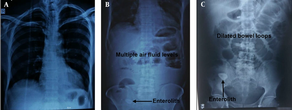
X-ray chest with dome of diaphragm 1A, showed no-gas-under-diaphragm, X-ray abdomen (erect and supine); 1B-C, showed dilated jejunum, proximal and mid ileum and multiple air-fluid levels. A radio-opaque-shadow (stony density) was seen in right iliac fossa (RIF). A, X-ray chest PA view; B, X-ray abdomen (erect); C, X-ray abdomen (supine).
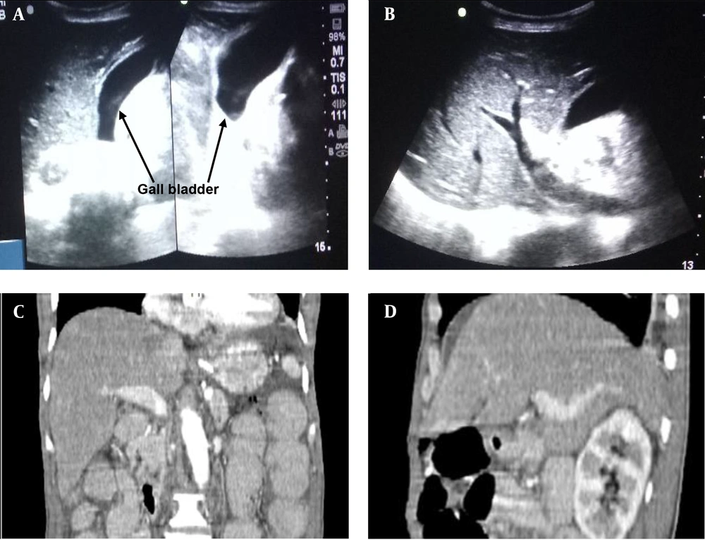
USG and CECT abdomen show normal hepatobiliary and portal anatomy without any evidence of obstruction, wall erosion or destruction, focal lesion or any bilioenteric fistula. These proved that the described stone in our case report was a primary enterolith and it did not originate from gall bladder; A, transabdominal USG showed empty GB lumen, normal CBD, normal portal vein and biliary radicals, no evidence of aerobilia or obstruction; B, transabdominal USG showed no direct or indirect evidence of bilio-enteric fistula (aerobilia), no evidence of erosion or destructions of wall; C, CECT abdomen (coronal plane) showed enlarged liver (17.5 cm). No evidence of any focal lesion was seen. GB distended. No intra luminal calcific density seen; D, CECT abdomen (sagittal plane) showed normal portal radicals and hepatic veins. Biliary radicals and CBD are not dilated. No aerobilia was observed.
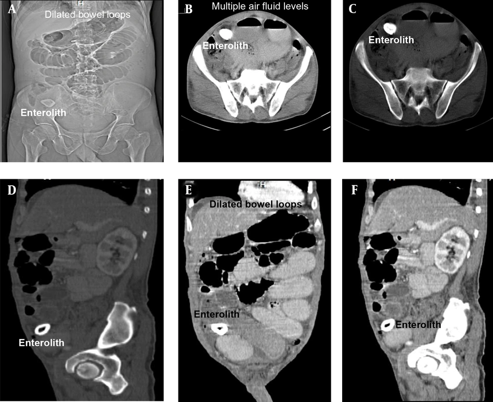
Images of CECT abdomen, showing small bowel obstruction with intra-luminal calculus in distal ileal loop right iliac fossa region in the zone of transition; A, scout film with stone in right-iliac-fossa; B, axial post-contrast CT: Intra-luminal stone with multiple air-fluid levels; C, axial post-contrast CT: Intra-luminal stone with multiple air-fluid levels; D, CECT abdomen (sagittal), showing enterolith with peripheral hyperdense calcification with hypodensity center; E, CECT abdomen (sagittal), showing dilated Jejunal, proximal and midileal loops (maximum caliber 4.5 cm); F, CECT abdomen (sagittal), showing collapsed distal ileal loops.
2.1. Management
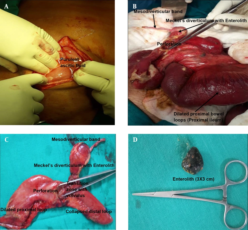
Intra-operative Images and specimen images showing enterolith in perforated Meckel’s diverticulum with mesodiverticular band. Meckel’s-diverticulum was found at 26 cm from ileocolic junction (unusual location). No bilio-enteric fistula was observed intra-operatively; A, purulent ascitic fluid on opening the peritoneum. The peritoneum was thickened; B, dilated proximal loops with perforation at the base of Meckel’s diverticulum with mesodiverticular band. Picture was taken after adhesiolysis and correction of volvulus; C, Meckel’s diverticulum with mesodiverticular band with perforation at its base; D, enterolith from Meckel’s diverticulum.
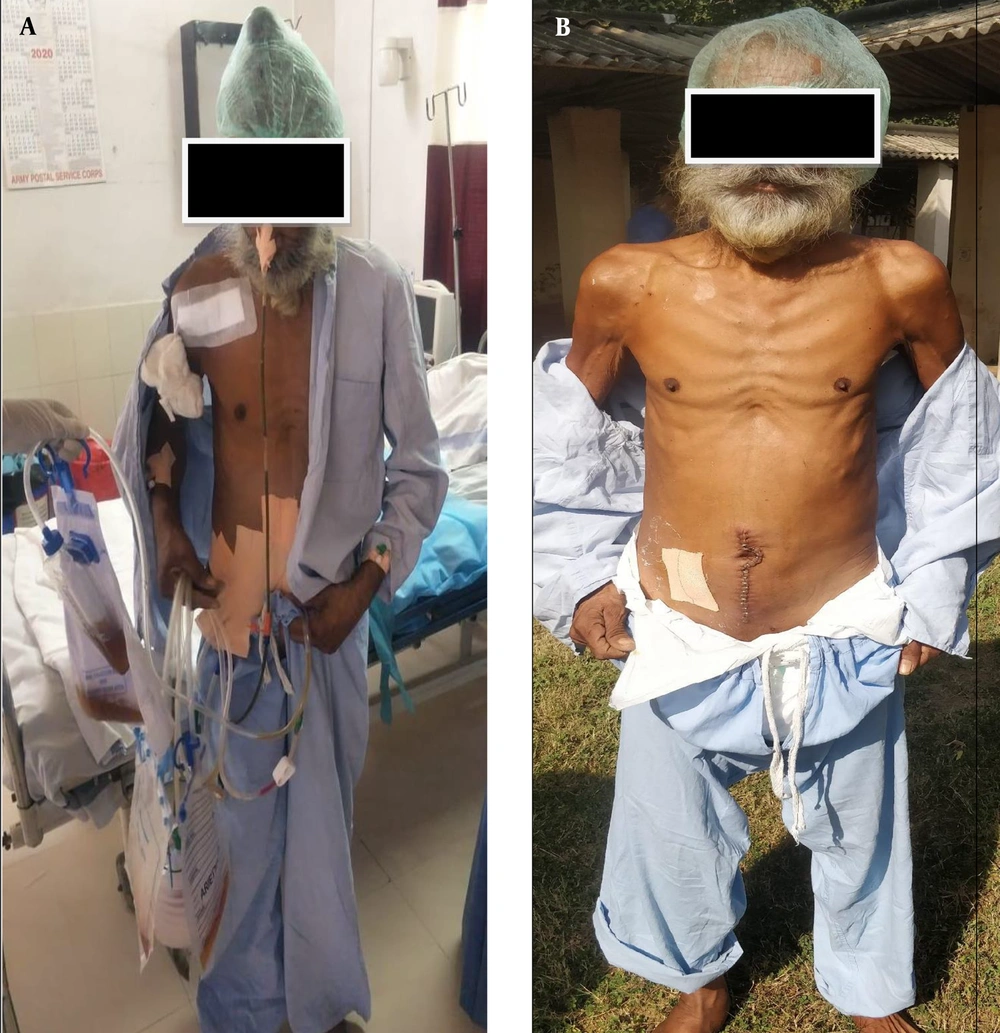
Postoperative images of the patient and wound. Postoperative period was uneventful; A, post-operation day 01. Patient started ambulation. Chest physiotherapy and incentive spirometer started; B, patient and wound on post-operation day 08. Wound was healthy and healing. Staples were in place.


