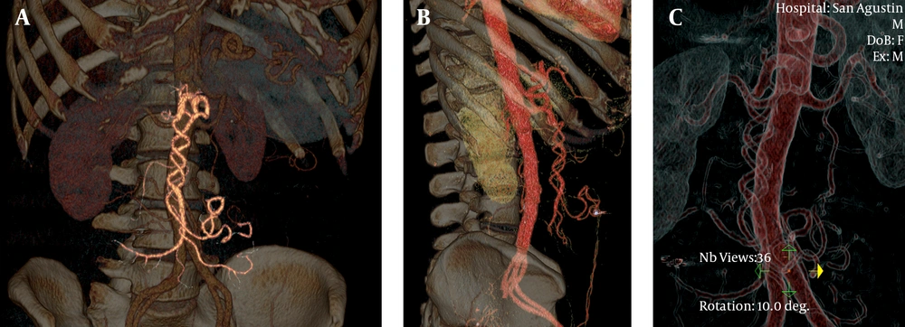
Midgut malrotation is a rare diagnosis, with a reported incidence lower than 0.2%, and is usually asymptomatic. When symptoms occur, normally present during the first year after birth, although a few of them may remain quiescent during childhood and appear in adult life, presenting with long-term unspecific abdominal complains such as pain, bloating and vomiting due to intermittent volvulation, a dynamic lesion with frequent spontaneous remissions and relapses. Less frequently, patients may present with acute bowel obstruction. Both its low incidence and unspecific symptoms make this pathologic entity rarely considered on clinical diagnosis. This explains often diagnostic delay, accounting for many years of abdominal complains before diagnosis (
4). Midgut malrotation is an embryological anomaly. From the fifth to the tenth week of fetal development, the embryonic gut undergoes rapid elongation exceeding the capacity of the abdominal cavity causing umbilical herniation. During this period, midgut goes through counter-clockwise rotation in a 270 degree, gradually returning to the abdominal cavity. Failure in this process can cause different degrees of anomalies in the location of small bowel, caecum, colon and appendix. Peritoneal fibrous bands fixing the duodenum, known as the Ladd´s bands, may persist and eventually compress the duodenum causing extrinsic obstruction. Malrotation results in malposition of the bowel and malfixation of the mesentery, resulting in a narrow pedicle at the mesenteric attachment, which facilitates volvulation.
Imaging techniques play an important role in the diagnosis of this pathologic condition. The actual widespread increase of diagnostic imaging studies may increase asymptomatic midgut malrotation diagnosis. In symptomatic patients, imaging techniques may reveal a non-suspected diagnosis, shortening the time gap between debut of symptoms and surgical treatment if required.
Diagnosis hints of midgut malrotation may be recognized by different imaging techniques. Plain abdominal radiography is often the first imaging study performed in these patients, but conventional radiography has low utility. Nevertheless, there are a few findings such as abnormal gas-bowel distribution, right sided jejunal loops and absent colon in the right lower quadrant, which may raise the suspicion of abdominal anomaly and lead to more specific imaging studies.
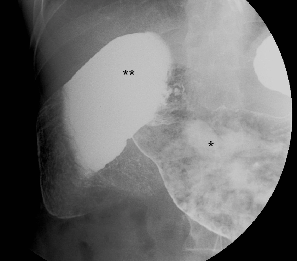
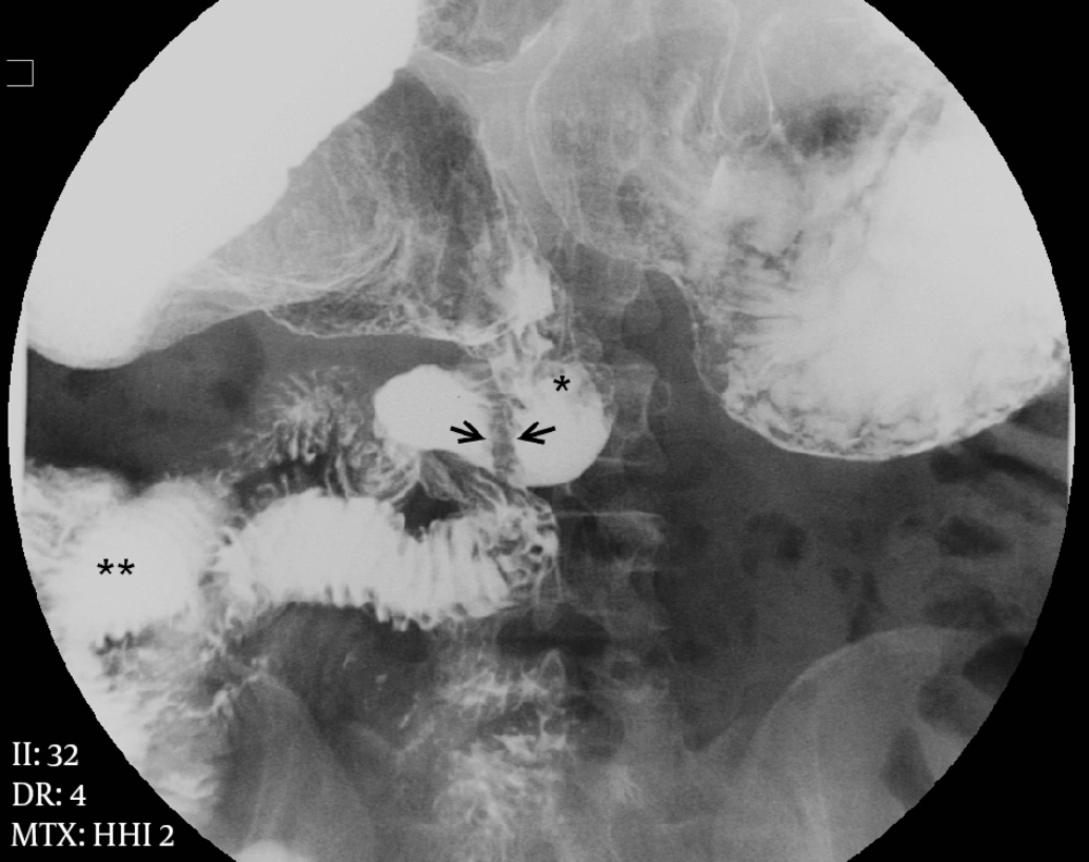
Upper gastrointestinal barium contrast study is highly sensitive for detection of midgut malrotation. It usually shows duodenum and duodenojejunal flexure in the right half of the abdomen, as well as abnormal location of jejunum loops. However, these studies have lower sensitivity for detecting associated complications and are not very specific; a normal barium study cannot rule out gut malrotation (
5).
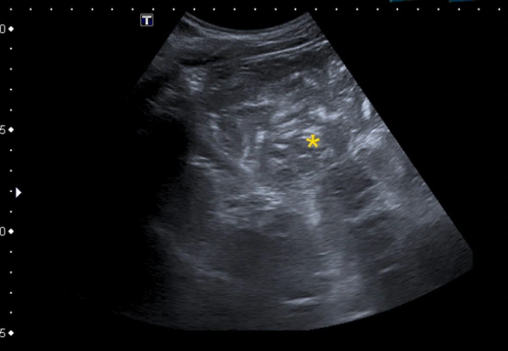
Ultrasound study, especially indicated in pediatric patients, may reveal abnormal location and caliber of bowel loops; in some cases a dilated duodenum with distal tapering is appreciable. Using color Doppler atypical situation of SMA may be shown as well as twisted mesentery around the SMA; these features are known as the “whirlpool” sign, however in adult patients, technical difficulties limit its use (
6).
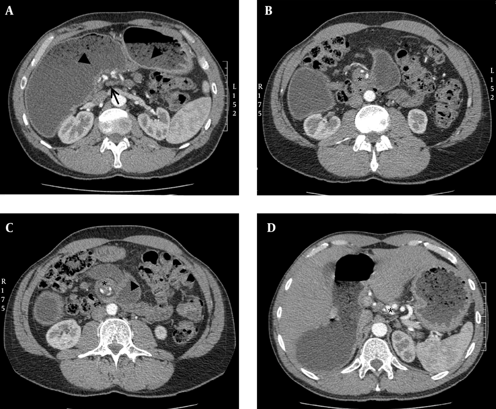
Nowadays, CT has been considered the most useful imaging technique for demonstrating adult midgut malrotation with or without volvulus; it can depict SMV in anomalous location posterior and left of the SMA (
7); this is a frequent finding but is not entirely diagnostic. Patients with malrotation may have a normal SMA-SMV orientation, and patients with abnormal SMA-SMV relationship may not have malrotation corresponding to normal variant (
8). In addition, abnormal disposition of midgut and duodenum not crossing the spine can be shown. In case of malrotation with volvulus, besides the above findings, CT can show small bowel and mesentery twisted around the narrow SMA pedicle showing the “whirl-pool sign”, first described by Fisher (
9). Also a range of abnormalities like dilatation of small intestine, circumferential bowel thickening, halo appearance, beak-like appearance or closed loops can be seen (
10). In specific circumstances, abdominal MRI may be performed (
11) revealing similar anatomical findings.
Mesenteric angiography has nowadays limited use in diagnostic management of suspected malrotation, because of invasive nature and high cost, but in the past, it was used to demonstrate abnormal mesenteric vessels, with typical corkscrew appearance of whirling SMA and its branches, showing the “barber pole sign” (
3). In addition to this finding, tapering or abrupt termination of mesenteric vessels, prolonged contrast transit time, absent venous opacification, or dilated SMV can be detected. Angiographic CT volume rendering reconstructions can depict similar findings helping in surgical approach (
12), although in articles previously cited, similar cases were reported, value of volume rendering reconstructions replacing diagnostic angiography is the outstanding point in this case report.
When symptomatic, midgut malrotation requires surgical intervention whether open or laparoscopic, habitually following the Ladd’s Procedure or any of its variations. Meanwhile, management of asymptomatic patients is controversial (
13); there are studies that reveal low risk of volvulus at follow-up (
14) as well as studies that recommend surgery to prevent catastrophic complications (
15). Opinion of patient, clinical condition and potential risks of the procedure weighted against benefits should be considered in each case.