
1. Context
Sepsis remains a leading cause of mortality among critically ill patients worldwide. Despite advances in antimicrobial therapy and intensive care management, it continues to impose a considerable burden on health care systems (1). Among its complications, sepsis-associated acute kidney injury (SA-AKI) is one of the most common and clinically significant manifestations, affecting approximately half of patients with severe sepsis and septic shock (2). The development of SA-AKI is associated with prolonged hospitalization, an increased need for renal replacement therapy, progression to chronic kidney disease, and markedly increased mortality (2, 3).
Unlike ischemic or toxic forms of acute kidney injury (AKI), SA-AKI arises from highly complex and dynamic interactions among systemic inflammation, microvascular dysfunction, endothelial injury, immune dysregulation, metabolic reprogramming, and mitochondrial impairment (3). Contemporary evidence suggests that renal dysfunction during sepsis cannot be explained solely by reductions in renal perfusion. Instead, excessive production of reactive oxygen species (ROS), mitochondrial damage, release of mitochondrial DNA, activation of innate immune signaling pathways, and multiple forms of regulated cell death—including apoptosis, ferroptosis, and pyroptosis—collectively contribute to progressive tubular injury and loss of renal function (3, 4). These interconnected processes create a self-amplifying cycle that perpetuates inflammation and cellular injury even after the initial infectious insult has been controlled (4).
Despite substantial progress in understanding the pathophysiology of SA-AKI, its management remains largely supportive. Current treatment strategies focus on infection control, hemodynamic optimization, fluid management, vasopressor therapy, and renal replacement therapy when indicated (4). However, no pharmacological intervention has consistently demonstrated efficacy in preventing or reversing kidney injury in patients with sepsis (3). Consequently, considerable attention has shifted toward agents capable of modulating multiple pathogenic pathways simultaneously rather than targeting a single molecular mechanism.
Melatonin (N-acetyl-5-methoxytryptamine) is an endogenous indoleamine synthesized primarily by the pineal gland, although substantial extrapineal production also occurs in numerous tissues, including the kidney (5). Beyond its established role in regulating circadian rhythms, melatonin has potent antioxidant, anti-inflammatory, immunomodulatory, and mitochondrial-protective properties (5). Unlike conventional antioxidants, which primarily scavenge reactive oxygen species, melatonin acts as a pleiotropic cytoprotective molecule capable of preserving mitochondrial function, enhancing endogenous antioxidant defenses, modulating innate immune responses, regulating mitophagy, and suppressing multiple forms of programmed cell death (5, 6). These diverse biological activities make melatonin an attractive therapeutic candidate for diseases characterized by complex and overlapping pathogenic mechanisms, including SA-AKI.
Recent experimental investigations have substantially expanded our understanding of the molecular mechanisms underlying melatonin-mediated renoprotection. In addition to reducing oxidative stress and inflammatory cytokine production, melatonin has been shown to enhance mitochondrial quality control through activation of the PTEN-induced kinase 1 (PINK1)/Parkin-mediated mitophagy pathway, preserve mitochondrial biogenesis via SIRT3-dependent deacetylation of mitochondrial transcription factor A (TFAM), inhibit cyclic GMP–AMP synthase–stimulator of interferon genes (cGAS–STING)-mediated innate immune activation, suppress NOD-like receptor protein 3 (NLRP3) inflammasome signaling, activate the nuclear factor erythroid 2-related factor 2 (Nrf2)/heme oxygenase-1 (HO-1) antioxidant pathway, and attenuate ferroptotic and apoptotic cell death (7-11). Collectively, these findings suggest that melatonin functions not merely as an antioxidant but also as a regulator of the interconnected molecular networks that drive septic kidney injury through its effects on mitochondrial quality control and innate immune signaling.
Although several recent studies have reported encouraging findings, the available evidence remains predominantly preclinical and varies considerably across animal models, experimental designs, treatment protocols, and mechanistic endpoints. Furthermore, the translational potential of melatonin has not been comprehensively evaluated.
Therefore, this narrative review critically examines current evidence on the renoprotective effects of melatonin in SA-AKI, integrates emerging mechanistic insights, discusses translational challenges, identifies limitations in the existing literature, and highlights future research priorities for successful clinical implementation.
2. Evidence Acquisition
2.1. Review Design
This structured narrative review was conducted to summarize current evidence on the renoprotective effects of melatonin and melatonergic agents in SA-AKI. In contrast to a systematic review, its objective was to provide an up-to-date overview of experimental and emerging clinical evidence, emphasizing underlying molecular mechanisms and translational relevance rather than quantitative evidence synthesis.
2.2. Literature Search
A structured, non-systematic literature search was performed in PubMed/MEDLINE and Google Scholar from database inception through January 31, 2026. Search terms combined keywords related to melatonin or melatonergic agents, sepsis, septic shock, and acute kidney injury. The PubMed search strategy included the following terms:
("melatonin"[Title/Abstract] OR "melatonergic"[Title/Abstract] OR "agomelatine"[Title/Abstract]) AND ("sepsis"[Title/Abstract] OR "septic shock"[Title/Abstract] OR "sepsis-associated"[Title/Abstract]) AND ("acute kidney injury"[Title/Abstract] OR "acute renal failure"[Title/Abstract] OR "renal injury"[Title/Abstract] OR "kidney injury"[Title/Abstract]).
Reference lists of relevant articles were also reviewed to identify additional eligible studies.
2.3. Study Selection
Original experimental studies investigating the effects of melatonin or melatonergic agents in animal or cellular models of SA-AKI were considered. Clinical studies evaluating melatonin in patients with sepsis-associated kidney injury were also included when such studies were available. Review articles, editorials, conference abstracts, duplicate publications, and studies unrelated to SA-AKI or melatonin were excluded. Studies using non-septic AKI models were considered only when they provided complementary mechanistic insights relevant to SA-AKI.
2.4. Data Synthesis
Relevant information from the selected studies was reviewed and synthesized narratively. Evidence was organized according to major research themes, including experimental evidence in SA-AKI models, molecular mechanisms of renoprotection, combination therapies, melatonergic analogues, and available clinical or translational findings. Because of substantial heterogeneity in study designs, experimental models, interventions, outcome measures, and mechanistic endpoints, no meta-analysis was performed.
2.5. Study Quality and Risk-of-Bias Assessment
The methodological quality and risk of bias of the included animal studies were assessed using the SYRCLE Risk of Bias tool (12). The assessed domains included random sequence generation, baseline comparability, allocation concealment, random housing, blinding of caregivers and investigators, random outcome assessment, blinding of outcome assessment, incomplete outcome data, selective outcome reporting, and other potential sources of bias. Each domain was classified as having a low, high, or unclear risk of bias based on the information reported in the original publication. Detailed assessments are provided in Table 1 in the Supplementary File.
3. Results
Ten reports were included in the primary SA-AKI evidence set. Of these, seven evaluated melatonin monotherapy, two evaluated melatonin-containing combination therapies, and one evaluated the melatonergic analogue agomelatine. The seven monotherapy studies constitute the direct melatonin evidence set. The two combination studies provide evidence regarding melatonin-containing adjunctive interventions but do not permit the independent effect of melatonin to be determined with certainty. The agomelatine study was considered indirect melatonergic evidence because agomelatine is a melatonergic analogue rather than native melatonin.
Studies involving nonseptic AKI models, including drug-induced nephrotoxicity, were excluded from the primary SA-AKI evidence count. These studies are discussed separately to provide mechanistic context and should not be interpreted as direct evidence of efficacy in SA-AKI. General reviews and mechanistic publications were used to support biological interpretation but were not counted as eligible primary studies (Table 1). Methodological reporting was incomplete across the included preclinical studies. Randomization, allocation concealment, blinding of investigators and outcome assessors, and justification of sample size were frequently unreported, resulting in an unclear risk of bias for several domains. The main concerns related to incomplete reporting of randomization procedures, allocation concealment, blinding, and selective outcome reporting. These limitations reduce confidence in the magnitude of the observed treatment effects, despite the generally consistent direction of the findings.
Table 1.
Primary Evidence Set of Studies Evaluating Melatonin or Melatonergic Agents in Sepsis-Associated Acute Kidney Injury a
| Author (Ref) | Type of Study | Animal Model | Sample Size | Duration (from Intervention to Endpoint; Minutes, Hours, and Days) | Intervention Type | Principal Findings | Proposed Mechanism | Overall Risk of Bias |
|---|---|---|---|---|---|---|---|---|
| Chen et al. (13) | Experimental animal study | CLP-induced SA-AKI in male Sprague-Dawley rats | 5 groups (Sham, CLP, CLP+Mel, CLP+A-ADMSC, CLP+Mel+A-ADMSC); n = 13 per group initially; mortality 0 - 46.2%; n = 7 for histological analyses | Melatonin administered at 30 min, 6 h, and 18 h after CLP; ADMSC administered at the same time points; outcomes assessed at 72 h after CLP | Melatonin + apoptotic adipose-derived mesenchymal stem cells | Improved renal histology, angiogenesis, and reduced fibrosis | Antioxidant, anti-inflammatory, and regenerative effects | Moderate to High |
| Dai et al. (9) | Experimental animal study | CLP-induced SA-AKI in male Sprague-Dawley rats | 3 groups (Control, Sepsis, Sepsis+Mel) | Melatonin administered 30 min before surgery, immediately after surgery, and 4 h after surgery; animals euthanized 8 h after CLP | Melatonin | Reduced renal dysfunction and apoptosis | PINK1/Parkin-mediated mitophagy | Moderate to High |
| Chen et al. (14) | Experimental animal study | CLP-induced SA-AKI in male C57BL/6 mice | 4 groups (Sham, Sham+Mel, CLP, CLP+Mel); n = 6 - 8 per group; total n ≈ 28 | Melatonin administered in drinking water for 14 days before CLP; mice euthanized 24 h after CLP | Melatonin | Improved renal function and histology | Reduced oxidative stress and inflammation | Moderate |
| Hamza et al. (15) | Experimental animal study | CLP-induced SA-AKI in male Swiss-albino mice | 4 groups (Sham, Sepsis, Vehicle, Mel); n = 6 per group; total n = 24 | Melatonin administered 30 min before CLP and 30 min, 4 h, and 8 h after CLP; animals euthanized at 8 h after CLP | Melatonin | Decreased cytokine production | Notch1/Jagged1 inhibition | Moderate |
| Qiu et al. (10) | Experimental animal study | Renal IR and FA-induced AKI in male C57BL/6 mice | IR: 5 groups (Sham, Sham+Mel, IR, IR+Mel, IR+Fer-1); n = 6 per group. FA: 5 groups (NC, NC+Mel, FA, FA+Mel, FA+Fer-1); n = 6 per group | IR model: melatonin administered 3 days and 1 h before IR; mice euthanized 24 h after reperfusion. FA model: melatonin administered 3 days before and 1 day after FA; mice euthanized 48 h after FA injection | Melatonin | Reduced ferroptosis | Activation of Nrf2/HO-1 | Moderate |
| Üstündağ et al. (16) | Experimental animal study | CLP-induced sepsis (heart and kidney injury) in male rats | 5 groups (Sham, CLP, Mel, ASA, Mel+ASA) | Melatonin and/or ascorbic acid administered after CLP; animals euthanized at endpoint | Melatonin + vitamin C | Synergistic protection | SIRT1 activation | Moderate to High |
| Deng et al. (8) | Experimental animal study + clinical cohort | CLP-induced SA-AKI in male C57BL/6 mice + septic patients | Animal: 4 groups (Control, CLP, CLP+Mel, CLP+3-TYP, CLP+Chloroquine); n = 10 per group for survival study. Clinical: 48 septic patients included | Mice: melatonin/3-TYP/chloroquine administered for 3 days before CLP; mice euthanized 24 h after CLP. Humans: blood/urine collected within 24 h after sepsis diagnosis | Melatonin | Enhanced mitophagy | SIRT3-TFAM signaling | Moderate |
| Başol et al. (17) | Experimental animal study | CLP-induced SA-AKI in male Sprague-Dawley rats | 4 groups (Normal, Sham, CLP+Saline, CLP+Agomelatine); n = 8 per group; 10 rats died in first 24 h and were excluded | Agomelatine administered within the first hour after surgery; study concluded after 24 h | Melatonergic analogue | Improved renal injury | Kynurenine pathway modulation | Moderate to High |
| Cao et al. (7) | Experimental animal study | LPS-induced SA-AKI in male C57BL/6 mice | 4 groups (Control, LPS, LPS+H-151, LPS+Mel) | Melatonin administered 3 h before and 6 and 12 h after LPS; mice euthanized 24 h after LPS injection | Melatonin | Reduced mitochondrial DNA release | cGAS-STING inhibition | Moderate |
| Potić et al. (18) | Experimental animal study | LPS-induced SA-AKI in male Wistar rats | 4 groups (Control, Mel, LPS, LPS+Mel); n = 6 per group; total n = 24 | Single melatonin dose 12 h before LPS; animals were euthanized 12 h after LPS administration | Melatonin | Reduced oxidative stress and cytokines | TLR4/NF-κB inhibition | Low to Moderate |
a Abbreviations: 3-TYP, 3-(trifluoromethyl)-N-(4-pyridinyl)benzamide (SIRT3 activator); ADMSC, Adipose-derived mesenchymal stem cells; A-ADMSC, apoptotic adipose-derived mesenchymal stem cells; AKI, acute kidney injury; CLP, Cecal ligation and puncture; FA, Folic acid; HO-1, heme oxygenase-1; IR, Ischemia-reperfusion; LPS, Lipopolysaccharide; Mel, Melatonin; SA-AKI, Sepsis-associated acute kidney injury; Sham, Sham-operated control group; Vehicle, Control solution (e.g., saline, ethanol in saline); ASA, ascorbic acid.
3.1. Improvement in Renal Function
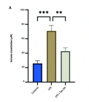
Most studies in the primary evidence set reported improved functional or structural renal outcomes following melatonin-containing interventions. However, because the evidence set also included combination therapies and one melatonergic analogue, the findings should not be interpreted as evidence of an identical effect attributable exclusively to native melatonin. Compared with untreated septic animals, melatonin consistently reduced serum creatinine and blood urea nitrogen concentrations, reflecting attenuation of renal dysfunction (7-9, 13-18). Several studies also reported restoration of electrolyte balance, including normalization of sodium and potassium concentrations (18). These functional improvements closely paralleled reductions in structural kidney injury.
3.2. Preservation of Renal Histology
Histopathological examination consistently demonstrated attenuation of tubular epithelial degeneration, tubular dilatation, glomerular injury, inflammatory cell infiltration, interstitial edema, hemorrhage, tubular cast formation, and renal fibrosis in melatonin-treated animals (7-9, 14, 15, 18). Ultrastructural analyses further revealed preservation of mitochondrial morphology, including reduced mitochondrial swelling and maintenance of cristae architecture, suggesting improved mitochondrial integrity (7, 8).
3.3. Suppression of Oxidative Stress
Reduced oxidative stress was one of the most reproducible findings across all included studies. Melatonin significantly decreased renal concentrations of malondialdehyde (MDA), thiobarbituric acid-reactive substances, advanced oxidation protein products, and ROS. Simultaneously, endogenous antioxidant defenses—including superoxide dismutase, catalase, glutathione peroxidase, glutathione reductase, HO-1, and NAD(P)H quinone oxidoreductase-1 (NQO1)—were consistently restored, indicating the reestablishment of intracellular redox homeostasis (10, 13, 14, 17, 18).
3.4. Attenuation of Inflammatory Responses
Melatonin markedly reduced both systemic and renal inflammatory responses. Most studies reported significant decreases in TNF-α, IL-1β, and IL-6, whereas one investigation observed increased expression of the anti-inflammatory cytokine IL-10, suggesting restoration of immune homeostasis rather than simple suppression of inflammation. Inhibition of nuclear factor kappa B (NF-κB) activation was consistently reported, accompanied by reductions in multiple downstream inflammatory mediators, including VEGF, MCP-1, TNFR1, MMP-9, RANTES, and MIP-1α (7, 9, 13, 15, 16, 18). These molecular changes were associated with reduced inflammatory cell infiltration and lower histological inflammation scores.
3.5. Regulation of Mitochondrial Quality Control
Recent investigations have identified mitochondrial preservation as a central mechanism underlying melatonin-mediated renoprotection. Multiple studies demonstrated activation of mitophagy through the PINK1/Parkin pathway and SIRT3-mediated TFAM deacetylation, resulting in enhanced clearance of dysfunctional mitochondria and improved mitochondrial quality control. Increased expression of PINK1/Parkin and LC3-II, together with reduced SQSTM1/p62 accumulation, indicated restoration of mitophagic flux. Consequently, mitochondrial DNA leakage into the cytoplasm was reduced, thereby inhibiting cGAS–STING signaling and downstream activation of IRF3 and NF-κB (7, 8).
3.6. Modulation of Regulated Cell Death
Melatonin significantly attenuated apoptosis, as indicated by reductions in TUNEL-positive cells, Bax expression, caspase-3 activation, caspase-9 activation, PARP cleavage, DNase activity, and DNA fragmentation. More recent studies further demonstrated inhibition of ferroptosis through activation of the Nrf2/HO-1 signaling pathway and suppression of NLRP3 inflammasome activation, indicating that melatonin modulates multiple forms of regulated cell death rather than apoptosis alone (7, 9, 10, 13, 18).
3.7. Combination Therapy
Two studies evaluated combination therapeutic strategies. Co-administration of melatonin with apoptotic adipose-derived mesenchymal stem cells produced greater reductions in oxidative stress, inflammation, fibrosis, and apoptosis than either intervention alone, while enhancing angiogenesis and preserving renal architecture (13). Similarly, combination therapy with melatonin and ascorbic acid synergistically enhanced antioxidant capacity, increased SIRT1 expression, and further improved biochemical and histopathological outcomes (16).
Collectively, the available evidence shows consistent findings across diverse experimental models. Rather than acting through a single molecular pathway, melatonin appears to interrupt multiple interconnected pathogenic processes, including mitochondrial dysfunction, oxidative stress, inflammatory signaling, inflammasome activation, ferroptosis, apoptosis, and tissue remodeling, ultimately preserving renal structure and function during sepsis.
3.8. Discussion
The findings synthesized in this review provide encouraging preclinical evidence for the biological plausibility of melatonin as a multi-target therapeutic candidate for SA-AKI. Although the included studies differed substantially in animal species, experimental models, treatment schedules, and dosing regimens, their conclusions were consistent. Rather than acting through a single signaling pathway, melatonin appears to interrupt the self-perpetuating cycle linking mitochondrial dysfunction, oxidative stress, innate immune activation, regulated cell death, and progressive tubular injury. However, the methodological quality of the included studies varied considerably. Reporting of randomization, allocation concealment, blinded outcome assessment, and sample-size calculation was frequently incomplete, limiting confidence in the estimated treatment effects.
The apparent consistency of the reported protective effects should therefore be interpreted in the context of incomplete methodological reporting. In particular, the frequent absence of information on allocation concealment, blinded outcome assessment, sample-size calculation, and attrition limits the ability to exclude performance, detection, and reporting bias. Accordingly, the current evidence supports biological plausibility and provides a rationale for further investigation, but it does not establish the efficacy of melatonin in clinical SA-AKI.
Among the molecular mechanisms identified, enhancement of mitochondrial quality control through activation of PINK1/Parkin-dependent mitophagy represents one of the most compelling findings (7, 9). Efficient removal of dysfunctional mitochondria reduces ROS generation, preserves ATP synthesis, limits mitochondrial DNA release, and interrupts activation of the cGAS–STING signaling cascade. Recent studies demonstrating SIRT3-mediated TFAM deacetylation further support the concept that melatonin promotes mitochondrial biogenesis and quality control rather than functioning solely as an antioxidant (7-9). These observations place mitochondrial preservation at the center of melatonin-mediated renoprotection and provide a unifying explanation for its antioxidant and anti-inflammatory actions.
This system-level mechanism distinguishes melatonin from many conventional antioxidant therapies. Although excessive ROS generation has long been considered a hallmark of septic organ injury, increasing evidence suggests that oxidative stress is not an isolated pathological event but rather a consequence of impaired mitochondrial quality control (19, 20). Damaged mitochondria generate excessive ROS, release mitochondrial DNA into the cytoplasm, and activate innate immune pathways that further amplify inflammation and cellular injury (19, 20). Consequently, therapies directed solely at free-radical scavenging have produced disappointing clinical results because they fail to correct the underlying mitochondrial dysfunction. In contrast, melatonin directly scavenges reactive oxygen and nitrogen species. It also preserves mitochondrial integrity, enhances endogenous antioxidant defenses, and restores mitochondrial homeostasis, thereby addressing the upstream drivers of oxidative injury (21).
Inflammation constitutes another major therapeutic target in SA-AKI. Activation of NF-κB signaling and excessive production of pro-inflammatory cytokines amplify endothelial dysfunction, leukocyte recruitment, vascular leakage, and tubular injury (18). Across virtually all included studies, melatonin consistently reduced TNF-α, IL-1β, IL-6, and multiple downstream inflammatory mediators, while increasing IL-10 in selected models (7, 9, 13, 15, 16, 18). Importantly, inhibition of cGAS–STING and NLRP3 inflammasome signaling suggests that melatonin not only suppresses cytokine production but also interrupts upstream innate immune activation triggered by mitochondrial injury (7, 9, 18). This integrated regulation of inflammatory pathways may explain why melatonin demonstrates protective effects across multiple organ systems during sepsis.
Recent evidence also indicates that melatonin regulates several forms of programmed cell death (22). Beyond classical apoptosis, ferroptosis has emerged as an important contributor to septic AKI because of its dependence on iron-mediated lipid peroxidation and oxidative stress (23). Activation of the Nrf2/HO-1 pathway by melatonin appears to limit ferroptotic cell death while simultaneously enhancing endogenous antioxidant defenses. Together with consistent reductions in caspase activation and DNA fragmentation, these findings suggest that melatonin broadly suppresses regulated cell death, regardless of the initiating pathway (10, 24). Such pleiotropic effects are particularly relevant in sepsis, where multiple cell death programs frequently coexist and interact.
An important observation emerging from this review is that melatonin should not be regarded merely as an antioxidant supplement, but rather as a system-level regulator of mitochondrial function, redox homeostasis, innate immunity, and cellular survival (25). The additive benefits observed when melatonin is combined with mesenchymal stem cells or ascorbic acid further support its potential role as an adjunctive therapy within multimodal treatment strategies (13, 16).
Although they fall outside the predefined evidence set, studies conducted in non-septic AKI models, such as cisplatin- and vancomycin-induced injury, provide complementary mechanistic evidence supporting the biological plausibility of melatonin-mediated renoprotection. In cisplatin-treated animals, melatonin lowers blood urea nitrogen and creatinine, restores renal glutathione, attenuates lipid peroxidation, and preserves proximal tubular architecture. These effects have been mechanistically linked to direct ROS scavenging, upregulation of endogenous antioxidant defenses, inhibition of caspase-3/PARP-1–dependent apoptosis, and preservation of mitochondrial energy homeostasis via PPARα-mediated fatty acid oxidation (16). For vancomycin, preclinical data and at least one retrospective human cohort similarly indicate reduced oxidative stress, less tubular injury, and a substantially lower incidence of AKI with concomitant melatonin, again pointing to its antioxidant and anti-inflammatory actions (26). However, these findings should not be considered equivalent to direct evidence in SA-AKI because the initiating injury, inflammatory environment, treatment context, and clinical pathophysiology differ substantially.
An important barrier to clinical translation is the substantial gap between experimental and clinically used melatonin doses. In the included animal studies, melatonin was administered at doses ranging from approximately 20 to 150 mg/kg, which are several orders of magnitude higher than those commonly used in humans for sleep disorders or circadian indications. Allometric scaling suggests that these rodent doses may correspond to very high human-equivalent doses, raising concerns about feasibility, safety, and tolerability in critically ill patients. In addition, melatonin undergoes extensive first-pass metabolism, has a relatively short plasma half-life, and exhibits considerable interindividual variability in absorption and clearance. These pharmacokinetic characteristics may be further altered during sepsis by changes in hepatic blood flow, cytochrome P450 activity, protein binding, renal function, and concomitant medications. Consequently, it is uncertain whether clinically achievable plasma and renal tissue concentrations would be sufficient to reproduce the protective effects observed in experimental models. These considerations highlight the need for carefully designed dose-finding and pharmacokinetic studies in patients with sepsis and AKI. Such studies should evaluate multiple dose levels, administration routes, and dosing schedules and incorporate measurements of plasma and, where feasible, urine melatonin concentrations alongside biomarkers of mitochondrial injury, oxidative stress, and tubular damage. Until these data are available, the high doses used in animal models should not be directly extrapolated to clinical practice.
Despite these promising findings, several important limitations hinder clinical translation. First, nearly all available evidence derives from experimental animal models, with only limited observational clinical data. Second, substantial heterogeneity in animal species, methods of sepsis induction, melatonin dosage, timing of administration, and treatment duration prevents identification of an optimal therapeutic strategy. Third, most experimental studies administered melatonin before or immediately after sepsis induction, conditions that may not accurately reflect routine clinical practice, in which treatment typically begins after organ dysfunction is established. Furthermore, relatively few investigations have evaluated aged or female animals or models incorporating common comorbidities such as diabetes or chronic kidney disease, thereby limiting external validity. Finally, reporting of randomization, allocation concealment, and blinded outcome assessment remains inconsistent across preclinical studies, increasing the risk of methodological bias.
Future research should prioritize well-designed translational investigations to define clinically achievable dosing regimens, optimal therapeutic windows, pharmacokinetic characteristics, and long-term renal outcomes. Randomized controlled trials evaluating melatonin as an adjunct to current standard-of-care therapy are urgently needed. Biomarker-guided approaches incorporating markers of mitochondrial injury, oxidative stress, and tubular damage may further facilitate patient selection and treatment monitoring. Additionally, exploration of chronotherapeutic strategies may be particularly valuable given melatonin’s physiological role in circadian regulation and the growing recognition that circadian disruption contributes to immune dysregulation in critically ill patients.
Based on the available preclinical evidence, a pragmatic phase II/III randomized controlled trial could be considered to evaluate melatonin as an adjunctive therapy in SA-AKI. The target population would comprise adult patients with sepsis or septic shock who meet consensus criteria for AKI or are at high risk of developing AKI within the first 24 - 48 hours of intensive care unit admission. Key exclusion criteria might include severe hepatic impairment, known hypersensitivity to melatonin, pregnancy, and concurrent use of investigational agents with overlapping mechanisms. The intervention could involve enteral or intravenous melatonin administered at predefined dose levels, with dose escalation guided by safety, tolerability, and preliminary pharmacokinetic data. The comparator would be standard care plus placebo. Stratification factors could include baseline severity of illness, presence of shock, need for vasopressors, baseline kidney function, and timing of AKI onset. Primary endpoints might include change in serum creatinine over 7 days, incidence and duration of AKI, and need for renal replacement therapy. Secondary endpoints could include mortality, ventilator-free days, vasopressor-free days, length of stay, and safety outcomes. Mechanistic biomarkers of mitochondrial injury, oxidative stress, inflammation, and tubular damage could be collected to explore dose–response relationships and identify potential responders. Such a trial would provide essential data on feasibility, safety, pharmacokinetics, and preliminary efficacy before larger confirmatory studies are undertaken.
Overall, current evidence suggests that melatonin is a promising investigational therapy with consistent preclinical renoprotective effects. However, definitive conclusions regarding clinical efficacy and safety cannot be drawn until adequately powered human studies are available. By simultaneously preserving mitochondrial function, restoring redox balance, suppressing innate immune activation, limiting regulated cell death, and maintaining renal structural integrity, melatonin interrupts the vicious cycle driving septic kidney injury. Although further clinical validation is required, these mechanistic insights provide a rationale for continued translational investigation and support the potential incorporation of melatonin into future multimodal therapeutic strategies for sepsis-associated acute kidney injury. The proposed mechanisms underlying melatonin’s potential renoprotective effects in sepsis-associated acute kidney injury, based predominantly on preclinical evidence, are summarized in Figure 1.
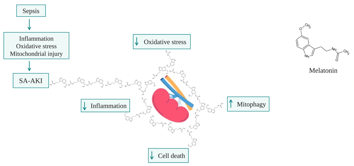
Figure 1.
Proposed mechanisms underlying the potential renoprotective effects of melatonin in sepsis-associated acute kidney injury. Melatonin may attenuate mitochondrial dysfunction, oxidative stress, innate immune activation, inflammation, ferroptosis, and apoptosis while enhancing mitochondrial quality control and antioxidant defenses. These mechanisms may collectively preserve renal tubular structure and function. The proposed effects are based predominantly on preclinical evidence and should not be interpreted as evidence of established clinical efficacy.
4. Conclusions
Preclinical studies consistently suggest that melatonin may attenuate multiple interconnected mechanisms involved in sepsis-associated acute kidney injury, including mitochondrial dysfunction, oxidative stress, innate immune activation, and regulated cell death. However, the current evidence is derived predominantly from animal studies using high doses and heterogeneous designs, and robust clinical data are lacking.
Future research should prioritize well-designed dose-finding and pharmacokinetic studies in patients with sepsis and AKI, followed by randomized controlled trials evaluating clinically relevant renal and survival outcomes. Biomarker-guided approaches and chronotherapeutic strategies may further refine patient selection and treatment timing. Until such evidence is available, melatonin should be regarded as a promising investigational therapy rather than an established treatment for SA-AKI.
Abbreviations: AKI: acute kidney injury; SA-AKI: Sepsis-associated acute kidney injury; ROS: reactive oxygen species; mtDNA: mitochondrial DNA; Nrf2: nuclear factor erythroid 2-related factor 2; HO-1: heme oxygenase-1; NF-κB: nuclear factor kappa B; NLRP3: NOD-like receptor protein 3; cGAS–STING: cyclic GMP–AMP synthase–stimulator of interferon genes; PINK1: PTEN-induced kinase 1; TFAM: mitochondrial transcription factor A.