
1. Background
Pressure ulcers, also referred to as pressure injuries, are localized areas of damage to the skin and underlying soft tissue that typically occur over bony prominences as a result of sustained mechanical loading. These lesions represent a significant clinical challenge in ICUs, where patients are particularly vulnerable because of prolonged immobility, hemodynamic instability, malnutrition, and impaired tissue perfusion (1). The prevalence of pressure ulcers among critically ill patients varies widely across studies, with reported rates ranging from 12% to more than 40%, depending on the patient population and assessment methodology (2).
The pathophysiology of pressure ulcer development involves a complex interplay of ischemia-reperfusion injury, sustained tissue deformation, and inflammatory cascades that ultimately lead to cell death and tissue necrosis (3). Once established, pressure ulcers are associated with substantial morbidity, including an increased risk of nosocomial infections, prolonged hospitalization, and higher health care costs (4). Furthermore, pressure ulcers in ICU patients have been independently associated with increased mortality, with adjusted odds ratios ranging from 2.0 to 4.0 in large observational cohorts (5).
Current standard-of-care strategies for pressure ulcer management focus primarily on pressure redistribution, moisture management, and local wound care. Despite adherence to evidence-based prevention bundles, a considerable proportion of ICU patients develop new pressure injuries or experience delayed healing of existing wounds (6). This therapeutic gap has prompted investigations into adjunctive nutritional interventions that may accelerate wound repair by providing specific substrates required for collagen synthesis and tissue remodeling (7).
Collagen is the most abundant structural protein in the extracellular matrix and plays a central role in all phases of wound healing, from hemostasis through proliferation and remodeling (8). Oral supplementation with hydrolyzed collagen peptides has been shown to increase circulating levels of hydroxyproline-containing dipeptides, which serve as both building blocks and bioactive signaling molecules that stimulate fibroblast proliferation and extracellular matrix deposition (9). Clinical studies in surgical and chronic wound populations have demonstrated that oral collagen supplementation can improve wound healing outcomes, including faster reduction in wound surface area and enhanced granulation tissue formation (10).
Bromelain is a mixture of cysteine proteases derived from the stem and fruit of Ananas comosus (pineapple), with well-documented anti-inflammatory, antiedematous, and fibrinolytic properties (11). The enzymatic debridement activity of bromelain facilitates the removal of necrotic tissue from the wound bed, thereby promoting a cleaner wound environment conducive to healing (12). In addition, bromelain has been shown to modulate inflammatory cytokine expression, reduce neutrophil migration to sites of inflammation, and enhance the absorption of coadministered therapeutic agents (13).
The biological rationale for combining collagen and bromelain is based on their complementary mechanisms: collagen provides the structural substrate necessary for tissue regeneration, whereas bromelain reduces inflammatory burden and facilitates debridement, potentially creating a synergistic effect on wound healing (14). Although each agent has been studied individually in various wound types, evidence regarding their combined use in critically ill patients with pressure ulcers remains limited (15).
To date, most clinical trials evaluating nutritional supplementation for pressure ulcer healing have been conducted in long-term care or community settings, and data specific to the ICU population, in which healing is often impaired by systemic inflammation, catabolism, and hemodynamic compromise, are scarce (16). Given the high prevalence, substantial morbidity, and limited adjunctive treatment options for pressure ulcers in critically ill patients, well-designed randomized controlled trials evaluating novel nutritional interventions in this population are needed (17).
2. Objectives
The present study aimed to evaluate the efficacy of combined oral collagen hydrolysate and bromelain supplementation, compared with placebo, on pressure ulcer healing outcomes in ICU patients over a 21-day follow-up period.
3. Methods
3.1. Study Design and Setting
This randomized, triple-blind, placebo-controlled, parallel-group clinical trial was conducted in the ICU at Labbafinejad Hospital, a tertiary referral center affiliated with Shahid Beheshti University of Medical Sciences, Tehran, Iran. The study was conducted from October 2024 to December 2025. The trial was prospectively registered in the Iranian Registry of Clinical Trials (IRCT20190215042716N7) on 2024 - 10 - 17, before enrollment of the first participant, and was approved by the Ethics Committee of Shahid Beheshti University of Medical Sciences (IR.SBMU.MSP.REC.1403.385; approved 2024 - 10 - 08). Written informed consent was obtained from patients or their legal guardians before enrollment.
4. Results
4.1. Participant Flow and Baseline Characteristics
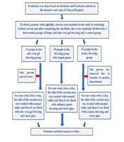
A total of 70 patients were assessed for eligibility; 10 were excluded before randomization (6 died before the intervention started, and 4 could not tolerate oral feeding). Sixty patients were randomized (30 per group), and all were included in the intention-to-treat analysis (Figure 1). The 2 groups were comparable with respect to age, sex, diabetes, APACHE II score, Braden Scale score, and BMI (all P > 0.05) (Table 1). However, statistically significant baseline differences were observed: hemoglobin was higher in the intervention group (12.0 ± 1.9 vs 10.5 ± 1.8 g/dL; P = 0.002), and albumin was also higher (3.1 ± 0.5 vs 2.7 ± 0.5 g/dL; P = 0.001). These imbalances were addressed in the post hoc adjusted ANCOVA.
Table 1.
Baseline Demographic and Clinical Characteristics of Participants a
| Characteristics | Intervention (n = 30) | Placebo (n = 30) | P-Value |
|---|---|---|---|
| Age, y | 62.4 ± 14.2 | 64.1 ± 13.8 | 0.627 |
| Male sex | 18 (60.0) | 17 (56.7) | 0.793 |
| APACHE II score | 22.7 ± 6.1 | 23.2 ± 5.8 | 0.742 |
| BMI, kg/m2 | 24.1 ± 4.2 | 23.8 ± 4.5 | 0.780 |
| Diabetes mellitus | 11 (36.7) | 12 (40.0) | 0.793 |
| Hemoglobin, g/dL c | 12.0 ± 1.9 | 10.5 ± 1.8 | 0.002 b |
| Albumin, g/dL c | 3.1 ± 0.5 | 2.7 ± 0.5 | 0.001 b |
| Braden Scale score | 10.2 ± 2.1 | 10.5 ± 2.3 | 0.571 |
| Pressure ulcer stage (II/III/IV) | 12/11/7 | 13/10/7 | 0.946 |
a Values are expressed as mean ± SD or No. (%).
b P < 0.05 was considered statistically significant.
c Baseline imbalances were addressed in the post hoc ANCOVA.
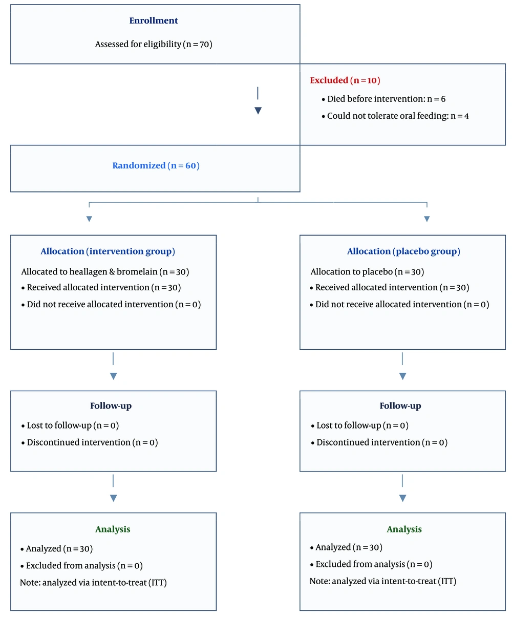
Figure 1.
CONSORT flow diagram
4.2. Changes in Wound Depth Over Time
At baseline (day 1), mean wound depth was similar between groups (27.3 ± 10.3 vs 26.6 ± 10.5 mm; P = 0.792). By day 6, a nonsignificant trend favored the intervention group (17.7 ± 10.2 vs 22.4 ± 10.4 mm; P = 0.055). From day 11 onward, wound depth was significantly lower in the intervention group: day 11, 10.7 ± 6.6 vs 17.4 ± 10.4 mm (P = 0.005); day 16, 4.7 ± 4.1 vs 8.5 ± 5.1 mm (P = 0.002); and day 21, 1.8 ± 1.6 vs 4.3 ± 3.8 mm (P = 0.001) (Table 2).
Table 2.
Comparison of Wound Depth Between Groups Over Time a
| Days | Intervention | Placebo | P-Value (Mann-Whitney U) |
|---|---|---|---|
| Day 1 | 27.3 ± 10.3 | 26.6 ± 10.5 | 0.792 |
| Day 6 | 17.7 ± 10.2 | 22.4 ± 10.4 | 0.055 |
| Day 11 | 10.7 ± 6.6 | 17.4 ± 10.4 | 0.005 b |
| Day 16 | 4.7 ± 4.1 | 8.5 ± 5.1 | 0.002 b |
| Day 21 | 1.8 ± 1.6 | 4.3 ± 3.8 | 0.001 b |
| Change from day 1 to day 21 | -25.5 ± 10.0 | -22.3 ± 10.2 | 0.001 b |
| Between-group difference (95% CI) | -3.2 mm (-8.1 to -1.7) | ||
| Post hoc ANCOVA adjusted for hemoglobin and albumin | B = -0.64 (95% CI, -0.97 to -0.30; P = 0.001) | ||
a Values are expressed as mean ± SD unless otherwise indicated. Between-group differences were tested using the Mann-Whitney U test. B is the regression coefficient representing the adjusted treatment effect estimated by ANCOVA after controlling for baseline hemoglobin and albumin.
b P < 0.05 was considered statistically significant.
The unadjusted mean reduction in wound depth from day 1 to day 21 was 25.5 ± 10.0 mm in the intervention group and 22.3 ± 10.2 mm in the placebo group; the between-group difference was -3.2 mm (95% CI, -8.1 to -1.7; P = 0.001). In the post hoc ANCOVA adjusted for baseline hemoglobin and albumin, the intervention remained significantly associated with a greater wound depth reduction than placebo (B = -0.64; 95% CI, -0.97 to -0.30; P = 0.001).
4.3. Changes in Wound Surface Area Over Time
Both groups showed a gradual reduction in wound surface area from day 1 to day 21, but no statistically significant between-group differences were observed at any time point (Table 3). On day 21, mean surface area was 1.7 ± 1.1 cm2 in the intervention group and 2.0 ± 1.1 cm2 in the placebo group; the between-group difference was -0.3 cm2 (95% CI, -0.8 to 0.2; P = 0.168). Repeated-measures analysis confirmed no significant group × time interaction for surface area.
Table 3.
Comparison of Wound Surface Area Between Groups Over Time a
| Days | Intervention | Placebo | P-Value (Mann-Whitney U) |
|---|---|---|---|
| Day 1 | 7.6 ± 1.8 | 6.8 ± 1.8 | 0.089 |
| Day 6 | 6.1 ± 1.7 | 5.5 ± 1.8 | 0.192 |
| Day 11 | 4.2 ± 1.5 | 4.3 ± 1.7 | 0.841 |
| Day 16 | 2.8 ± 1.2 | 3.2 ± 1.4 | 0.312 |
| Day 21 | 1.7 ± 1.1 | 2.0 ± 1.1 | 0.168 |
| Change from day 1 to day 21 | -5.8 ± 2.9 | -5.2 ± 3.1 | 0.168 |
| Between-group difference (95% CI) | -0.3 cm2 (-0.8 to 0.2); P = 0.168 | ||
a Values are expressed as mean ± SD. No significant between-group differences were observed at any time point.
4.4. Within-Group Changes in Wound Depth and Surface Area
In both groups, wound depth and surface area decreased significantly between each pair of consecutive time points (Wilcoxon signed-rank test, all P < 0.001), confirming progressive healing throughout the 21-day follow-up (Table 4).
Table 4.
Comparison of Mean Wound Depth and Surface Area Between Consecutive Time Points by Group
| Groups and Items and Time Differences | Mean Difference | P-Value |
|---|---|---|
| Heallagen and bromelain | ||
| Depth | ||
| Day 1 - 6 | -2.9 | < 0.001 |
| Day 6 - 11 | -5.9 | < 0.001 |
| Day 11 - 16 | -7.0 | < 0.001 |
| Day 16 - 21 | -9.6 | < 0.001 |
| Surface area | ||
| Day 1 - 6 | -1.5 | < 0.001 |
| Day 6 - 11 | -1.9 | < 0.001 |
| Day 11 - 16 | -1.4 | < 0.001 |
| Day 16 - 21 | -1.0 | < 0.001 |
| Placebo | ||
| Depth | ||
| Day 1 - 6 | -4.2 | 0.014 |
| Day 6 - 11 | -8.9 | 0.004 |
| Day 11 - 16 | -5.0 | < 0.001 |
| Day 16 - 21 | -4.2 | < 0.001 |
| Surface area | ||
| Day 1 - 6 | -1.3 | < 0.001 |
| Day 6 - 11 | -1.2 | < 0.001 |
| Day 11 - 16 | -1.1 | < 0.001 |
| Day 16 - 21 | -1.1 | < 0.001 |
4.5. Safety and Exploratory Outcomes
No serious adverse events attributable to the intervention were reported. Diarrhea occurred in 4 of 30 patients (13.3%) in the intervention group and 2 of 30 patients (6.7%) in the placebo group (P = 0.67, Fisher exact test); all cases were mild (grade 1) and self-limiting.
ICU mortality and length of stay were reported as exploratory, descriptive outcomes. ICU mortality was 23.3% (7/30) in the intervention group and 30.0% (9/30) in the placebo group (P = 0.791). ICU length of stay was 10.3 ± 2.9 vs 10.6 ± 2.7 days (P = 0.665). The study was not powered to detect mortality differences; therefore, no inference regarding a survival benefit should be drawn.
4.6. Repeated-Measures Analysis of Wound Depth and Surface Area
Repeated-measures analysis of variance was performed to assess the effects of group, time, and the group x time interaction on wound depth and wound surface area (Table 5). For wound depth, there were significant effects of group (P < 0.001) and time (P < 0.001). In addition, the group x time interaction was significant (F = 4.87; P = 0.002), indicating that the pattern of change in wound depth over time differed significantly between the 2 groups.
Table 5.
Repeated-Measures ANOVA: Effects of Group, Time, and Group X Time Interaction a
| Effects | df | F | P-Value | Note |
|---|---|---|---|---|
| Wound depth | ||||
| Group | 1 | - | < 0.001 b | Between-subjects |
| Time | 3.2 (G-G) | - | < 0.001 b | Within-subjects |
| Group x time | 3.2, 185.6 | 4.87 | 0.002 b | G-G correction applied |
| Wound surface area | ||||
| Group | 1 | - | 0.874 | Not significant |
| Time | 3.4 (G-G) | - | < 0.001 b | Within-subjects |
| Group x time | 3.4, 197.2 | 1.12 | 0.34 | Not significant |
a Abbreviation: G-G, Greenhouse-Geisser. Mauchly test for wound depth: P = 0.01 (sphericity violated). Compound symmetry was assumed as the covariance structure.
b P < 0.05 was considered statistically significant.
For wound surface area, the main effect of time was significant (P < 0.001), whereas the main effect of group was not significant (P = 0.874). The group × time interaction was also not significant (F = 1.12; P = 0.34), indicating no significant between-group difference in the pattern of change in wound surface area over time.
5. Discussion
This randomized, triple-blind, placebo-controlled trial evaluated the efficacy of oral collagen hydrolysate (Heallagen) combined with bromelain (Anaheal) as an adjunctive nutritional intervention for pressure ulcer healing in critically ill patients. The principal finding was that collagen hydrolysate, 26 g twice daily, combined with bromelain, 500 GDU twice daily, for 21 days significantly accelerated wound depth reduction compared with placebo, whereas no statistically significant between-group difference was observed in wound surface area.
Of note, a recent study by Ardeshiri et al., published in the Journal of Cellular and Molecular Medicine, investigated a niosomal hydrogel loaded with bromelain in patients with scleroderma, demonstrating bromelain’s capacity to reduce skin collagen, a mechanistically distinct but conceptually relevant application that underscores the tissue-remodeling potential of bromelain in cutaneous pathology (17).
The observed effect on wound depth is biologically plausible. Oral collagen hydrolysate provides bioavailable peptides, particularly prolyl-hydroxyproline and hydroxyprolyl-glycine, that are absorbed intact into the bloodstream and accumulate in dermal tissue (9). These peptides stimulate fibroblast proliferation, enhance collagen synthesis, and promote extracellular matrix deposition in the wound bed (8, 14). Bromelain, a mixture of cysteine proteases derived from pineapple stem, may contribute complementary mechanisms, including enzymatic debridement of necrotic tissue, reduction of edema and inflammation, and enhancement of local microcirculation (11-13). However, these are plausible mechanisms rather than effects demonstrated in this study.
Our findings regarding wound depth are consistent with prior evidence supporting collagen supplementation in pressure ulcer healing. Lee et al. (10) demonstrated that a concentrated, fortified collagen protein hydrolysate supplement significantly improved healing rates in patients with stage II-IV pressure ulcers compared with standard care. Cereda et al. (7) conducted a systematic review and meta-analysis confirming the efficacy of disease-specific nutritional formulas containing collagen and other micronutrients for pressure ulcer healing. However, these studies were predominantly conducted in long-term care or community settings, and evidence from ICU populations has been notably scarce (16). The present trial addresses this gap by demonstrating that the benefits of collagen-based supplementation may extend to critically ill patients with high disease severity and substantial nutritional compromise.
The lack of a significant effect on wound surface area warrants discussion. Surface area reduction depends primarily on epithelialization and wound contraction, processes influenced by wound-edge keratinocyte migration and myofibroblast activity (14). It is possible that the 21-day follow-up period was insufficient to capture the full effect of improved granulation and depth healing on subsequent epithelialization. In addition, ICU patients face persistent challenges to wound healing, including hemodynamic instability, vasopressor use, and systemic inflammation (1, 16), which may disproportionately impair lateral wound closure. Future studies with extended follow-up may clarify whether the depth advantage observed in the intervention group ultimately translates into accelerated surface area reduction.
Regarding safety, no serious adverse events attributable to the intervention were reported during the 21-day treatment period.
Regarding baseline imbalances in hemoglobin and albumin, both nutritional parameters were higher in the intervention group (hemoglobin, P = 0.002; albumin, P = 0.001). The post hoc ANCOVA-adjusted analysis confirmed that the treatment effect on wound depth remained significant after adjustment for these covariates, although the possibility of residual confounding cannot be fully excluded. Future trials should ensure balance in nutritional parameters at randomization or prespecify stratified randomization.
ICU mortality was 23.3% (7/30) in the intervention group and 30.0% (9/30) in the placebo group. This difference was not statistically significant, and the study was not powered to detect mortality differences; therefore, no inference regarding a survival benefit should be drawn.
5.1. Limitations
First, despite post hoc ANCOVA adjustment, statistically significant baseline imbalances in hemoglobin (P = 0.002) and albumin (P = 0.001), with higher values in the intervention group, represent a potential confounding factor. Because the original statistical plan did not include covariate adjustment, the possibility that the observed treatment effect on wound depth was partially influenced by higher baseline nutritional status in the intervention group cannot be excluded.
Second, the sample size was relatively small (n = 30 per group), which limits statistical power for secondary outcomes such as wound surface area and precludes definitive conclusions regarding subgroup effects. The sample size calculation was based on a medium effect size (Cohen d = 0.75) for the primary outcome, and the study achieved statistical significance for wound depth; however, larger trials are needed to confirm these findings.
Third, the study was conducted at a single center, Labbafinejad Hospital, Tehran, Iran, which may limit the generalizability of the results to other ICU populations with different case mixes, nutritional practices, or wound care protocols.
Fourth, the follow-up period of 21 days, although adequate for detecting differences in wound depth, may have been insufficient to capture the full trajectory of wound surface area healing and complete wound closure. Longer follow-up would provide more comprehensive information on the durability of the treatment effect.
Fifth, laboratory outcomes, including albumin, prealbumin, and hemoglobin, were measured only at baseline and were not reassessed at the end of the intervention period, precluding evaluation of the intervention effect on nutritional biomarkers over time.
Sixth, the PUSH tool score was not included as a composite outcome measure in the analysis, which would have provided a validated, standardized assessment of overall healing progress.
Despite these limitations, the strengths of this study include its randomized, triple-blind, placebo-controlled design; the use of matched placebos for both supplements; complete follow-up of all randomized patients; and the focus on an understudied ICU population.
5.2. Conclusions
Oral supplementation with collagen hydrolysate (Heallagen, 26 g twice daily) combined with bromelain (Anaheal, 500 GDU twice daily) for 21 days significantly reduced wound depth in ICU patients with stage II-IV pressure ulcers over a 21-day follow-up period compared with placebo. No significant effect was observed on wound surface area. Post hoc ANCOVA-adjusted analysis confirmed the treatment effect after accounting for baseline nutritional imbalances. These findings suggest that collagen-bromelain supplementation may serve as a safe and feasible adjunctive intervention to standard wound care in critically ill patients. Larger multicenter trials with prespecified covariate-adjusted analyses and extended follow-up are warranted.