
1. Background
Diabetes mellitus (diabetes mellitus (DM)) is a major global public health concern, with its prevalence and severity continuing to rise (1). According to the International Diabetes Federation (IDF) Diabetes Atlas, DM is projected to affect 853 million adults by 2050 (1). It is associated with microvascular complications, including nephropathy and retinopathy, and macrovascular complications, including cardiovascular disease, stroke, and peripheral arterial disease (1-3). These complications adversely affect clinical outcomes and impair quality of life (1-3).
Diabetic foot ulcer (DFU) is a major chronic complication of DM and carries substantial risks of infection, hospitalization, lower-extremity complications, and mortality (4, 5). The lifetime risk of DFU has been estimated to be as high as 34% among individuals with DM (4, 5). Despite advances in diabetes care, DFU remains highly prevalent worldwide and causes substantial disability (4, 5). Epidemiological studies in Iran have similarly identified DFU as a common complication (6, 7).
The pathogenesis of DFU is multifactorial and involves peripheral neuropathy, peripheral arterial disease, and repetitive mechanical stress (8). Loss of protective sensation, foot deformities, excessive skin dryness, and tissue ischemia increase susceptibility to skin breakdown, potentially leading to ulcer formation and impaired healing (4, 8-10).
The perfusion, extent/size, depth/tissue loss, infection, and sensation (PEDIS) acronym comprises five clinical domains: perfusion (assessment of arterial blood supply), extent (wound surface area in cm2), depth (tissue loss ranging from the superficial dermis to bone or joint involvement), infection (graded according to local and systemic clinical manifestations), and sensation (loss of protective sensation assessed using a Semmes-Weinstein 10-g monofilament). Complementary methods, such as vascular imaging, are also needed to assess tissue perfusion and guide revascularization (10-14). Other systems, such as the Wound, Ischemia, and foot Infection (WIfI) system, can be used to assess wound size, ischemia severity, and foot infection. This study used the PEDIS classification system, developed by the International Working Group on the Diabetic Foot for clinical and research applications (14). Diabetic foot ulcers are a serious clinical problem and may lead to amputation and death. Most patients with DFU develop disabilities and have poor clinical outcomes (15-19). A multidisciplinary approach involving regular debridement, infection management, optimization of glycemic control, pressure relief, and revascularization is essential (12-14, 18). Regular treatment with targeted adjunctive interventions can reduce the risk of amputation and promote wound healing (13, 14). It may also prevent ulcer recurrence (4).
Biochemical and nutritional factors, particularly vitamin D levels, are involved in the development of DFU (20-25). Clinical studies have shown that vitamin D supplementation reduces the occurrence of foot ulcers, primarily by improving glycemic control and reducing inflammation and oxidative stress, thereby accelerating tissue repair (23, 24, 26).
Vitamin D is a pleiotropic secosteroid hormone that may influence serotonergic pathways. Its effects are mediated primarily through the vitamin D receptor (VDR), a ligand-activated transcription factor involved in cell proliferation, differentiation, immune function, and tissue repair (27). Vitamin D may therefore contribute to skin wound healing by promoting keratinocyte migration and re-epithelialization while modulating inflammatory and antioxidant pathways (24, 28, 29). However, evidence from human studies remains insufficient to support this hypothesis. For example, a cross-sectional study found higher serum 25(OH)D levels in patients with active DFU than in those with healed DFU (30). Existing studies have found no consistent relationship between serum 25(OH)D levels and ulcer severity, persistence, or associated symptoms (30, 31). in vitro studies using 1,25-dihydroxyvitamin D3 demonstrated upregulation of antimicrobial peptides, including LL-37 and human beta-defensin-2, which could enhance keratinocyte migration. These findings provide a biologically plausible explanation for impaired skin wound healing resulting from deficient innate antimicrobial defense and epithelial repair (23, 24, 27-29, 32-36). A double-blind trial showed that vitamin D supplementation improved both metabolic parameters and wound healing in patients with DFU (26). Collectively, these findings provide a plausible biological rationale for the therapeutic potential of vitamin D in wound repair and tissue regeneration. Recent studies have reported a significant association between increased vitamin D supplementation and a lower risk of DFU, as well as improved healing through the regulation of glycemia, inflammation, and oxidative stress. Nevertheless, evidence remains insufficient regarding the ability of circulating vitamin D levels to predict disease severity.
2. Objectives
This study aimed to evaluate and compare demographic, clinical, and laboratory profiles, particularly serum 25(OH)D levels, in patients with type 2 diabetes mellitus (T2DM), with and without DFU.
3. Methods
3.1. Study Population
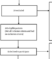
This hospital-based case-control study was conducted during 2024 - 2026 at Imam Hossein Educational and Therapeutic Hospital, affiliated with Shahid Beheshti University of Medical Sciences in Tehran, Iran. Adult patients diagnosed with T2DM who were admitted to inpatient wards, including internal medicine, endocrinology, and general medical units, were identified from the same hospital-based source population. Potentially eligible patients were screened consecutively using the hospital information system and daily admission records. Exclusion criteria were type 1 diabetes mellitus; New York Heart Association class III or IV heart failure; active inflammatory or infectious diseases other than DFU-related infection; autoimmune or rheumatic diseases; hematological disorders; pregnancy; end-stage renal disease or hepatic disease; immunosuppressant use; vitamin D supplementation; and incomplete medical records. Eligible patients were recruited into two groups. Cases were patients with T2DM admitted with active, chronic DFU. Controls were patients with T2DM who had no current or previous DFU and were admitted for other medical reasons, such as glycemic control, elective cardiovascular risk assessment, or acute nonseptic care. Frequency matching (group matching) was implemented during recruitment, with a target ratio of approximately 1:1, to balance age distribution within 5-year strata and sex proportions between groups.
3.2. Clinical and Laboratory Measurements
Demographic data, including sex, age, smoking status, and medication use, were collected using a standardized questionnaire. Weight was measured with participants wearing light clothing, to the nearest 0.1 kg, using a calibrated digital scale. Standing height was measured without shoes, to the nearest 0.5 cm, using a wall-mounted stadiometer. Body mass index (BMI) was calculated as weight in kilograms divided by height in meters squared (kg/m2). Blood pressure (BP) was measured twice in the seated position after at least 10 - 15 minutes of rest using a calibrated sphygmomanometer (Beurer GmbH, Ulm, Germany). The average of the two consecutive readings was recorded.
Venous blood samples were collected in the morning within the first 24 hours of hospital admission, following an 8 - 10-hour overnight fast. Samples were processed at the Central Laboratory of Imam Hossein Hospital. Total serum 25(OH)D was measured using an automated electrochemiluminescence immunoassay on a Cobas e 411 analyzer (Roche Diagnostics GmbH, Mannheim, Germany), using the Elecsys Vitamin D Total II assay kit. Serum glycated hemoglobin (HbA1c) was measured by high-performance liquid chromatography. Serum creatinine and blood urea nitrogen (BUN) were measured using standard enzymatic colorimetric methods on an automated clinical chemistry analyzer (Cobas c 311, Roche Diagnostics, Mannheim, Germany).
3.3. PEDIS Classification System
The severity and clinical characteristics of DFUs were evaluated using the PEDIS classification system. Each domain was graded semiquantitatively according to International Working Group on the Diabetic Foot guidelines. To quantify overall ulcer severity for statistical comparisons, subscores across the five domains were summed to generate a cumulative PEDIS composite score. Higher scores reflected greater severity, more complex pathology, and a higher risk of poor outcomes, including lower-extremity amputation. Further details are presented in Table 1.
Table 1.
Clinical Characteristics of Diabetic Foot Ulcers According to the PEDIS Classification System in Case Patients (N = 102) a
| PEDIS Domain and Grade | Category Definition | Values |
|---|---|---|
| Perfusion (P) | ||
| Grade 1 | No symptoms or signs of PAD: palpable pedal pulses (dorsalis pedis and posterior tibial), ABI 0.90 - 1.10, TBI > 0.60, or tcpO2 > 60 mm Hg. | 48 (47.1) |
| Grade 2 | Signs or symptoms of PAD without critical limb ischemia: intermittent claudication, ABI < 0.90 (ankle pressure > 50 mm Hg), TBI < 0.60 (toe systolic pressure > 30 mm Hg), or tcpO2 30 - 60 mm Hg. | 41 (40.2) |
| Grade 3 | Critical limb ischemia (CLI): systolic ankle pressure < 50 mm Hg, systolic toe pressure < 30 mm Hg, or tcpO2 < 30 mm Hg. | 13 (12.7) |
| Extent/size (E) | Measured after debridement (cm2) using planimetry, a grid technique, or maximum perpendicular diameters; reported as continuous area and quartiles. | |
| Quartile 1 | ≤ 25th percentile | 25 (24.5%) |
| Quartile 2 | > 25th to 50th percentile | 26 (25.5%) |
| Quartile 3 | > 50th to 75th percentile | 26 (25.5%) |
| Quartile 4 | > 75th percentile | 25 (24.5%) |
| Depth (D) | ||
| Grade 1 | Superficial: full-thickness skin ulcer confined to the dermis, without penetration into deeper structures. | 31 (30.4) |
| Grade 2 | Deep: penetration below the dermis into subcutaneous tissues, involving fascia, tendon, or muscle. | 52 (51.0) |
| Grade 3 | Bone/joint involvement: penetration through all layers, with exposed bone, probing to bone, joint involvement, or osteomyelitis. | 19 (18.6) |
| Infection (I) | ||
| Grade 1 | Uninfected: no clinical symptoms or signs of inflammation. | 14 (13.7) |
| Grade 2 | Mild: involvement of skin and subcutaneous tissue only, with ≥ 2 local signs (erythema 0.5 - 2.0 cm, induration, tenderness, warmth, or purulent discharge), without systemic signs. | 49 (48.0) |
| Grade 3 | Moderate: erythema > 2.0 cm or deep tissue involvement (abscess, osteomyelitis, septic arthritis, or fasciitis), without systemic signs. | 30 (29.4) |
| Grade 4 | Severe: foot infection accompanied by systemic inflammatory response syndrome, defined as ≥ 2 criteria: temperature > 38 °C or < 36 °C; heart rate > 90 beats/min; respiratory rate > 20 breaths/min; white blood cell count > 12,000 or < 4,000/μL, or > 10% bands. | 9 (8.8) |
| Sensation (S) | ||
| Grade 1 | Intact: normal protective sensation in the affected foot. | 18 (17.6) |
| Grade 2 | Loss of protective sensation: absent pressure perception at ≥ 2 of 3 plantar sites using a Semmes-Weinstein 10-g monofilament, or absent vibration sensation (128-Hz tuning fork/vibration perception threshold > 25 V). | 84 (82.4) |
| Composite score, Ulcer area (cm2) | Total PEDIS score (sum of domain scores) | 3.4 ± 2.1 |
a Values are expressed as No. (%) or mean ± SD. Abbreviations: PEDIS, perfusion, extent/size, depth/tissue loss, infection, and sensation; PAD, peripheral arterial disease; ABI, ankle-brachial index; TBI, toe-brachial index; tcpO2, transcutaneous oxygen pressure; SD, standard deviation.
3.4. Ethical Considerations
The study protocol was approved by the Research Committee and the Ethics Committee of the School of Medicine, Shahid Beheshti University of Medical Sciences, before study initiation (ethics approval code: IR.SBMU.MSP.REC.1403.412). All patients were informed of the study purpose and procedures, and written informed consent was obtained from each participant. No additional costs were imposed for laboratory tests or research-related procedures. Participants were free to withdraw at any stage without negative consequences.
3.5. Statistical Analysis
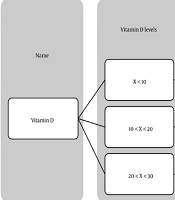
Normally distributed continuous variables, assessed by visual inspection of Q-Q plots, were expressed as mean ± standard deviation (SD) and compared using independent-samples t tests. Categorical variables were presented as frequencies and percentages and analyzed using the chi-square test or Fisher's exact test, as appropriate. Univariable and multivariable logistic regression analyses were performed to evaluate the association between serum 25(OH)D levels and DFU presence, with DFU status (1 = case, 0 = control) as the binary dependent variable. Serum 25(OH)D was modeled as a continuous variable (per 1 ng/mL decrease) and as a categorical variable according to standard Endocrine Society clinical cutoffs: < 20 ng/mL for deficiency versus 20 ng/mL or higher. Hierarchical multivariable models were constructed to control for potential confounding. Model 1 was unadjusted; Model 2 was adjusted for age, sex, BMI, and diabetes duration; and Model 3 was additionally adjusted for HbA1c, systolic BP, and serum creatinine. Multicollinearity was assessed before modeling using variance inflation factor diagnostics; all values were well below 2.5. Odds ratios (ORs) and corresponding 95% confidence intervals (CIs) were reported. All analyses were performed using SPSS version 26.0 (IBM Corp., Armonk, NY, USA). A two-sided P < 0.05 was considered statistically significant.
4. Results
A total of 200 patients with T2DM were evaluated, including 102 with DFU (cases) and 98 without DFU (controls). Ulcer characteristics in the case group, classified by PEDIS domain, are presented in Table 1. The mean composite PEDIS score was 7.27 ± 1.45 (range: 5 - 10). Peripheral arterial disease (grades 2 and 3) was evident in 52.9% (n = 54) of ulcerated extremities, with severe limb ischemia in 12.7%. Deep tissue involvement (depth grades 2 and 3) was identified in 69.6% (n = 71) of ulcers, including confirmed bone or joint involvement in 18.6%. Clinical infection was present in 86.3% (n = 88) of ulcers; mild and moderate infections accounted for 48.0% and 29.4%, respectively. Loss of protective sensation was observed in 82.4% (n = 84) of cases.
Baseline demographic, clinical, and laboratory characteristics of the 102 cases and 98 controls are summarized in Table 2. The groups were well matched for age, with a mean age of 63.6 ± 3.5 years in cases and 63.1 ± 3.7 years in controls (P = 0.16). Sex distribution was also comparable (P = 0.21), with men accounting for 59.8% of cases and 50.0% of controls. Patients with DFU had a significantly higher mean BMI than controls (28.0 ± 1.2 vs. 26.2 ± 0.9 kg/m2; P < 0.01). Diabetes duration was also significantly longer in cases (10.0 ± 1.5 vs. 6.5 ± 1.2 years; P = 0.02).
Table 2.
Demographic, Clinical, and Laboratory Characteristics of the Case and Control Groups a
| Variables | Cases with Chronic DFU (n = 102) | Controls Without Chronic DFU (n = 98) | P value |
|---|---|---|---|
| Continuous variables | |||
| Age (y) | 63.6 ± 3.5 (56 - 70) | 63.1 ± 3.7 (57 - 71) | 0.16 |
| BMI (kg/m2) | 28.0 ± 1.2 (25.0 - 30.0) | 26.2 ± 0.9 (25.0 - 28.2) | < 0.01 |
| Diabetes duration (y) | 10.0 ± 1.5 (8 - 13) | 6.5 ± 1.2 (25.0 - 28.2) | 0.02 |
| Systolic blood pressure (mm Hg) | 164.7 ± 3.5 (160 - 170) | 150.2 ± 2.3 (148 - 153) | 0.02 |
| Diastolic blood pressure (mm Hg) | 92.5 ± 2.1 (88 - 96) | 84.0 ± 1.8 (81 - 87) | < 0.01 |
| Serum creatinine (mg/dL) | 2.24 ± 0.20 (2.0 - 2.5) | 1.50 ± 0.10 (1.4 - 1.6) | 0.05 |
| BUN (mg/dL) | 23.81 ± 2.90 (20 - 28) | 18.12 ± 1.70 (15 - 20) | 0.04 |
| Serum 25(OH)D (ng/mL) | 19.82 ± 3.10 (15 - 25) | 25.08 ± 2.40 (22 - 29) | < 0.001 |
| HbA1c (%) | 9.44 ± 0.3 (8.5 - 10) | 7.52 ± 0.2 (7 - 8) | < 0.001 |
| PEDIS score | 7.27 ± 1.0(5 - 10) | NA | NA |
| Categorical variables | |||
| Gender | 0.21 | ||
| Male | 61 (59.8) | 49 (50.0) | |
| Female | 41 (40.2) | 49 (50.0) | |
| Current smoker, yes | 54 (52.9) | 17 (17.3) | 0.01 |
| Medication regimen | 0.01 | ||
| Oral agents only | 4 (3.9) | 22 (22.5) | |
| Insulin only | 29 (28.4) | 26 (26.5) | |
| Insulin plus oral hypoglycemic agents | 69 (67.7) | 50 (51.0) | |
| Hypertension, yes | 59 (57.84) | 49 (50.00) | 0.27 |
| Ischemic heart disease, yes | 37 (36.27) | 34 (34.69) | 0.81 |
| Chronic kidney disease, yes | 27 (26.47) | 37 (37.75) | 0.09 |
| Cancer, yes | 5 (5.0) | 4 (4.0) | 0.05 |
a Values are expressed as mean ± SD (min - max) or No. (%). Abbreviations: DFU, diabetic foot ulcer; BMI, body mass index; BUN, blood urea nitrogen; 25(OH)D, 25-hydroxyvitamin D; HbA1c, glycated hemoglobin; PEDIS, perfusion, extent/size, depth/tissue loss, infection, and sensation; SD, standard deviation; NA, not applicable. P values were calculated using the independent-samples t test for continuous variables and the χ2 (chi-square) test or Fisher exact test for categorical variables, as appropriate.
Systolic blood pressure and renal function markers were higher in the DFU group. Systolic blood pressure (164.7 ± 3.5 vs. 150.2 ± 2.3 mm Hg; P = 0.02) and BUN (23.81 ± 2.90 vs. 18.12 ± 1.70 mg/dL; P = 0.04) were significantly higher in cases. Serum creatinine was also higher (2.24 ± 0.20 vs. 1.50 ± 0.10 mg/dL; P = 0.05).
Glycemic control and laboratory profiles differed between groups. Mean HbA1c was higher in patients with DFU than in controls (9.44 ± 0.30% vs. 7.52 ± 0.20%; P < 0.001). Conversely, serum 25(OH)D levels were significantly lower in cases (19.82 ± 3.10 vs. 25.08 ± 2.40 ng/mL; P < 0.001).
Current cigarette smoking was significantly more prevalent in patients with DFU than in controls (52.9% vs. 17.3%; P = 0.01). Medication regimens also differed significantly (P = 0.01), with a higher proportion of cases receiving combination therapy with insulin and oral hypoglycemic agents (67.7% vs. 51.0%). The prevalence of hypertension (57.8% vs. 50.0%; P = 0.27) and ischemic heart disease (36.3% vs. 34.7%; P = 0.81) was comparable, whereas differences in the prevalence of CKD and cancer were nonsignificant or marginal.
The association between serum 25(OH)D levels and the odds of chronic DFU is presented in Table 3. In unadjusted univariable logistic regression (Model 1), each 1-ng/mL decrease in serum 25(OH)D was significantly associated with a 28% increase in the odds of DFU (crude OR = 1.28; 95% CI, 1.18 - 1.39; P < 0.001). This association remained significant after adjustment for age, sex, BMI, and diabetes duration in Model 2 (adjusted OR = 1.25; 95% CI, 1.15 - 1.37; P < 0.001). In Model 3, which additionally accounted for HbA1c, systolic blood pressure, and serum creatinine, the association remained significant (aOR = 1.19; 95% CI, 1.08 - 1.32; P = 0.001). Vitamin D deficiency, defined as serum 25(OH)D < 20 ng/mL, was associated with approximately threefold higher odds of DFU in Model 3 (aOR = 2.84; 95% CI, 1.42 - 5.68; P = 0.003).
Table 3.
Univariable and Multivariable Binary Logistic Regression for the Association Between Serum 25(OH)D and Chronic DFU Presence (Total N = 200) a
| Variables | Model 1 | Model 2 | Model 3 |
|---|---|---|---|
| Serum 25(OH)D, per 1 ng/mL decrease | 1.28 (1.18 - 1.39) | 1.25 (1.15 - 1.37) | 1.19 (1.08 - 1.32) |
| Serum 25(OH)D category, ng/mL | |||
| ≥ 20 | 1.00 (Reference) | 1.00 (Reference) | 1.00 (Reference) |
| < 20 | 4.15 (2.30 - 7.50) | 3.65 (1.95 - 6.82) | 2.84 (1.42 - 5.68) |
a Values are expressed as OR (95% CI). Model 1: Unadjusted comparison of DFU presence groups. Model 2: Model 1 adjusted for age, sex, BMI, and diabetes duration. Model 3: Model 2 additionally adjusted for HbA1c, systolic blood pressure, and serum creatinine. Abbreviations: CI, confidence interval; DFU, diabetic foot ulcer; HbA1c, glycated hemoglobin; OR, odds ratio; SBP, systolic blood pressure; 25(OH)D, 25-hydroxyvitamin D; BMI, Body Mass Index.
5. Discussion
In this hospital-based case-control study of patients with T2DM, serum 25(OH)D concentrations were substantially lower in those with DFU than in those without ulcers. Vitamin D deficiency remained associated with DFU presence after adjustment for major clinical and metabolic covariates, including diabetes duration, adiposity, glycemic control, BP, and renal function. These observational findings suggest that serum 25(OH)D may serve as an important metabolic indicator of disease severity.
Our finding of lower circulating 25(OH)D levels in patients with active DFU is consistent with observational studies, umbrella reviews, and meta-analyses documenting an inverse relationship between vitamin D status and foot complications (22, 23, 25, 31). A 2024 umbrella review reaffirmed that profound 25(OH)D deficiency correlates with higher odds of DFU (23). A 2025 pathophysiological review described the multidimensional biological roles of vitamin D, including immunomodulatory, neuroprotective, and angiogenic effects and the induction of antimicrobial peptides (24). Nevertheless, conflicting reports exist. For example, Afarideh et al. reported higher serum 25(OH)D concentrations in patients with active DFU (30). Such discrepancies may reflect methodological heterogeneity, including differences in ulcer chronicity and depth, baseline nutritional status, seasonal variation in blood collection, unrecorded prior supplementation, and population-specific genetic variation in vitamin D-binding proteins and receptor sensitivity (23-25, 30). Despite these heterogeneous findings, the broader evidence supports an inverse relationship between circulating vitamin D levels and DFU vulnerability (22, 23, 25, 31).
At the cellular and molecular levels, vitamin D signaling extends beyond calcium and mineral homeostasis. Ligand binding to the nuclear VDR regulates transcriptional networks involved in innate and adaptive immunity, oxidative stress, keratinocyte differentiation, fibroblast proliferation, and local angiogenesis (24, 27-29, 33-36). In chronic DFU, sustained proinflammatory signaling, impaired cellular migration, excessive reactive oxygen species, and disrupted progression through the proliferative and remodeling phases collectively impede physiological wound repair (4, 8, 9, 16, 24, 29). Through its actions on keratinocytes, dermal fibroblasts, monocytes, and macrophages, VDR activation promotes local tissue homeostasis, re-epithelialization, and suppression of excessive inflammatory cytokine signaling (24, 27-29, 33-36). Supporting this biological rationale, preclinical models and selected randomized clinical trials indicate that targeted vitamin D repletion may improve local wound healing and metabolic indices, as demonstrated by Razzaghi et al. (26, 29).
However, lower 25(OH)D levels in clinical practice should not be interpreted solely as an isolated upstream cause of ulceration. Patients with active DFU experience severe mobility limitations, prolonged bed rest, and extended hospitalization, all of which reduce direct cutaneous exposure to solar ultraviolet B radiation. Chronic diabetes complications are also frequently accompanied by nutritional deterioration and progressive renal dysfunction. Because the kidneys regulate 1-alpha-hydroxylation and vitamin D-binding protein clearance, the elevated BUN and serum creatinine levels observed in our DFU group suggest that impaired renal reserve and microvascular damage may directly lower circulating 25(OH)D concentrations. Hypovitaminosis D may therefore be a consequence or epiphenomenon of advanced metabolic morbidity, systemic inflammation, and physical confinement rather than an independent cause.
Our study also demonstrated an inverse correlation between 25(OH)D concentrations and glycemic indices, consistent with pathways linking VDR signaling to pancreatic beta-cell function and peripheral insulin sensitivity, although clinical interventional findings remain heterogeneous (27). Suboptimal glycemic control, chronic low-grade inflammation, and hypovitaminosis D may mutually reinforce one another, impairing peripheral vascular resistance and repair capacity (4, 8, 16, 22-24, 33-36). Cigarette smoking was also substantially more prevalent in patients with DFU. These adverse vascular effects are compounded by vitamin D deficiency, which further impairs endothelial nitric oxide synthase expression and cellular repair (24, 37, 38), creating a tissue microenvironment that delays wound closure.
Priyanto et al. found no statistically significant correlation between vitamin D levels and clinical parameters such as ulcer duration or PEDIS scores (31). Smoking accelerates tissue hypoxia, endothelial dysfunction, vasoconstriction, and microvascular thrombosis (39, 40). Todorova et al. documented profound hypovitaminosis D in patients with diabetes and peripheral neuropathy (41). Because loss of protective sensation is an integral component of PEDIS scoring, neurovascular impairment may explain the close correlation between low 25(OH)D levels and higher PEDIS scores (41, 42). Serum 25(OH)D concentrations also showed an independent inverse correlation with increasing clinical severity, as assessed using PEDIS classification (42). These associations should nevertheless be interpreted cautiously because observational data cannot establish whether hypovitaminosis D directly accelerates tissue destruction or reflects severe, longstanding systemic neuropathy and immobility.
5.1. Study Limitations
Several limitations should be acknowledged. This study used a hospital-based case-control design with consecutive sampling at a single center. Although recruiting controls from the same clinical wards ensured comparable diagnostic workups and satisfied the study-base principle (43), hospitalized controls often have a greater burden of metabolic comorbidities than community-dwelling patients. This may introduce Berkson bias, an admission-rate bias that could attenuate or distort the observed effect estimates (44, 45). Consequently, the findings may not be fully generalizable to outpatients with well-managed T2DM or patients with minor ulcers treated outside hospitals. We excluded conditions and medications known to influence 25(OH)D levels, including end-stage renal disease, hepatic disease, immunosuppressants, and vitamin D supplementation. However, the clinical registry data did not permit comprehensive assessment of or adjustment for several exogenous determinants of circulating 25(OH)D concentrations, including the timing of DFU diagnosis, seasonal variation, individual sun exposure, dietary intake, and history of vitamin D supplementation. Although serum assays were standardized, the absence of these data limits our ability to exclude residual confounding. Future multicenter, population-based prospective cohorts or seasonal tracking of vitamin D intake are therefore needed to confirm these findings across broader clinical and demographic populations.
5.2. Conclusions
Serum 25(OH)D deficiency was significantly and independently associated with DFU presence and clinical severity, as assessed using the PEDIS classification. Lower vitamin D levels were also associated with an unfavorable metabolic and clinical profile characterized by impaired glycemic regulation, higher BP, subclinical renal impairment, and cigarette smoking. Rather than being a confirmed causal factor, circulating 25(OH)D appears to be a surrogate marker of systemic inflammation, advanced microvascular compromise, and impaired physical mobility.
Although routine assessment and targeted correction of vitamin D deficiency constitute a safe, biologically plausible, and inexpensive adjunct to multidisciplinary diabetes management, our observational data do not establish therapeutic efficacy. Adequately powered prospective multicenter cohorts and randomized controlled trials are needed to determine whether correcting hypovitaminosis D independently improves ulcer healing, preserves sensory function, or reduces the risk of lower-extremity amputation in patients with T2DM.