
1. Background
Aspergillus species are ubiquitous saprophytic fungi commonly found in soil, air, and decaying organic matter and are capable of causing a wide range of infections, particularly in immunocompromised individuals (1). Among the numerous species within this genus, A. fumigatus, A. flavus, A. terreus, and A. niger are considered the most clinically significant (2) and are responsible for diverse manifestations of aspergillosis, including allergic, superficial, chronic, and invasive diseases (3, 4). A. flavus has been reported to cause infections in both humans and animals (5, 6). The increasing prevalence of A. flavus infections in certain geographic regions underscores the need for reliable and rapid laboratory identification (7). Reports from different regions have also indicated increasing isolation of A. flavus from clinical specimens, further emphasizing the importance of reliable and timely methods for its identification (8). Conventional identification based on colony morphology and microscopic characteristics remains useful in routine mycology; however, it may not always provide sufficient resolution because phenotypic features can overlap among closely related species. In addition, isolates that appear morphologically similar may differ genetically, which can be relevant for epidemiological studies and antifungal susceptibility patterns (9). Consequently, molecular approaches such as internal transcribed spacer (ITS) sequencing, analysis of protein-coding genes (including calmodulin and β-tubulin), PCR–RFLP, and random amplified polymorphic DNA (RAPD) PCR have been introduced to enhance diagnostic precision (10, 11). The β-tubulin (BenA) gene contains species-specific polymorphic regions that provide suitable targets for restriction-based molecular identification of Aspergillus species. The restriction enzyme AlwI recognizes defined nucleotide sequences within the BenA locus and generates distinctive fragment patterns that facilitate differentiation among clinically relevant species. Nasri et al. reported that β-tubulin PCR–RFLP using AlwI (BspPI) can reliably distinguish clinical Aspergillus isolates in a rapid and cost-effective manner (11). Based on these findings, AlwI was used in the present study to generate diagnostic RFLP profiles for the identification of clinical A. flavus isolates. Assessment of virulence-related characteristics is equally important for elucidating pathogenic potential (12). Extracellular hydrolytic enzymes, including proteases and phospholipases, contribute to host tissue invasion, inflammatory responses, and microbial colonization (12, 13). Nevertheless, studies integrating β-tubulin PCR–RFLP using AlwI with phenotypic evaluation of virulence-associated traits in clinical A. flavus isolates remain scarce.
2. Objectives
Therefore, this study was conducted to identify clinical isolates of A. flavus using β-tubulin PCR–RFLP with the restriction enzyme AlwI as a preliminary approach and to investigate selected virulence factors, particularly protease and phospholipase activities, in A. flavus isolates obtained from different anatomical sites.
3. Methods
3.1. Clinical Isolates and Specimen Sources
This retrospective study analyzed 45 clinical isolates of A. flavus obtained from routine diagnostic specimens, including bronchoalveolar lavage (BAL), sinus discharge, and nail scrapings, collected between October 2021 and March 2025. Each isolate represented a different patient. A total of 51 isolates were initially assessed for eligibility; of these, 6 were excluded because of duplication, contamination, or incomplete documentation regarding specimen origin. Consequently, 45 isolates were retained for further analysis. To reduce selection bias, all eligible isolates identified during the study period were enrolled consecutively, irrespective of specimen category.
Eligible isolates were required to demonstrate consistent morphological features, positive findings on direct microscopic examination and culture, and adequate viability after subculture. Isolates were excluded if they represented repeat sampling from the same patient, showed evidence of contamination, or lacked reliable information on specimen source. Ethical approval for this retrospective anonymized study was granted by the institutional ethics committee (IR.QUMS.REC.1399.544). Because all data were analyzed in de-identified form and no direct patient contact was involved, the requirement for informed consent was waived.
For primary laboratory evaluation, clinical samples were examined directly using 10% KOH and then cultured on Sabouraud dextrose agar (SDA) and potato dextrose agar (PDA) (Merck, Germany) at 28 °C for 7 days. Initial identification of the isolates was based on macroscopic colony characteristics and microscopic examination using lactophenol cotton blue stain. All isolates were preserved in 10% glycerol at −80 °C until further use.
3.2. Genomic DNA Extraction and PCR
A fresh colony from each isolate was disrupted in 200 µL of lysis buffer containing sterile glass beads by vigorous vortexing, followed by incubation at 70 °C for 30 minutes. DNA was then purified by adding 200 µL phenol–chloroform–isoamyl alcohol (25:24:1) and centrifugation at 14,000 rpm for 5 min; the aqueous phase was collected and used as the PCR template. The β-tubulin (BenA) locus was amplified with primers Bt2a (5′ GGTAACCAAATCGGTGCTGCTTTC 3′) and Bt2b (5′ ACCCTCAGTGTAGTGACCCTTGGC 3′). Reactions (25 µL) contained PCR master mix, 0.5 µM of each primer, and 2 µL of template DNA. Thermal cycling consisted of 95 °C for 5 min, 35 cycles of 94 °C for 45 s, 60 °C for 45 s, and 72 °C for 1 min, followed by a final extension at 72 °C for 6 min. Amplicons were resolved on a 1.5% agarose gel and documented under UV illumination, and selected products were verified by Sanger sequencing.
3.3. Restriction Digestion of PCR Products (AlwI/BspPI)
PCR amplicons of the β-tubulin gene were subjected to restriction digestion using AlwI (BspPI). For each reaction, 10 µL of the amplified product was mixed with 1 µL AlwI enzyme, 2 µL of 10× reaction buffer, and nuclease-free water to a final volume of 30 µL. The mixture was incubated at 55 °C for 4 h to allow enzymatic cleavage. The resulting restriction fragments were resolved on a 2% agarose gel and examined under ultraviolet illumination.
3.4. Extracellular Enzyme Production
Extracellular enzyme production was expressed as an Enzyme Activity Index (EAI), adapted from the approach of Balakrishnan Sangeetha et al. (2020) (14). For each culture plate, EAI was defined as the ratio of the diameter of the visible enzyme-related halo to the diameter of the fungal colony:
Diameters were recorded in millimeters using a millimeter-scale ruler. To improve accuracy, two orthogonal (perpendicular) diameters were measured for both the colony and the surrounding zone, and the mean of these two readings was used for calculations. Measurements were performed by one trained assessor who was blinded to the anatomical origin of each isolate. Each enzymatic assay was repeated three times on independent plates to reduce measurement variability. For each isolate, EAI was calculated per replicate and summarized as the mean ± SD. If any replicate deviated by > 10% from the isolate’s mean EAI, the readings were rechecked and confirmed.
Based on EAI, enzyme production was categorized as high (EAI ≥ 1.75), moderate (1.25 ≤ EAI < 1.75), low (1.10 ≤ EAI < 1.25), or negligible/absent (1.00 ≤ EAI < 1.10).
3.4.1. Phospholipase Activity
To evaluate phospholipase production, the isolates were inoculated onto egg yolk medium. This medium was prepared by supplementing Sabouraud dextrose agar (SDA) with 8% sterile egg yolk emulsion, 1 M NaCl, and 0.005 M CaCl_2. A 10 µL aliquot of a standardized conidial suspension (1 × 10⁶ conidia/mL) was spot-inoculated onto the agar. After incubation at 37 °C for 5 days, the formation of a distinct precipitation halo around the colony was considered indicative of enzyme secretion. Enzyme activity was quantified using the EAI as described above. All assays were performed in triplicate to determine the mean EAI (MEAI) for each isolate.
3.4.2. Protease Activity
Protease production was assessed on bovine serum albumin (BSA) agar. A 10 µL aliquot of the standardized conidial suspension (1 × 10⁶ conidia/mL) was applied as a spot inoculum, and the plates were incubated at 37 °C for 5 - 7 days. Proteolytic activity was indicated by a distinct clearing zone surrounding the colony. Enzyme activity was determined using the EAI described above. Each experiment was performed in triplicate. Aspergillus flavus ATCC 204304 served as a positive control, and uninoculated plates served as negative controls.
3.5. Statistical Analysis
Data were analyzed using IBM SPSS Statistics software (version 26; IBM Corp., Armonk, NY, USA). The Shapiro–Wilk test was used to assess the normality of the data distribution. Given the non-normal distribution of the results, the Kruskal–Wallis test was used to compare enzyme activity indices across clinical sources. When the overall test was significant, Dunn’s pairwise test with Bonferroni adjustment was applied. The magnitude of group differences was quantified using Kruskal–Wallis eta-squared (ηH2), interpreted as small (0.01 to < 0.06), medium (0.06 to < 0.14), or large (≥ 0.14). Outcomes are summarized as mean EAI values for descriptive reporting, whereas inferential statistics are presented as the corresponding test statistics and P values. For all analyses, a two-sided P value < 0.05 was considered statistically significant.
4. Results
4.1. Preliminary Identification of Clinical A. Flavus Isolates
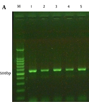
Forty-five clinical isolates identified as A. flavus were obtained from various anatomical sites, including bronchoalveolar lavage (17 isolates; 37.8%), sinus discharge (15 isolates; 33.3%), and nail scrapings (13 isolates; 28.9%). Initial identification based on colony morphology and microscopic features suggested A. flavus, which was subsequently confirmed by PCR-RFLP analysis and sequencing of the β-tubulin gene. Amplification yielded a single DNA fragment of approximately 550 base pairs in all isolates (Figure 1A). Subsequent digestion with AlwI produced two fragments measuring 296 and 254 base pairs, corresponding to the characteristic RFLP profile of A. flavus (Figure 1B). No additional or nonspecific bands were detected.
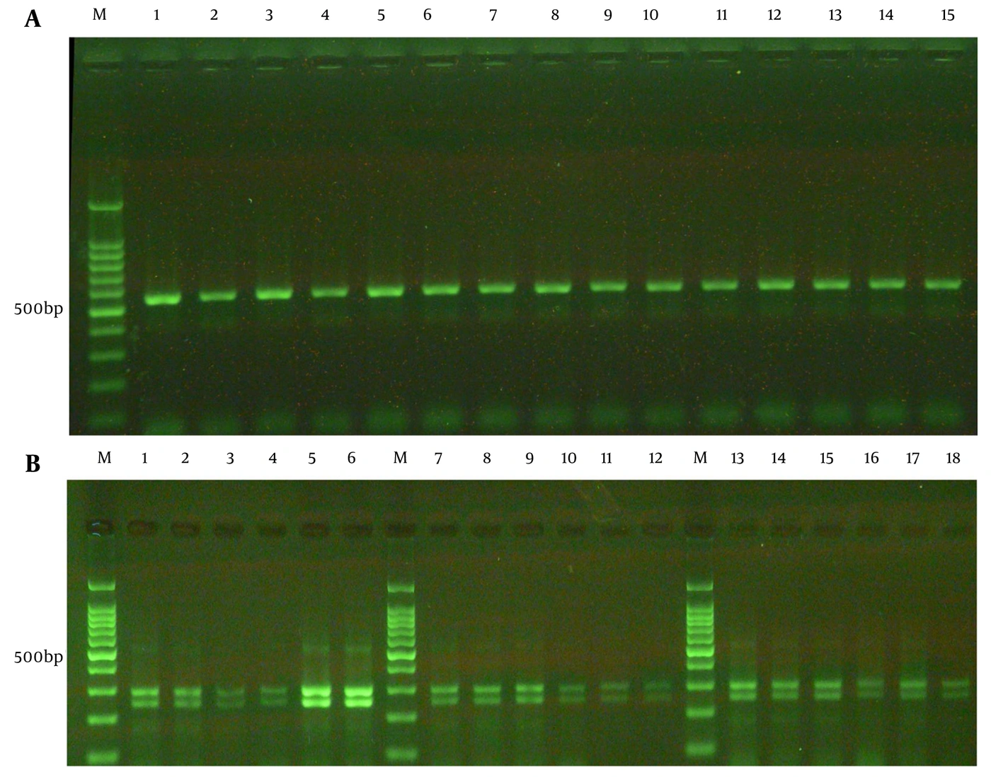
Figure 1.
Molecular identification of clinical Aspergillus flavus isolates. (A) PCR amplification of the β-tubulin gene showing a single ~550-bp product. Lane M: 100-bp DNA ladder; Lanes 1 - 15: clinical isolates. (B) RFLP pattern of the β-tubulin PCR products digested with AlwI (BspPI), yielding two fragments of ~296 bp and ~254 bp. Lane M: 100-bp DNA ladder; Lanes 1 - 18: clinical isolates.
To further confirm species identification, 15 isolates (33.3%), randomly selected to represent each specimen type (BAL, sinus, nail), underwent Sanger sequencing of the β-tubulin gene. Sequence analysis revealed a high degree of similarity between the analyzed isolates and existing A. flavus sequences in the GenBank database. The newly acquired sequences have been deposited in GenBank (NCBI) under accession numbers PX260964–PX260978.
4.2. Production of Virulence Factors
For each isolate, extracellular enzyme activity was measured in three independent experiments. Results are reported as mean ± SD to reflect variation among replicates. Each isolate was then classified as strong, moderate, weak, or none, based on the predefined EAI cutoffs.
4.2.1. Phospholipase Production
Phospholipase activity was observed in 26 of 45 isolates (57.8%). The overall phospholipase EAI was 1.38 ± 0.36, ranging from 1.00 to 1.99. Using the predefined thresholds, 7 isolates (15.6%) were categorized as high producers, 12 (26.7%) as moderate, 7 (15.6%) as low, and 19 (42.2%) showed no measurable activity (Table 1).
Table 1.
Enzymatic Activity Indices (EAI) and Distribution of Activity Levels for Phospholipase and Protease Among Clinical A. Flavus Isolates from Different Anatomical Sources a
| Enzyme and Clinical Source | EAI | Activity Level | |||
|---|---|---|---|---|---|
| High | Moderate | Low | Non-producer | ||
| Phospholipase | |||||
| BAL (n = 17) | 1.50 ± 0.40 | 5 (29.4) | 4 (23.5) | 4 (23.5) | 4 (23.5) |
| Sinus (n = 15) | 1.37 ± 0.32 | 1 (6.7) | 6 (40.0) | 3 (20.0) | 5 (33.3) |
| Nail (n = 13) | 1.23 ± 0.33 | 1 (7.7) | 2 (15.4) | 0 (0.0) | 10 (76.9) |
| Total (n = 45) | 1.38 ± 0.36 | 7 (15.6) | 12 (26.7) | 7 (15.6) | 19 (42.2) |
| Protease | |||||
| BAL (n = 17) | 1.32 ± 0.34 | 3 (17.6) | 3 (17.6) | 4 (23.5) | 7 (41.2) |
| Sinus (n = 15) | 1.66 ± 0.31 | 6 (40.0) | 6 (40.0) | 1 (6.7) | 2 (13.3) |
| Nail (n = 13) | 1.49 ± 0.37 | 3 (23.1) | 5 (38.5) | 1 (7.7) | 4 (30.8) |
| Total (n = 45) | 1.48 ± 0.36 | 12 (26.7) | 14 (31.1) | 6 (13.3) | 13 (28.9) |
a Values are expressed as mean ± SD or No. (%). Abbreviation: EAI, enzymatic activity index.
When stratified by specimen type, mean phospholipase activity was highest among BAL isolates (1.50 ± 0.39), followed by sinus isolates (1.37 ± 0.32) and nail isolates (1.23 ± 0.33). However, group comparisons revealed no statistically significant differences (Kruskal–Wallis: H (2) = 4.965, P = 0.084, η2H = 0.071) (Table 2).
Table 2.
Statistical Comparison of Protease and Phospholipase Activities Among Clinical Sources a
| Enzymes and Comparisons | Test Statistic | Raw P-Value | Adjusted P-Value |
|---|---|---|---|
| Phospholipase | |||
| BAL vs sinus vs nail | H = 4.965 | 0.084 | - |
| Protease | |||
| BAL vs sinus vs nail | H = 6.909 | 0.032 | - |
| BAL vs sinus | Z = −2.585 | 0.010 | 0.029 |
| BAL vs nail | Z = −0.739 | 0.460 | 1.000 |
| Sinus vs nail | Z = 1.698 | 0.090 | 0.269 |
a H, Kruskal–Wallis test statistic; Z, standardized test statistic from Dunn’s pairwise tests. Adjusted P values were calculated using Bonferroni adjustment.
4.2.2. Protease Production
Protease secretion was more frequent than phospholipase production and was detected in 32/45 isolates (71.1%). The mean protease EAI was 1.48 ± 0.36 (range: 1.00 - 1.96). Based on the activity categories, 12 isolates (26.7%) showed high protease activity, 14 (31.1%) were moderate, 6 (13.3%) were low, and 13 (28.9%) had no detectable protease activity (Table 1).
Across clinical sources, sinus-derived isolates had the highest mean protease activity (1.66 ± 0.31), followed by nail isolates (1.48 ± 0.37) and BAL isolates (1.32 ± 0.34). In contrast to phospholipase, protease activity differed significantly across clinical specimen groups (Kruskal–Wallis H (2) = 6.909, P = 0.032, η2H = 0.117). Dunn’s pairwise test with Bonferroni adjustment revealed that sinus isolates exhibited significantly higher protease activity than BAL isolates (Z = –2.585, adjusted P = 0.029). No significant differences were detected between BAL and nail isolates (Z = –0.739, adjusted P = 1.000) or between sinus and nail isolates (Z = 1.698, adjusted P = 0.269). Corresponding descriptive values and mean ranks are presented in Table 2.
5. Discussion
Extracellular enzymes, particularly phospholipases and proteases, are widely regarded as key virulence-associated traits in Aspergillus because they can facilitate tissue penetration and modulate host defenses (12). In this study, we assessed these activities in clinical A. flavus isolates obtained from three anatomical sources. The isolates were recovered from BAL, sinus discharge, and nail scrapings, supporting the view that A. flavus is encountered across diverse clinical settings. This observation is consistent with reports from Iran and other regions with hot, dry climates, where A. flavus is frequently detected in a broad spectrum of specimens (15, 16). These findings underscore both the environmental fitness of this species and its clinical relevance in these geographic areas. For species-level screening, β-tubulin PCR–RFLP followed by AlwI (BspPI) digestion yielded the same two-fragment profile in every isolate (≈296 and 254 bp). This uniform pattern aligns with previous work suggesting that β-tubulin-based PCR–RFLP can improve identification reliability compared with morphology alone, which may be affected by phenotypic overlap and culture conditions (11, 17). From a practical perspective, the consistency of the restriction pattern indicates that this assay can serve as a rapid and inexpensive preliminary method, particularly in laboratories in which routine sequencing is not available. At the same time, the molecular approach used here has important constraints. Only one restriction enzyme was applied, and sequencing was performed for 15 isolates (33.3%). Therefore, the overall discriminatory ability of PCR–RFLP in our dataset is inherently limited. Members of the A. flavus complex (e.g., A. oryzae and A. nomiae) can be genetically similar and may not be fully distinguished by single-enzyme RFLP patterns (16). For this reason, sequencing remains the most reliable option when definitive identification is required. Regarding virulence traits, phospholipase activity was detected in 57.8% of isolates, which is comparable to earlier studies on A. flavus (11). Although BAL isolates had a higher mean phospholipase EAI than sinus and nail isolates, the difference among specimen types was not significant (P = 0.084). Similar results have been reported previously (12), suggesting that phospholipase production may not be strongly determined by anatomical source. The variability we observed may instead reflect isolate-to-isolate differences, local host conditions, or limited statistical power. In contrast, protease activity was more frequent (71.1%) and differed significantly by source (P = 0.032). Sinus isolates showed higher protease activity than BAL isolates, and this difference remained significant after Bonferroni adjustment (Z = −2.585, adjusted P = 0.029; η2H = 0.117). Although this pattern may suggest that protease expression varies across anatomical niches, it should be interpreted cautiously. Subgroup sizes were modest (BAL, n = 17; sinus, n = 15; nail, n = 13), and the study did not include patient-level clinical metadata such as disease severity, infection versus colonization status, treatment, or outcomes. Therefore, these findings should be considered exploratory and primarily hypothesis-generating rather than evidence of clinical or pathogenic associations.
The higher protease activity in sinus-derived A. flavus isolates is consistent with reports linking proteolysis to chronic rhinosinusitis and other upper-airway presentations. Proteases have been proposed to contribute to epithelial damage and persistence within sinonasal tissues; however, the mechanisms and their in vivo relevance remain incompletely defined. In our work, the isolates were archived and lacked patient identifiers and detailed clinical information. As a result, we could not relate enzyme activity to disease severity, colonization versus infection, treatment, or clinical outcomes. Future studies combining enzyme phenotyping with robust clinical datasets are needed to determine whether elevated protease activity has prognostic or therapeutic implications in sinonasal aspergillosis (18).
Despite the value of these findings, several limitations should be considered. Although combining phenotypic assays with molecular identification strengthens the study, sequencing was performed for only a subset of isolates because of cost constraints. Consequently, the present data do not permit firm estimates of the diagnostic performance of PCR–RFLP (e.g., sensitivity, specificity, or predictive values). In addition, incomplete sequencing limits our ability to fully distinguish all members of the A. flavus complex. Larger, preferably multicenter, studies with sequence confirmation for all isolates, broader virulence profiling, and more comprehensive molecular analyses are needed to better clarify the pathogenic potential and clinical significance of these isolates.
5.1. Conclusions
Our findings indicate that clinical A. flavus isolates produce variable levels of virulence-related extracellular enzymes. Although phospholipase activity was detected in over half of the isolates regardless of specimen source, protease production was significantly higher in sinus-derived samples than in those from BAL. Furthermore, β-tubulin PCR–RFLP with the AlwI enzyme yielded consistent patterns across all isolates, supporting its value as a rapid, practical, and cost-effective tool for preliminary species identification in routine diagnostic settings. Nevertheless, because sequencing remains necessary for precise differentiation within the species complex and was performed for only a subset of samples, these results should be considered exploratory. Future research involving larger cohorts, more extensive virulence profiling, and detailed clinical correlations is essential to clarify the role of these enzymes in the pathogenesis of Aspergillus infections.