1. Context
Multiple sclerosis (MS), recognized as one of the most prevalent multifactorial disorders affecting the nervous system in young adults, is defined by its chronic inflammatory demyelinating characteristics that arise from a complex and largely unknown pathogenesis. The MS is a chronic autoimmune and inflammatory condition marked by the degeneration of myelin in the white matter of the brain, optic nerve, and spinal cord, which ultimately leads to nerve damage (1). The onset and progression of MS are shaped by various factors, including age, gender, physical activity, obesity, and dietary habits. While the exact cause of MS remains unclear, it appears to result from a complex interaction of autoimmune mechanisms, genetic susceptibility, and environmental influences (2, 3).
Today, the primary focus of MS treatment strategies revolves around preventing inflammation in the central nervous system (CNS). Interferon beta (IFN-beta) was the first effective drug approved for MS treatment in 1993, followed by the introduction of other therapeutic agents such as glatiramer acetate, alemtuzumab, natalizumab, and fingolimod (2). While these therapies have shown some efficacy, long-term safety and effectiveness remain limited. Recently, herbal therapies have emerged as a promising approach for treating MS (2). Medicinal plants have demonstrated therapeutic potential in various disorders, including diabetes, cancer, and neurodegenerative diseases (4). Increasing evidence suggests that certain herbal compounds can enhance myelin repair and suppress inflammation (5).
Natural compounds exhibit a variety of biological activities, such as antioxidant, anti-inflammatory, anti-apoptotic, and other advantageous properties, which render them a potentially safe and dependable choice for treating neurodegenerative diseases. One such example is saffron, derived from the flower Crocus sativus L., which has been traditionally used in medicine worldwide (6). Saffron is rich in secondary metabolites, including anthocyanins, flavonoids, terpenes, and carotenoids, with crocin and crocetin identified as the primary carotenoids (7). Pharmacological studies have confirmed various properties of crocin, including its antidepressant (8), anti-atherosclerotic (9), anti-platelet aggregation, renoprotective (10), anti-tumor (11), and antioxidant effects (12). Crocin has also demonstrated protective effects against neurodegenerative diseases such as Alzheimer's and Parkinson's (13). Additionally, crocin has been shown to suppress microglial activation and reduce inflammation-induced neurotoxicity in animal models (14).
In preclinical studies using the experimental autoimmune encephalomyelitis (EAE) model in mice, which mimics MS, crocin administration has been shown to significantly reduce clinical severity scores, mitigate neuroinflammation, and decrease demyelination in the CNS (2). This is achieved through mechanisms such as attenuating T-cell proliferation, shifting cytokine profiles toward anti-inflammatory states by suppressing pro-inflammatory cytokines like interleukin (IL)-17 and IFN-γ while promoting IL-10 and TGF-β, and enhancing regulatory T-cell (Treg) responses via modulation of transcription factors (15). Additionally, as part of the carotenoid family, crocin contributes to neuroprotection by deactivating reactive oxygen species (ROS), preserving mitochondrial integrity, preventing lipid peroxidation, suppressing nuclear factor-kappa B (NF-κB) activation, and activating the Nrf2 pathway, which collectively help maintain blood-brain barrier (BBB) integrity and protect oligodendrocytes essential for remyelination in MS (16). These findings underscore crocin's potential to address the core pathophysiological features of MS, including oxidative stress and immune dysregulation.
Clinical trials further support crocin's therapeutic applications in MS management, particularly in alleviating inflammation and improving associated symptoms like cognitive impairment and mental health issues. A randomized, double-blinded trial involving MS patients demonstrated that 30 mg/day of crocin supplementation over 8 weeks significantly reduced high-sensitivity C-reactive protein (hs-CRP) levels, indicating a reduction in systemic inflammation, and also decreased anxiety scores, suggesting benefits for psychiatric comorbidities common in MS (15, 17). Another study using crocin-selenium nanoparticles (Cor@SeNs, containing 5.74 mg crocin) for 12 weeks showed improvements in total antioxidant capacity and cognitive functions, including verbal learning, delayed recall, and information processing speed, highlighting its role in combating oxidative stress and neurodegeneration (16, 18). Reviews of antioxidant therapies, including crocin, emphasize its potential to mitigate cognitive decline by addressing oxidative stress, inflammation, and secondary symptoms like depression and fatigue, though results can vary and call for larger, longer-term studies to optimize dosing and confirm efficacy (19, 20). Overall, these recent investigations position crocin as a safe, natural adjunct therapy for MS, warranting further research to translate preclinical promise into clinical practice. Therefore, this review aims to evaluate the potential applications of crocin for the management of MS.
2. Multiple Sclerosis
The MS is a chronic inflammatory disease of the CNS and is more prevalent in women than in men (21, 22). The MS is caused by axonal degeneration, which results in the formation of demyelinating lesions in the white matter of both the brain and spinal cord. The etiology of these lesions is not fully understood, but studies have shown that an inflammatory infiltrate consisting of a small number of autoreactive T-cells and a large number of nonspecific pathogenic mononuclear cells occurs in areas of demyelination, axonal loss, and severe glial scarring (21, 23). Contributing to axonal pathology, in an autoimmune mechanism, T-lymphocytes specific for myelin antigens initiate an inflammatory attack causing degeneration of CNS axons, leading to disseminated demyelinating lesions accompanied by axonal degeneration (22).
Microglia are the resident immune cells found within the CNS. Under typical circumstances, these cells facilitate synaptogenesis by locally producing neurotrophic factors and regulating synaptic transmission and remodeling. When activated, microglial cells express and secrete inflammatory cytokines, which subsequently result in the generation of nitric oxide (NO) and ROS (24). Microglia, along with macrophages, gather at the locations of active demyelination and neurodegeneration in the brains of individuals with MS, and they are believed to play a crucial role in the progression of the disease (25).
The MS causes various types of damage, such as cognitive impairment, which affects about half of the patients with this disorder (26). Studies have reported that learning, remembering short-term, and spatial memory are affected by demyelination (26-28). According to the findings, serum neurotrophic and neurodevelopment levels, which play a major role in cognitive function, are impaired in patients with MS (29). Neurotrophins play a significant role in promoting cell survival and proliferation within the nervous system, and they are also found in the peripheral blood mononuclear cells (PBMCs) of the immune system (30). Additionally, they enhance synaptic transmission and plasticity, which are essential for memory and regenerative processes. Furthermore, the levels of neurotrophins fluctuate in relation to axonal damage (22).
The MS causes other clinical presentations such as fatigue, weakness, ataxia, numbness, imbalance, pain, visual disturbances (double vision, blurred vision), spastic paralysis, tremor, bladder disorder, impotence, speech impairment, epilepsy, depression, memory loss, and ultimately loss of quality of life (31-33). Other symptoms of MS include inflammation of the nerves and secretion of cytokines and inflammatory and immune cells. Thus, local zones of myelin degradation in MS occur during the inflammatory process due to the penetration of T lymphocytes, hematogenous macrophages, local microglia activity, and the relatively lower presence of B or plasma lymphocytes. This inflammatory reaction is due to increased levels of cytokines in MS lesions (34).
Cytokines serve as messengers between cells and are crucial in the immune system's reaction to trauma, inflammation, infection, and cell signaling. Among them are tumor necrosis factor alpha (TNF-α), interferons (IFNs) such as IFN-γ, and ILs such as IL-1, IL-2, IL-4, IL-6, IL-10, and IL-12 (35, 36). During lesions, levels of inflammatory factors increase significantly. In addition, T-cells are activated in MS and stimulate the secretion of cytokines such as TNF-α, IFN-γ, and ILs (37). T-cells and microglia are also stimulated during this process and are instrumental in the degradation of myelin nerves. Microglia and macroglia are summoned during the demyelination process to help repair damaged myelin. This process of myelin restoration is very important in stopping inflammation and initiating the process of remyelination in the CNS, especially in the early stages of the disease (38).
3. Crocin
Saffron, derived from the stigmata of the saffron crocus (C. sativus) belonging to the Iridaceae family, is naturally distributed in Mediterranean regions, East Asia, and Iran. The most significant constituents of saffron include crocin, crocetin, safranal, and picrocrocin (39). Crocin and crocetin, as active components of saffron, have demonstrated various pharmacological effects, such as cardiovascular protection, inhibition of cancer cell proliferation, and preservation of gastrointestinal, neural, and liver cells. The antioxidant capacity of these compounds is attributed to their anti-inflammatory and analgesic actions (40).
Crocin, a carotenoid pigment found in saffron, exhibits neuroprotective, antidiabetic, antioxidant, and anti-nociceptive activities. Several studies have documented the anti-inflammatory properties of crocin in inflammatory diseases (41). Numerous investigations have highlighted the beneficial role of saffron compounds, particularly crocin, in the improvement of neurological disorders, including pain relief (42), antioxidant activity, anti-inflammation, spasticity reduction, restoration of ethanol-impaired memory, enhancement of memory function, and amelioration of symptoms in EAE by reducing oxidative stress and inhibiting leukocyte infiltration into the brain (43). The administered doses of crocin varied from 50 - 200 mg/kg in these studies (44).
Saffron has also demonstrated lipid-lowering effects, anti-inflammatory properties, antioxidant activity, free radical scavenging abilities, and anti-cancer actions. Its aqueous extract, as well as crocin, have shown utility in preventing oxidative damage resulting from blood ischemia and reperfusion injury in rats (45). Goyal et al. demonstrated that saffron's crocin protects rat hearts against the toxic effects of isoproterenol by modulating oxidative stress (46). Moreover, saffron extract has been shown to protect primary hepatocytes from oxidative damage and mitigate the toxicity of aflatoxin B1, reducing liver lesions (47).
Crocin, a water-soluble carotenoid pigment, is a vibrant red compound found in the red stigmatic lobes of saffron crocus (C. sativus L.) flowers (39). Chemically, crocin is classified as a crocetin digentiobiose ester (C20H24O4), forming crystals with a melting point of 186°C and undergoing hydrolysis in emulsion. Crocins encompass various structural analogues, including crocin-I, crocin-II, crocin-III, crocin-IV, and others. Among these compounds, crocin-I constitutes more than 70% of the total crocin content (48). Recent studies have examined the properties of 25 different crocins from Italy and Iran, revealing a variation in the total dry mass concentration of crocin, ranging from 302 to 548 mg/g, depending on the geographical origin (49). Due to the presence of carboxyl groups at both ends of its polyene chain, crocin can function as an anionic dye for biological staining (50). Figure 1 shows two structures of crocin.
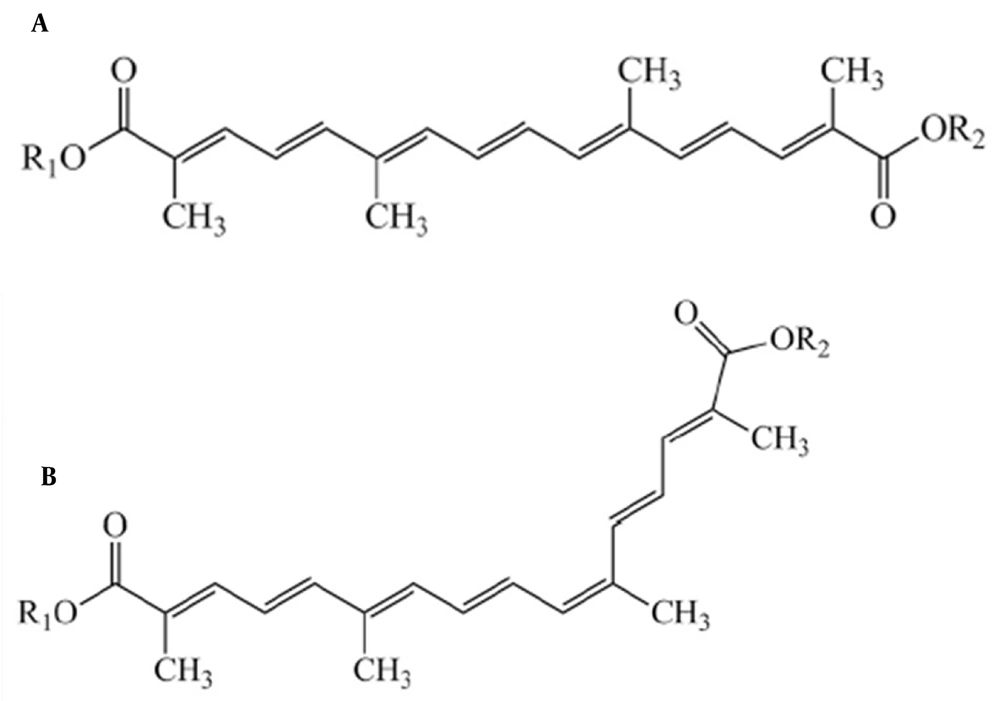
Figure 1.
Structures of crocins: A, trans-form of crocins; and B, cis-form of crocins
The isolated constituents of saffron, including crocin, have garnered increasing interest for their potential impact on human health. This is attributed to their potent antioxidant capacity and robust anti-inflammatory effects, which involve the inhibition of diazinon (DZN), reduction of lipid peroxidation, suppression of inflammatory cytokines, and mitigation of neuronal damage markers. These activities are primarily attributed to the free radical scavenging properties of crocin.
4. Crocin in Neurodegenerative Diseases
Crocin has been found to possess numerous protective effects against neurodegenerative diseases, primarily due to its anti-apoptotic, anti-inflammatory, and antioxidant properties. Research has highlighted crocin's efficacy in addressing conditions such as cerebral ischemia, traumatic brain injury (TBI), spinal cord injury (SCI), and cognitive impairments associated with Alzheimer's disease (AD) and Parkinson's disease (PD).
In the context of cerebral ischemia and ischemic stroke, crocin has demonstrated significant neuroprotective effects. For instance, a study by Oruc et al. indicated that administering crocin at a dosage of 40 mg/kg/day for 10 days resulted in decreased oxidative stress markers, including total antioxidant status (TAS), total oxidant status (TOS), and Oxidative Stress Index (OSI) (51). Additionally, crocin treatment led to reduced levels of hypoxia-inducible factor-1 alpha (HIF-1α), TUNEL-positive cells, and caspase-3 activity in models of brain injury induced by four-vessel occlusion (52). Similarly, another study showed that crocin pretreatment (20 mg/kg) improved neurological outcomes following TBI by reducing brain edema, microglial activation, and the release of pro-inflammatory cytokines, as well as decreasing cell apoptosis. Furthermore, crocin has been shown to protect the BBB from damage in aged rats following cerebral ischemia, preserving BBB function and maintaining tight junction integrity (51).
Crocin's beneficial effects extend to spinal cord injuries, where it has been observed to alleviate chronic pain and enhance locomotor function. In one study by Karami et al., treatment with crocin (150 mg/kg) improved behavioral outcomes in rats with spinal cord damage, potentially linked to a decrease in calcitonin gene-related peptide (CGRP), a key mediator of pain and inflammation (53). Cognitive impairments, often associated with neurodegenerative diseases, have also been addressed through crocin administration. Research has indicated that crocin can ameliorate memory deficits induced by chronic cerebral hypoperfusion, as demonstrated by improved performance in behavioral tests (54). Additionally, crocin has shown promise in enhancing cognitive performance in diabetic rats, suggesting its role as an antioxidant and antidiabetic agent. In models of AD, crocin has been effective in protecting against cognitive decline, with studies showing that it can improve cognitive function and reduce oxidative stress markers in models induced by streptozotocin (55).
The neuroprotective effects of crocin in AD are particularly noteworthy. Studies have demonstrated that crocin can mitigate cognitive deficits induced by intracerebroventricular injection of streptozotocin, with treatment leading to improvements in cognitive performance and reductions in malondialdehyde (MDA) levels, alongside increased activity of antioxidant enzymes (56). Moreover, crocin has been shown to counteract acrolein toxicity, a by-product of lipid peroxidation implicated in neurodegenerative disorders, by modulating signaling pathways associated with neuroinflammation and oxidative stress (57).
In the realm of PD, crocin has exhibited protective effects against dopaminergic neuron degeneration. Research indicates that crocin can ameliorate cell injury and apoptosis induced by neurotoxic agents, as well as improve locomotor function in Drosophila models of PD (57). Additionally, crocin has been shown to reduce oxidative stress levels and improve mitochondrial function in models of PD (58).
Overall, crocin's multifaceted neuroprotective effects make it a promising candidate for the treatment of various neurodegenerative diseases. Its ability to reduce oxidative stress, inflammation, and apoptosis, combined with its positive impact on cognitive function, highlights its potential as a therapeutic agent.
5. Crocin in Multiple Sclerosis
Among neurological disorders, MS is acknowledged as a chronic and debilitating condition affecting the CNS. It is characterized by autoimmune disturbances, inflammation, demyelination, axonal and neuronal damage, as well as the disruption of the BBB, which occurs during relapsing-remitting phases. Crocin has shown significant neuroprotective effects in MS through mechanisms that encompass antioxidant, anti-inflammatory, and anti-apoptotic properties. These effects are mediated by its interactions with various transcription factors, enzymes, inflammatory cytokines, proteins, growth factors, and receptors. The various mechanisms that contribute to the therapeutic benefits of crocin in MS are detailed in the subsequent sections.
5.1. Crocin in Oxidative Stress
Numerous studies have explored the antioxidant activity of crocin in combating oxidative stress. In both in vivo and in vitro investigations, crocin has been shown to exert its antioxidant effects through the modulation of various molecular mechanisms within cells. One prominent mechanism involves the alteration of gene expression levels associated with the redox system, such as GST, GPx, superoxide dismutase (SOD), and catalase (CAT). Additionally, crocin has demonstrated interactions with cellular enzymes, including SOD proteins and peroxidase, contributing to its antioxidant activity (9, 11).
Another mechanism underlying the antioxidant activity of crocin involves the modification of stress marker genes within the endoplasmic reticulum (ER) system. Crocin has been found to modulate the expression of genes such as XBP-1/s, BiP, PERK, and CHOP, which are relevant to ER oxidative stress markers (47). Furthermore, crocin has been implicated in epigenetic alterations and changes in telomerase activity, suggesting its ability to induce molecular-level modifications in cells and elicit antioxidant effects (4). The molecular mechanism of antioxidant and anti-inflammatory activity of crocin is shown in Figure 2.
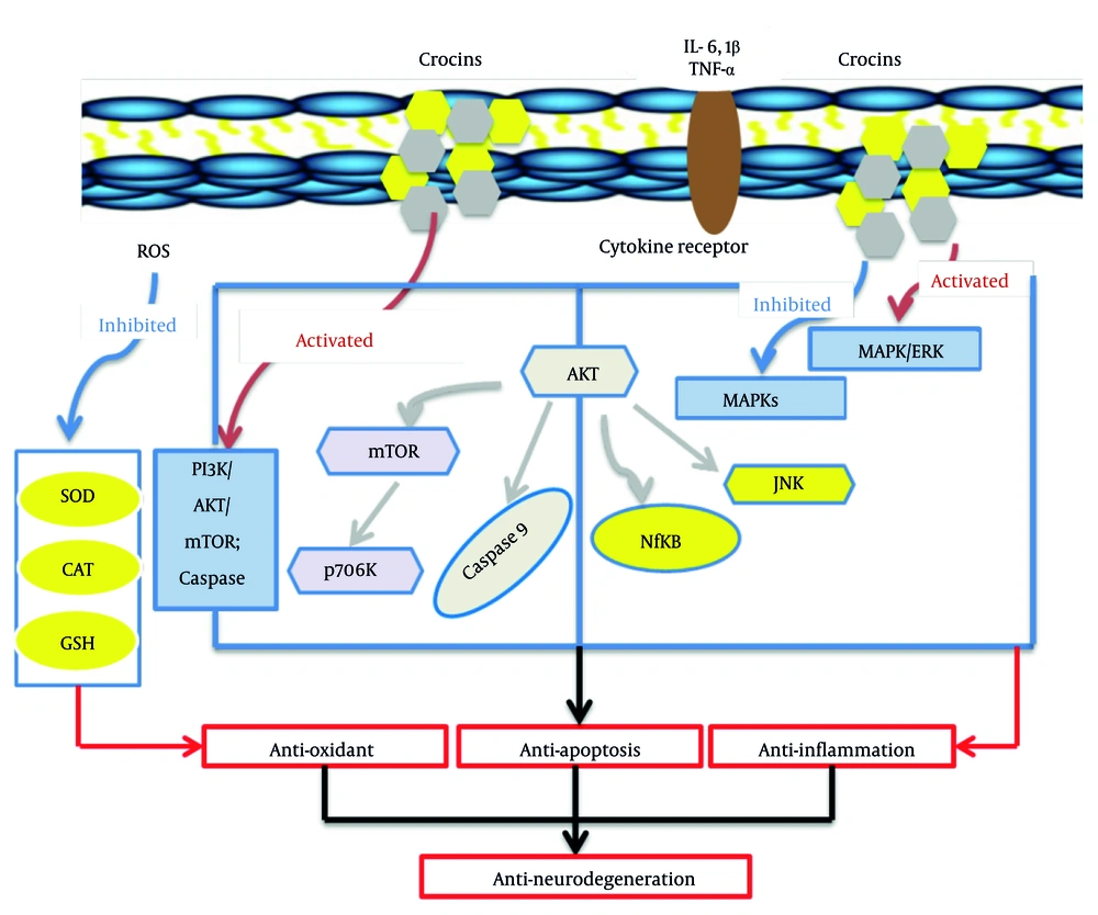
Figure 2.
Molecular mechanism of antioxidant and anti-inflammatory activity of crocin [activation of phosphoinositide 3-kinase (PI3K)/Akt/mammalian target of rapamycin (mTOR) axis; inhibiting mitogen-activated protein kinases (MAPKs) signaling pathway; increasing PI3K/Akt and ERK/MAPK pathways activity; (abbreviations: ROS, reactive oxygen species; SOD, superoxide dismutase; CAT, catalase; GSH, glutathione; TNF, tumor necrosis factor; IL, interleukin; JNK, c-Jun N-terminal kinase; NF-κB, nuclear factor-kappa B).
In a study conducted by Ghaffari et al., saffron extract, containing crocin, was directly injected into the hippocampus of demyelinated rats. The results demonstrated that saffron extract, through its antioxidant properties, effectively neutralized ROS and eliminated free radicals. This led to a significant reduction in the activity of GPx and MDA enzymes, as well as an increase in CAT activity (59). Another study by Kamyar et al. revealed that the administration of saffron extract strengthened the antioxidant system of the liver in Wistar rats, preventing significant changes in antioxidant activity and MDA levels in liver tissues. The study also found that crocin, in particular, showed positive effects on Antioxidant Index levels, with the dose of 100 mg/kg displaying the most significant improvements (60, 61).
Moreover, Rezaeimanesh et al. in 2024, in a triple-blind randomized clinical trial, investigated the effects of Corcin on cognitive function and oxidative stress markers in 60 MS patients. Participants received capsules containing 5.74 mg crocin and 55 mcg selenium or placebo for 12 weeks. The results showed significant improvements in total antioxidant capacity and cognitive function, particularly in verbal learning and symbol digit modalities tests, in the Cor@SeNs group compared to the placebo group (18). Interestingly, the antioxidant activity of crocin was found to be comparable to Tekfidra, a standard drug used in the treatment of MS, with an efficacy ranging from 35% to 45%.
Furthermore, the antioxidant properties of crocin have been reported in other neurodegenerative diseases. Vakili et al. demonstrated that saffron extract increased the concentration of GPx, SOD, and MDA enzymes in ischemic brain tissue (62). Sheng et al. observed the positive effects of crocin (100 mg/kg) in reducing lipid peroxidation in the brains of rats. However, a similar study using the intraventricular injection of picrocrocin did not yield the same effects (63).
Moreover, crocin has demonstrated a protective effect in EAE by down-regulating the expression of oxidative stress in the spinal cord. The EAE-induced upregulation of ER stress genes, including XBP-1/s, was found to be mitigated by crocin administration on day 7 post-EAE induction. This led to a reduction in ER stress and inflammatory gene expression in the spinal cord, as well as decreased expression of ER stress genes XBP-1/s. Crocin was found to regulate the ER, thereby preventing neuronal demyelination (64).
Furthermore, Fathi-Moghadam et al. reported that administration of crocin (at a dosage of 100 mg/kg) for 21 days following ethidium bromide (EB)-induced demyelination led to a significant decrease in brain levels of GPx, MDA, and SOD in EAE rats (61). Based on the aforementioned findings, it can be concluded that crocin, as a potent antioxidant, holds promise for the management and treatment of patients with MS. It achieves this by reducing lipid peroxidation activity and enhancing the antioxidant activity of enzymes involved in oxidative mechanisms.
5.2. Crocin in Neuroinflammation
Although the exact mechanisms behind MS are still not fully understood, chronic inflammation and oxidative stress are key factors in its pathogenesis. The inflammatory response in MS is primarily driven by the activation of microglia/macrophages and T-cells, which contributes to demyelination and neurodegeneration (65). Furthermore, the inflammatory environment generates free radicals, which amplify the inflammatory reaction, thereby exacerbating disease progression (66).
Crocin possesses the capability to modulate the expression of genes that encode inflammatory cytokines (such as IL-1, IL-2, IL-6, and TNF-α) (64), adhesion molecules (including ICAM, VCAM, E-selectin), inducible enzymes (like COX-2 and iNOS) (2), chemokines, and acute phase proteins (67). These components are essential in the regulation of inflammatory processes within the immune system (Figure 2). Research has shown that crocin not only reduces the production of ROS and inhibits the release of pro-inflammatory cytokines but also alleviates inflammation in various organs, including the lung, heart, kidney, and brain. This is primarily achieved through the modulation of the NF-κB pathway and the translocation of NF-κBp65 into the cell nucleus (68). In this regard, the regulation of the phosphoinositide 3-kinase (PI3K)/Akt pathway seems to be a significant target for crocin, aiding in the suppression of the NF-κB pathway (69).
Although clinical data regarding the anti-inflammatory and anti-nociceptive effects of crocin in humans is limited to a single relevant study, the findings from preclinical studies are promising and warrant further investigation in clinical settings (48, 67).
In neurodegenerative diseases, including epilepsy and PD, crocin demonstrated its ability to reduce the expression of inflammatory cytokines such as IL-1β, IL-18, NLRP1, and AIM2 (70, 71). These findings suggest that crocin's anti-inflammatory effects are mediated through the downregulation of pro-inflammatory factors and the inhibition of inflammasome activation, making it a promising therapeutic agent for managing inflammatory conditions associated with MS.
The effects of crocin on reducing inflammation and oxidative stress in patients with MS were evaluated in a double-blind, randomized placebo-controlled trial by Ghiasian et al. in 2019. Forty patients were assigned to receive either two crocin capsules (30 mg/day) or a placebo per day for 28 days. The results showed that crocin treatment significantly decreased levels of TNF-α and IL-17 in the serum compared to the placebo group (72).
A recent study by Kouchaki et al. in 2024 investigated the effects of crocin on MS patients, focusing on its anti-inflammatory, antioxidant, and mental health benefits. Patients with MS were randomized into two groups: One receiving 30 mg/day of crocin and the other a placebo for 8 weeks. Results showed that crocin significantly reduced hs-CRP levels and anxiety but did not affect MDA and NO levels. The findings suggest that crocin has potential in attenuating inflammation and improving mental health in MS patients, although longer treatment durations and larger sample sizes are needed to confirm its efficacy and safety (17).
In EAE models, crocin has shown a protective role by decreasing the expression of inflammatory genes within the spinal cord. It also inhibits T-cell infiltration and macrophage activation. Furthermore, treatment with crocin has been associated with lower levels of TNF-α in the spinal cord, highlighting its potential protective effects against SCI (64).
Furthermore, Banaeeyeh et al. in 2024 evaluated the protective effects of trans sodium crocetinate (TSC), a synthetic carotenoid, against EAE in mice (73). The results showed that TSC treatment (50 and 100 mg/kg) reduced oxidative stress markers, microgliosis, demyelination, and inflammatory factors (IL-1β and TNF-α) in spinal cord tissues. Additionally, TSC modulated the mitophagy pathway by decreasing PINK1 and Parkin protein levels. These results indicate that TSC protects against EAE-induced MS through its anti-inflammatory, antioxidant, and anti-mitophagy effects.
Crocin, a bioactive carotenoid derived from saffron, exerts its therapeutic effects on MS primarily through immunomodulatory, anti-inflammatory, and antioxidant mechanisms. In EAE models mimicking MS, crocin reduces clinical severity by attenuating T-cell proliferation and shifting cytokine profiles toward anti-inflammatory states, suppressing pro-inflammatory cytokines such as IL-17 and IFN-γ while enhancing IL-10 and TGF-β production. It also modulates transcription factors to favor Treg differentiation, thereby mitigating neuroinflammation and demyelination in the CNS.
As part of the broader carotenoid family, crocin deactivates ROS, preserves mitochondrial function, prevents lipid peroxidation, suppresses NF-κB activation, and activates the Nrf2 pathway to combat oxidative stress, which is a key driver of MS pathology. These actions collectively help maintain BBB integrity and protect oligodendrocytes, promoting remyelination and reducing neurodegeneration (15, 16).
The utility of crocin in MS management stems from its ability to address the multifaceted pathophysiology of the disease, including immune dysregulation, chronic inflammation, and oxidative damage, offering a safer, natural alternative or adjunct to conventional therapies. By promoting immune balance — shifting from pro-inflammatory Th1/Th17 responses to Treg dominance — crocin can potentially halt disease progression and alleviate symptoms like neurological dysfunction and cognitive impairment (18, 74). Its antioxidant properties further enhance neuroprotective effects, reducing CNS damage and improving overall quality of life for MS patients, as evidenced in preclinical models where it significantly lowered inflammation and demyelination scores (19).
With a favorable safety profile and potential for oral supplementation, crocin holds promise for clinical translation, though further human trials are needed to optimize dosing and confirm long-term efficacy in managing MS relapses and progression.
5.3. Crocin in Autoimmunity
The well-established anti-inflammatory properties of crocin have been demonstrated to have therapeutic effects in various health disorders, including autoimmune diseases (4), COVID-19 (75), osteoarthritis (76), bone and cartilage diseases (77), atherosclerosis (78), rheumatoid arthritis (79), and MS (72). Crocin is known for its ability to reduce inflammation and oxidative stress, acting as an antioxidant with potential neuroprotective effects, particularly in MS (4). It regulates the pathogenic stress of the ER, protects oligodendrocytes, and consequently reduces inflammatory demyelination.
Further investigation into the ER response to MS reveals that the unfolded protein response (UPR) occurs when the ER is under stress from unfolded or misfolded proteins, resulting in the upregulation of several response components in various cell types, including oligodendrocytes, T-cells, microglia/macrophages, and astrocytes. Crocin modulates the reactivity of BV2 microglia by promoting filopodia formation, enhancing microglial phagocytosis, and inhibiting NO production (80). This effect on microglial homeostasis is likely attributed to the antioxidant properties of crocin, as supported by research on crocin and its derivative crocetin in mesenchymal stem cells (81).
Here is the revised text with grammatical corrections:
Moreover, in a double-blind trial, crocin has been shown to reduce DNA damage, inflammation, and oxidative stress in MS patients. Recent experiments on demyelinated Wistar rats indicate that crocin attenuates pro-inflammatory cytokines and lipid peroxidation in brain tissue (60). The modulation of beta-adrenergic receptors' activity is another potential mechanism associated with crocin-mediated changes in inflammatory mediator levels. Crocin may decrease the number of beta-adrenergic receptors through negative self-regulation, thereby reducing the secretion of inflammatory cytokines by adipocytes (82).
Collectively, these findings highlight the significant anti-inflammatory properties of crocin and its potential as a beneficial therapeutic compound for MS. Studies have also reported a positive effect of crocin on cellular and humoral immune responses, including increased neutrophil cementation, stimulation of neutropenic severity, and reduced demyelination in rats.
In a study by Mohajeri and Doustar, crocin was shown to significantly decrease total bilirubin levels and increase total serum albumin and protein levels in rats receiving cisplatin. Additionally, saffron extract and silymarin decreased lipid peroxidation and increased antioxidant levels in a dose-dependent manner (83).
5.4. Crocin in Neurotrophic Factors
The MS is associated with various forms of damage, including cognitive impairment that affects approximately half of all patients. Demyelination in MS has been shown to impact learning, short-term memory, and spatial memory (27). Neurotrophins, such as brain-derived neurotrophic factor (BDNF) and nerve growth factor (NGF), are polypeptides belonging to the neurotrophic factor family and play a protective role towards specific neuronal populations in the adult CNS (21). Studies have indicated that serum levels of neurotrophic factors, which play a crucial role in cognitive function, are impaired in individuals with MS (29).
In a recent study by Fatemi et al. in 2024, researchers investigated the effects of crocin on brain neurotrophins, cognition, and sensory and motor dysfunction in female Wistar rats with toxic-induced demyelination using EB. The results showed that crocin improved cognitive variables, sensory and motor nerve conduction velocity, tail flick latency, and clinical scores. Moreover, crocin administration led to greater improvements in BDNF and NGF1 (84).
The effects of crocin on brain neurotrophins have been proven by the same researchers in another study of the EAE rat model (85). In this study, crocin and fingolimod were administered for 21 days, and the results indicated that sensory and motor nerve conduction velocity and the expression of neurotrophic factors were increased following co-administration of crocin with fingolimod.
Chronic administration of crocin at doses of 25 and 50 mg/kg has been reported to increase the expression of BDNF, VGF, CREB, and p-CREB in the rat hippocampus, similar to serotonergic agents (86). The regulation of synaptic plasticity in the hippocampus is associated with the VGF neuropeptide, and its mechanism closely resembles that of BDNF through a tyrosine receptor kinase B (TrkB)-dependent pathway. The expression of these proteins in the rat hippocampus has been identified as an antidepressant mediator, suggesting that crocin has an antidepressant-like action by increasing these neurotrophins.
In addition, the protein levels of BDNF, CREB, and p-CREB in the rat hippocampus were investigated in another study to evaluate the molecular mechanism of the aqueous extract of saffron as an antidepressant factor. The results showed that the application of the aqueous extract of saffron increased the protein levels of BDNF, CREB, and p-CREB, while the expression of the protein level VGF was also increased, but not significantly (87).
Existing studies reporting the positive effects of crocin in neurodegenerative diseases further support our results. Farkhondeh et al. demonstrated that crocin enhances BDNF secretion in the hippocampus by influencing the electrical ignition of epilepsy (13). However, Razavi et al. in 2017 reported no significant changes in BDNF, VGF, and CREB levels following long-term administration of crocin in the rat cerebellum. They concluded that the antidepressant activity of crocin might be partially associated with CREB, rather than BDNF (88). The function of crocin on molecular factors in EAE and MS models is summarized in Table 1.
Table 1.
The Function of Crocin on Molecular Factors in Experimental Autoimmune Encephalomyelitis and Multiple Sclerosis Models
| Models | Administration Routes | Markers | Results | Ref. |
|---|---|---|---|---|
| EAE | 30 mg/kg | MDA, GPx, SOD, and TAC | Crocin reduced MDA level and elevated the levels of GPx, GR, SOD, and TAC. | (89) |
| 10 ng/mL | NSC differentiation | Crocin enhanced NSC differentiation into oligodendrocytes. | (90) | |
| 5, 10, and 20 mg/kg; 21 d | MDA, GPX, and SOD | Crocin significantly decreased MDA levels, and increased GPx and SOD levels. | (61) | |
| 5, 10, and 20 mg/kg; 21 d | BDNF, IL-10, and learning and memory | Crocin improved the level of brain neurotrophins, exploratory behavior. | (85) | |
| 5, 10, and 20 mg/kg; 21 d | TNF-α, IL-1, and IL-6 | Crocin reduced the level of brain TNF-α, IL-1, and IL-6. | (60) | |
| 10 μg/mL | ER stress and inflammatory gene expression | Crocin protected oligodendrocytes exposed to cytotoxins. Crocin also suppressed ER stress and inflammatory gene expression in spinal cords. | (74) | |
| MS | 30 mg/d; 4 wk | Inflammation, oxidative damage, and DNA damage | Crocin significantly decreased lipid peroxidation, DNA damage, TNF-α, and interleukin 17. | (72) |
| 100 mg/kg; 5 wk | MDA, GPX, SOD, and TAS | Crocin significantly decreased MDA levels, and increased GPx, SOD, and TAC levels. | (91) | |
| 100 mg/kg; 21 d | MDA, GPX, and SOD | Crocin decreased the levels of GPx and SOD significantly as well as MDA level. | (92) |
Abbreviations: EAE, experimental autoimmune encephalomyelitis; MDA, malondialdehyde; SOD, superoxide dismutase; BDNF, brain-derived neurotrophic factor; IL, interleukin; TNF-α, tumor necrosis factor alpha; ER, endoplasmic reticulum; MS, multiple sclerosis; TAS, total antioxidant status.
6. Conclusions
Crocin has demonstrated numerous beneficial effects on autoimmune diseases. Its primary positive effect on MS and EAE outcomes is attributed to its anti-inflammatory, antioxidant, and anti-apoptotic properties as a natural-based agent. The results of this comprehensive review indicate that crocin leads to significant improvements in the immune system, oxidative stress levels, and neurotrophin expression in individuals with MS or EAE. These improvements have the potential to positively impact cognition, behavior, and ultimately enhance the overall mood and quality of life in this population. However, further studies are necessary to elucidate the precise and detailed mechanisms through which crocin alleviates the impairments associated with MS.
The results and data from studies on crocin's therapeutic potential in MS provide valuable insights for medical applications by demonstrating its ability to modulate key pathological processes, offering a foundation for developing safer, natural interventions. For instance, preclinical data from EAE models show that crocin reduces neuroinflammation and immune dysregulation, which could translate to clinical benefits in slowing MS progression and preserving neurological function.
Clinical trials further reveal that crocin supplementation lowers inflammatory biomarkers like hs-CRP and improves mental health outcomes, such as reduced anxiety, in MS patients, suggesting its utility in managing systemic inflammation and psychiatric comorbidities that exacerbate the disease. As part of carotenoid-based therapies, these findings highlight crocin's antioxidant mechanisms, including the deactivation of ROS and suppression of NF-κB pathways, which protect against oxidative stress-induced neurodegeneration, potentially aiding in the preservation of cognitive functions and overall quality of life.
In terms of treatment strategies, the data support crocin's integration as an adjunct therapy in MS management, enhancing conventional treatments by addressing unmet needs like cognitive impairment and chronic inflammation with minimal side effects. Reviews of antioxidant supplementation, including crocin, indicate improvements in cognitive domains such as verbal learning and information processing speed, positioning it as a promising option for mitigating MS-related cognitive decline.
Saffron-derived compounds like crocin also exhibit anti-apoptotic and anti-inflammatory effects in neurodegenerative contexts, which could inform personalized medicine approaches for MS subtypes, potentially reducing relapse rates and supporting remyelination efforts. Overall, these evidence-based results advocate for larger clinical trials to optimize dosing and combinations, ultimately advancing MS treatment paradigms toward more holistic, evidence-driven therapies.