
1. Background
Microbial spoilage and foodborne diseases remain critical challenges in the food industry (1). Each year, millions of deaths are linked to foodborne illnesses caused by contaminated food, highlighting a severe public health crisis. The substantial economic burden further drives the food industry to implement strict measures against spoilage microorganisms to minimize disease outbreaks (2). Conventional food preservation methods, such as heat treatment and disinfection, often fail to eliminate microbes and can alter product quality, sensory properties, and nutritional value, falling short of consumer satisfaction (3-6). The increase in antimicrobial resistance in bacteria and the emergence of multi-drug-resistant bacteria are among the most significant global health threats, projected to be the leading cause of human mortality by 2050 (7, 8).
Bacteriophages, as protective microorganisms, can meet consumer expectations by preserving food without affecting its physical properties and without disrupting the consumer's gut microbiota (9). Studies show that using phages in combination, especially in phage cocktails, can prevent the emergence of phage-resistant bacteria and enhance their activity (10, 11). Salmonella-specific phages can also reduce bacterial counts in contaminated fresh fruits, proving more effective than chemical sanitizers (12, 13). Beyond Salmonella, phages have also been successful in controlling other significant pathogens such as Listeria monocytogenes (14, 15), Escherichia coli O157 (16, 17), and Shigella.
2. Objectives
Our study focuses on the isolation and application of the regional phage PhSal, specifically targeting Salmonella spp. in meat. By applying this local phage, we aim to evaluate its effectiveness in controlling Salmonella spp. contamination and compare its performance to other phages and traditional methods. This research not only explores the potential of lytic PhSal as a biocontrol agent but also contributes to the development of tailored solutions for food safety in regional contexts.
3. Methods
3.1. Isolation, Identification, and Antimicrobial Susceptibility Testing of Salmonella spp. in Meat Products
Six Salmonella strains were isolated from fifty different meat samples, including raw chicken and raw beef. The samples were safely collected in sterile bags, labeled, and maintained at 4°C using specialized transport containers. They were then transported to the Microbiology Laboratory at Ayatollah Amoli Branch University. Under sterile conditions, 2.50 g of the sample was transferred into a 50 cc Falcon tube containing 22.50 mL of peptone water, vortexed for 1 minute to ensure thorough mixing, and incubated at 37°C for 24 hours. Following incubation, 10 mL of the homogenate was transferred to a sterile Erlenmeyer flask containing 90 mL of Rappaport-Vassiliadis enrichment medium and incubated at 37°C for an additional 24 hours. A loopful of the enriched homogenate was streaked onto a SalmonellaShigella (SS) agar plate to isolate single colonies (18, 19).
Single colorless colonies with black centers were selected from SS agar and transferred to triple sugar iron (TSI) agar and urea agar for further analysis. Bacterial species identification was performed using a series of biochemical tests with the Microbact 12E rapid detection system, specifically designed for gram-negative bacteria (GNB; Oxoid Ltd., Basingstoke, UK). Additionally, Salmonellaenterica PTCC 1230, obtained from the Iranian Fungi and Industrial Bacteria Collection Center, was used as a reference strain to evaluate the presence and characteristics of Salmonella spp. in meat products.
The antimicrobial sensitivity of the tested bacterial strains was assessed using the Kirby-Bauer disk diffusion method, with the results presented in Table 1. The disks used (Pad Tan Teb Company, Iran) included ampicillin (10 μg), gentamicin (10 μg), amikacin (15 μg), ceftazidime (30 μg), and cefixime (5 μg) to determine antimicrobial sensitivity (20-23). In this study, aminoglycosides (amikacin, gentamicin) and first- and second-generation cephalosporins (cefixime, ceftazidime) were tested to assess Salmonella spp. resistance patterns. Although not recommended as first-line treatments due to widespread resistance, these data help understand strain resistance and do not indicate clinical use. Salmonella enterica Sal03 was chosen for further analysis due to its resistance to cefixime, ceftazidime, sulfamethoxazole, and amoxicillin.
Table 1.
Antibiotic Susceptibility Profiles of Salmonella Strains
| Strains | Amikacin (1) | Cefixime (2) | Ceftazidime (3) | Sulfamethoxazole (4) | Gentamicin (5) | Amoxicillin (6) |
|---|---|---|---|---|---|---|
| Sal01 | S | S | I | S | I | R |
| Sal03 | I | R | R | R | S | R |
| Sal04 | I | I | S | S | S | R |
| Sal05 | S | I | S | I | I | I |
| Sal07 | S | S | R | S | S | R |
| PTCC 1230 | S | S | I | S | S | I |
Abbreviations: S, susceptible; I, intermediate; R, resistance.
3.2. Phylogenetic Analysis of 16S rRNA Gene Sequences in Salmonella spp.
In this study, DNA of S. enterica Sal03 was extracted using the boiling method, followed by PCR amplification using specific primers for the 16S rRNA gene (27F and 1525-R). The PCR program included an initial denaturation at 94°C for 2 minutes, followed by 30 cycles of 94°C for 1 minute, 60°C for 1 minute, and 72°C for 3 minutes, with a final extension step at 72°C for 10 minutes (24). The PCR products were sequenced using the ABI 3100 instrument and the Dye Terminator Cycle Sequencing Kit. The obtained sequences were subjected to BLAST analysis in the National Center for Biotechnology Information (NCBI) database to identify and download closely related strains. Subsequently, multiple sequence alignment of the 16S rRNA sequences was performed using Molecular Evolutionary Genetics Analysis (MEGAX). The phylogenetic tree was constructed using the Neighbour-Joining method and the Kimura 2-parameter model. After the analysis, we submitted it to NCBI with accession number SUB15307646.
3.3. Isolation and Purification of Bacteriophages from Hospital Pool Water
Twelve water samples were collected in 200 cc sterile containers from various hospital pool sites in Mazandaran province for the isolation of bacteriophages targeting specific Salmonella bacterial strains. The samples were immediately transported to the Microbiology Laboratory at Ayatollah Amoli Branch University under controlled conditions at 4°C. Upon arrival, the samples were centrifuged at 6000 × g for 20 minutes at -4°C, and the resulting supernatant was filtered through a 0.45-μm syringe filter (25, 26). For enrichment, 10 mL of the filtered liquid, 5 mL of Salmonella bacterial cultures in the logarithmic growth phase, and 10 mL of tryptic soy broth (Merck, Germany) were mixed and then incubated at 37°C for 24 hours with gentle agitation. Following incubation, the supernatant was collected and filtered again through a 0.45-μm syringe filter to remove any remaining bacteria (25, 26). The culture was centrifuged at 8000 × g for 15 minutes and filtered through a 0.22-μm filter to obtain a purified phage stock. The purified bacteriophages were stored at 4°C for future experiments. The double-layer agar method was used for further purification (27, 28).
3.4. Lytic Activity Assessment of the Isolated Bacteriophage
3.5. Transmission Electron Microscopy Sample Preparation Protocol
The isolated phages were examined using a transmission electron microscope (Hitachi H-7000FA, Tokyo, Japan). A 10 µL phage solution (> 10 log10 PFU/mL) was applied to a carbon-coated copper grid, dried under a lamp, and negatively stained with 2% uranyl acetate or 0.5% phosphotungstic acid. After drying, the grids were imaged under the microscope, and the images were analyzed using the Digital Micrograph Demo 3.9.1 program (Gatan, Inc., Pleasanton, USA) (32, 33).
3.6. One-Step Growth Curve Assay for Bacteriophages
For this assay, 1 mL of bacterial suspension at a final concentration of 106 CFU/mL was combined with 1 mL of phage extract at a titer of 105 PFU/mL and incubated at 37°C for 15 minutes to allow phage particles to adsorb. The solution was then centrifuged at 12000 × g for 1 minute to remove unabsorbed phage particles, and the supernatant was discarded. The remaining pellet was cultured again and incubated at 37°C. Then, 100 μL samples were collected every ten minutes for one hour. The PFU was determined using the double-layer agar method (25).
3.7. Evaluating Phage Multiplicity of Infection
To assess the phage infection of the studied bacteria at a concentration of approximately 107 CFU/mL, phages were added at different dilutions (105, 106, 107, and 108 PFU/mL) and incubated at 37°C for six hours. Bacterial growth was measured at 30-minute intervals for 60 minutes at OD600nm (33, 34).
3.8. Assessment of Thermal and pH Stability of Bacteriophage Suspensions
To assess the thermal stability of phage particles, 100 µL of a phage suspension with a titer of 109 PFU/mL was added to 900 µL of TSB medium. The mixture was incubated at temperatures of 30, 40, 50, 60, 70, and 80°C (35). At each temperature, the samples were incubated for two time intervals: Thirty minutes and 60 minutes. After each interval, the phage titer was determined using the double-layer agar method (Table 2).
Table 2.
Assessment of Thermal and pH Stability of Bacteriophage Suspensions
| Parameters | Conditions Tested | Incubation Time (s) | Measurement Method (PFU/mL) |
|---|---|---|---|
| Thermal stability | 30°C, 40°C, 50°C, 60°C, 70°C, 80°C | 30 min, 60 min | Double-layer agar method |
| pH stability | pH 2, 3, 4, 5, 6, 7, 8, 9, 10, 11, 12, 13 | 60 min at 37°C | Double-layer agar method |
To assess the stability of bacteriophages at different pH levels, 100 µL of the phage suspension was added to 900 µL of TSB medium adjusted to pH values ranging from 2 to 13 using NaOH or HCl. The samples were incubated at 37°C for 60 minutes, and then the phage titer was measured again to evaluate the effect of pH variations on phage particle stability. Remaining phage activity was measured using the double-layer agar method (Table 2). This version condenses the information while retaining the key details (12).
3.9. Preparation and Application of a Phage Cocktail
For applications such as food treatment or biofilm control, a phage cocktail was prepared by combining two individual phages (Sal04 and Sal06) with strong activity against the target bacteria at a 1:1 ratio, each with a titer of 9 log10 PFU/mL. The phage cocktail was diluted in sterile SM buffer (10 mM NaCl, 10 mM MgSO4, 50 mM Tris-HCl, pH 7.5) to achieve the desired concentration for treatment (19).
3.10. Assessment of Antimicrobial Activity of the Bacteriophage Cocktail in Culture Media
In this phase, the antimicrobial activity of the bacteriophage cocktail was evaluated independently, without the presence of other agents. For this purpose, 10 µL of the bacteriophage cocktail was added to the culture medium containing the target bacteria. The plates were incubated at 37°C for 24 hours. Following the incubation period, the lytic activity was assessed by examining the clear zones on the plates (24, 35).
3.11. Bacteriophage Application Procedure in Meat Samples
Meat samples were aseptically collected and cut into uniform 2 × 2 cm sections under sterile conditions. Each sample was placed in sterile Petri dishes and screened on TSA to verify the absence of contamination. Uncontaminated samples were then sterilized by immersion in 75% ethanol for 5 minutes, followed by UV irradiation for 40 minutes to eliminate any residual microorganisms (18). Following sterilization, biocontrol assays were performed at two temperatures: Four centigrade degrees and 25°C. Meat surfaces were inoculated with 100 µL of Sal03 at a concentration of 104 CFU/cm2, allowed to air dry for 30 - 40 minutes, and subsequently treated with 100 µL of phage PhSal06 at a concentration of 108 PFU/cm2. The phage suspension was evenly distributed over the meat surface and incubated under the specified temperatures. At predetermined intervals (0, 3, 12, 24, 48, 72, and 96 hours), 1 cm2 sections of treated and control samples were collected. Each section was placed in 1 mL of sterile PBS, homogenized, and centrifuged at 3000 × g for 10 minutes. The bacterial pellet was retained, and the supernatant containing phages was discarded. The bacterial pellet was then resuspended in 1 mL of fresh PBS, vortexed, serially diluted, and plated on TSA to quantify viable bacterial counts. Bacterial enumeration was conducted for both control and treatment groups at each time point to assess the phage’s antibacterial efficacy (16-18, 36).
3.12. Statistical Analysis
The lytic activity data of five individual bacteriophages (PhSal01, PhSal03, PhSal04, PhSal05, and PhSal06), as well as a phage cocktail (PhSal06 + PhSal04), were converted into numerical values (0 - 3) as follows: (+++ = 3, ++ = 2, + = 1, and - = 0). A one-way analysis of variance (ANOVA) was performed to compare the lytic activities among individual phages and the phage cocktail, with a significance level set at 0.05. Post-hoc analysis was conducted using Tukey’s Honest Significant Difference (HSD) test. All statistical analyses were carried out using RStudio version 4.3.0.
4. Results
4.1. Isolation of Salmonella Strains in Meat
Over six months, five types of Salmonella strains (Sal01, Sal03, Sal04, Sal05, and Sal07) were identified from various food product samples, mainly chicken.
4.2. Antimicrobial Susceptibility and Phylogenetic Analysis of Salmonella Strains
The antimicrobial susceptibility of six Salmonella strains (Sal01 to Sal07) was evaluated (Table 1). Table 1 highlights the varying antibiotic susceptibility patterns among the Salmonella strains, emphasizing the need for targeted antibiotic therapy based on strain-specific resistance profiles. All strains, except Sal05 and PTCC 1230, were resistant to amoxicillin. All strains were sensitive or intermediate to gentamicin and amikacin. Cefixime and sulfamethoxazole were effective, while resistance to ceftazidime was observed in Sal03 and Sal07. After performing phenotypic tests and microbiological comparisons, the S. enterica Sal03 isolate was selected as the representative sample for genetic sequencing. The genetic data obtained from sequencing were analyzed using the BLAST tool in the NCBI database to identify the bacterial strains with the closest genetic similarity. For phylogenetic analysis, a tree was constructed using the Neighbor-Joining method, and bootstrap analysis with 1000 repetitions was conducted. In this phylogenetic tree, the S. enterica Sal03 isolate, marked in red, clusters with the PV489022.1 S. enterica HS52 strains, demonstrating 100% genetic similarity (Figure 1).
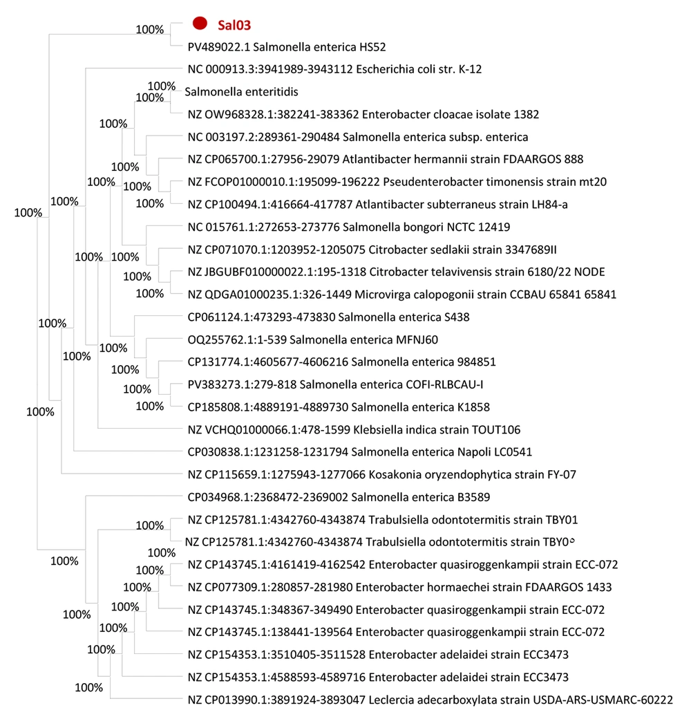
Figure 1.
Phylogenetic tree constructed using MEGA X based on genetic sequences of Sal03 and related strains – the Sal03 isolate (marked in red) clusters with PV489022.1 Salmonellaenterica HS52, showing 100% genetic similarity
4.3. Enhanced Lytic Activity of Phages Against Salmonella Strains and the Synergistic Efficacy of Their Combined Phage Cocktail
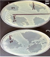
Based on the data presented in Table 3, PhSal04 and PhSal06 exhibited significant lytic activity against most of the Salmonella strains tested (Sal03, Sal04, Sal05, PTCC 1230, and Sal07). Specifically, PhSal04 displayed strong lytic activity (+++) against Sal04, while showing moderate activity (++) against Sal03, Sal05, PTCC 1230, and Sal07, with no activity observed (-) against Sal01. PhSal06 demonstrated robust lytic activity (+++) against Sal03, Sal04, Sal05, and Sal07, and moderate activity (++) against PTCC 1230. In contrast, the other bacteriophages tested (PhSal01, PhSal03, and PhSal05) showed limited or inconsistent activity across the Salmonella strains. This suggests that PhSal04 and PhSal06 are more effective in targeting a broad range of Salmonella strains, with PhSal04 showing particular strength against Sal04 and PhSal06 having a strong effect on multiple strains. PhSal04 also reduced the growth of Sal05 and Sal07, although to a lesser degree. Remarkably, the phage cocktail composed of PhSal06 and PhSal04 exhibited even greater lytic activity (+++) against all tested Salmonella strains, except Sal01. This indicates that combining PhSal06 and PhSal04 significantly enhances the bactericidal efficacy compared to using each phage individually, as shown in Figure 2C.
Table 3.
Lytic Activity of Individual Bacteriophages and Phage Cocktail Against Salmonella Strains
| Strains | PhSal01 | PhSal03 | PhSal04 | PhSal05 | PhSal06 | Cocktail Sal06 + Sal04 |
|---|---|---|---|---|---|---|
| Sal01 | - | ++ | - | - | - | - |
| Sal03 | +++ | - | ++ | - | +++ | +++ |
| Sal04 | - | - | +++ | - | +++ | +++ |
| Sal05 | + | - | ++ | +++ | +++ | +++ |
| Sal07 | ++ | - | ++ | - | +++ | +++ |
| PTCC 1230 | - | - | ++ | - | ++ | +++ |
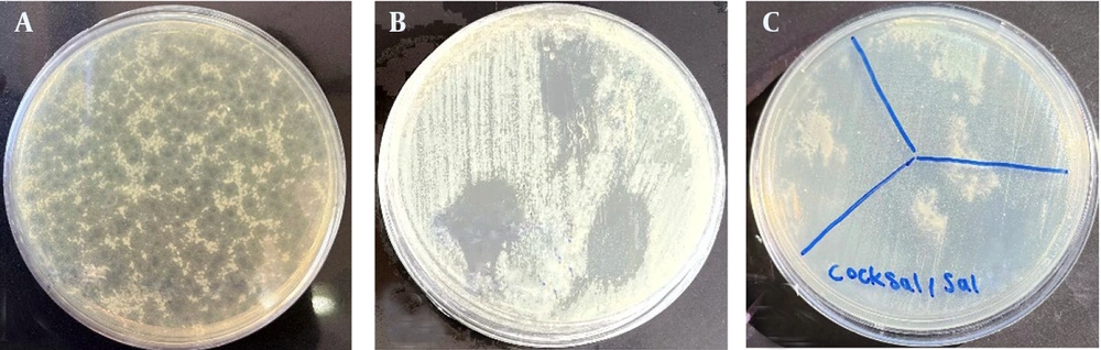
Figure 2.
Plaque formation and lytic activity of phage PhSal04 and phage cocktail: The figure highlights the lytic potential and specificity of PhSal04 alone and in combination against targeted bacterial strains – A, plaques formed by phage PhSal04 on a double-layer agar plate; B, the activity range of bacteriophage PhSal04 against the bacterium PTCC 1230; and C, the lytic effects of the phage cocktail on the bacterium Sal03.
Table 3 presents the qualitative lytic activity of individual bacteriophages (PhSal01, PhSal03, PhSal04, PhSal05, and PhSal06) as well as a phage cocktail (PhSal06 + PhSal04) against various Salmonella strains (Sal01 to Sal07). The lytic effects were evaluated based on spot test results and categorized as follows: “+++” indicates clear and translucent plaques, “++” indicates slightly turbid plaques, “+” indicates turbid plaques, and “-” indicates no visible plaques.
4.4. Bacteriophage Isolation and Characterization
From 12 water samples collected from hospital pools in Mazandaran province, five distinct bacteriophages, designated as PhSal01, PhSal03, PhSal04, PhSal05, and PhSal06, were successfully isolated, all specifically targeting Salmonella strains (Figure 2A - C). The morphology of isolated bacteriophages was examined using transmission electron microscopy (TEM). PhSal01 (Siphoviridae) had an elongated, noncontractile tail measuring 7 × 10² nm and a head with dimensions of 64 × 71 nm; PhSal04 (Siphoviridae) had a head diameter of 37 × 40 nm and a thin tail length of 8 × 153 nm; PhSal06, classified under the Myoviridae family, is characterized by its robust, contractile tail measuring 15 × 152 nm and a head diameter of 71 × 72 nm, resulting in a distinct and durable structure; and PhSal05 (Siphoviridae) had a head diameter of 25 × 30 nm and a thin tail length of 7 × 70 nm (Figure 3A - D).

Figure 3.
Morphological analysis of phages PhSal01, PhSal04, PhSal06, and PhSal05 using transmission electron microscopy (TEM) – TEM images showing A, PhSal01 (Siphoviridae) with an elongated, noncontractile tail measuring 7 × 10² nm and a head with dimensions of 64 × 71 nm; B,PhSal04 (Siphoviridae) with a head diameter of 37 × 40 nm and a thin tail length of 8 × 153 nm; C, PhSal06 classified under the Myoviridae family, characterized by its robust, contractile tail measuring 15 × 152 nm and a head diameter of 71 × 72 nm; and D, PhSal05 (Siphoviridae) with a head diameter of 25 × 30 nm and a thin tail length of 7 × 70 nm.
4.5. One-Step Growth Curve for Bacteriophages
The one-step growth curves of phages PhSal01, PhSal04, and PhSal06 were analyzed. The latency periods for PhSal01, PhSal04, and PhSal06 were determined to be 25, 20, and 10 minutes, respectively. PhSal06, with a shorter latency period, exhibits a higher replication and release rate compared to the other two phages (Figure 4A - C).

Figure 4.
One-step growth curves of phages PhSal01, PhSal04, and PhSal06: One-step growth curves showing the latency periods of A, PhSal01 (25 minutes); B, PhSal04 (20 minutes); and C, PhSal06 (10 minutes).
4.6. Optimizing Multiplicity of Infection for Enhanced Antibacterial Activity of Bacteriophages
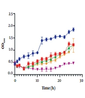
The antibacterial activity of bacteriophages PhSal01, PhSal04, and PhSal06 was tested at multiplicity of infection (MOI) 0.01, 0.1, 1, and 10. Higher MOI significantly enhanced bacterial growth inhibition. For example, at MOI 10, PhSal06 limited bacterial density to 0.4 over six hours, compared to the control (1.1). Lower MOI (0.1) resulted in increased bacterial density (PhSal06: 0.52 vs. control: 1.1), highlighting the importance of optimizing MOI for effective phage therapy (Figure 5A - C).
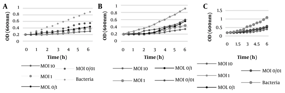
Figure 5.
Multiplicity of infection (MOI) of phages PhSal01, PhSal04, and PhSal06: The MOI results for ; A, PhSal01; B, PhSal04; and C, PhSal06.
4.7. Bacteriophage Stability Against Heat and pH
The thermal stability of phages PhSal06, PhSal04, and PhSal01 was tested at 30 - 80°C for 30 and 60 minutes (Figure 6A - C). The pH stability was assessed between pH 4 - 12, with optimal activity at pH 7 (log PFU/mL 7.55 - 8.25). Activity dropped below detection limits at pH ≤ 3 and ≥ 13, demonstrating 100% stability within pH 4 - 12 (Figure 7A - C).

Figure 6.
Thermal stability of ; A, PhSal01; B, PhSal04; and C, PhSal06 at temperatures ranging from 30°C to 80°C after 30 minutes (red) and 60 minutes (black) of exposure – phage activity remained stable (log PFU/mL 8 - 9) at ≤ 60°C but was lost at ≥ 70°C.

Figure 7.
The stability of phages ; A, PhSal01; B, PhSal04; and C, PhSal06 against pH were assessed over a wide range, between pH 4 and 12
4.8. Antimicrobial Activity of Bacteriophages in Meat
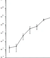
The assessment of live cells in meat artificially contaminated with the bacterium Sal03 showed that after treatment with PhSal06 at 25°C, the number of live cells decreased from log CFU/mL 3.51 at zero time to log CFU/mL 2.91 after three hours. However, bacterial regrowth was observed after 24 hours, reaching log CFU/mL 5.33 after 96 hours. In the control group, the live cell titer increased from log CFU/mL 3.72 at zero time to log CFU/mL 6.12 after 96 hours.
When meat contaminated with Sal01 was treated with PhSal06 at 4°C, the number of live cells decreased from log CFU/mL 3.57 at zero time to log CFU/mL 2.82 and log CFU/mL 2 after three and twelve hours, respectively. No bacterial cells were detectable after 24 hours, and no bacterial cells were observed even after 96 hours. In contrast, in the control group, the bacterial cell titer increased to log CFU/mL 5.32 after 96 hours. These results indicate complete lysis of host bacteria by the studied bacteriophages (Figure 8A and B).
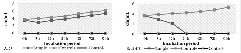
Figure 8.
Antimicrobial activity of bacteriophage PhSal06 against Salmonella enterica Sal03 in meat: A, reduction of 2.91 log CFU/mL of S. enterica Sal03 cells after 3 hours at 25°C; B, reduction of 2.82 log CFU/mL of S. enterica Sal03 cells after 3 hours and complete elimination after 24 and 96 hours at 4°C (abbreviations: MOI, multiplicity of infection; CFU/mL, colony forming units per mL).
4.9. Statistical Analysis of Lytic Activity in Bacteriophages and Phage Cocktail
A one-way ANOVA was performed to assess the mean lytic activity of the phages (PhSal01, PhSal03, PhSal04, PhSal05, and PhSal06) and the phage cocktail (PhSal06 + PhSal04) against Salmonella strains (Sal01 to Sal07). The null hypothesis assumed no significant difference in lytic activity across the groups. The calculated F-statistic was 6.25, with a P-value of 0.004, which is below the significance threshold of 0.05. This indicates a statistically significant difference in lytic activity between the phages and the phage cocktail. To identify specific group differences, Tukey’s HSD test was conducted. The results revealed that PhSal04 and PhSal06 exhibited significantly stronger lytic activity against Salmonella strains compared to the other phages. Additionally, the phage cocktail (Sal06 + Sal04) demonstrated comparable or superior lytic activity to most individual phages.
5. Discussion
Bacteriophages are effective biocontrol agents due to their high specificity, stability across temperatures and pH, and recognized safety by regulatory agencies such as the United States Food and Drug Administration (FDA) and Department of Agriculture (USDA) (37-39). In our study, we evaluated the thermal stability of bacteriophage PhSal06, which retained lytic activity between 30°C and 60°C, though its effectiveness declined at higher temperatures. Our findings are consistent with previous reports, demonstrating that the phage remains stable at common food storage temperatures, making its use in food safety both practical and effective (35). Our phages also demonstrated broad pH tolerance, enhancing their applicability across various food products, provided exposure to extreme pH is avoided. PhSal06, in particular, showed strong antibacterial performance in contaminated meat stored at 4°C, where it significantly reduced bacterial load over time. These results align with earlier findings on the effectiveness of phages against Salmonella strains in refrigerated poultry products (35, 40).
Comparative analysis with the study “Application of Phage LPSE1 in Controlling Foodborne S. enterica” further supports our observations. LPSE1 showed good activity in milk at 28°C and in sausage and lettuce at both 4°C and 28°C, although reduced efficacy was noted at 4°C in milk due to slower bacterial metabolism (27). Our findings show that the isolated phages, especially PhSal06, are highly stable under diverse environmental conditions, supporting their potential for industrial use. Their stability enables long-term storage, transport, and effective application in food production, offering a safer and more sustainable alternative to conventional chemical methods. These results underline the promise of phage-based strategies for controlling Salmonella in food products.
The findings of the three recent studies (41-43), together with our results, demonstrate that phage cocktails exhibit significantly higher efficacy in controlling Salmonella compared to individual phages. In the present study, the combination of PhSal04 and PhSal06 was able to inhibit a broader spectrum of Salmonella strains, with the synergistic interaction between the phages leading to the suppression of almost all tested strains. These results not only enhanced the overall bactericidal efficiency but also reduced the likelihood of bacterial resistance, which is consistent with previous reports highlighting the superiority of multi-phage formulations over single-phage applications. Torkashvand et al. (43) reported that a three-phage cocktail, in addition to being safe in human cell lines, significantly reduced Salmonella in various food products and in a broiler chicken model, while also promoting growth performance.
This finding suggests that phage cocktails may serve not only as antibacterial agents but also as potential biological growth promoters in the poultry industry. Similarly, Islam et al. (42) demonstrated that a cocktail of three phages belonging to different families (Siphoviridae, Ackermannviridae, and Podoviridae) effectively reduced the bacterial load in milk and chicken meat at both 4°C and 25°C, and significantly disrupted Salmonella biofilms on microplates and stainless-steel surfaces. This emphasizes the importance of phage cocktails in industrial hygiene and the control of secondary contamination in food processing environments.
Furthermore, Gvaladze et al. (41) showed that phage cocktails can also be highly effective under cold-chain conditions. In their study, the cocktail successfully reduced five major Salmonella serotypes even at low temperatures (down to 8°C) and at very low MOIs. Such a feature is particularly relevant for refrigerated food storage, where Salmonella can persist and serve as a potential source of foodborne infections.
Notably, when compared with conventional chemical sanitizers such as chlorine and peracetic acid, our phage cocktail (PhSal06 + PhSal04) achieved comparable or even greater reductions in Salmonella populations. While chemical sanitizers typically reduce contamination by only 1 - 3 log CFU/mL under commercial conditions, their application may alter the flavor, odor, or texture of meat and may also leave undesirable chemical residues (44, 45). In contrast, our cocktail achieved a 2.91 log CFU/mL reduction at 25°C and complete elimination of Salmonella at 4°C within 24 hours, with no regrowth over 96 hours. Moreover, unlike chemical agents, bacteriophages maintain activity during storage, providing prolonged protection against bacterial proliferation without compromising product quality or safety (46, 47).
Overall, our findings support the potential of phages PhSal01, PhSal04, and PhSal06 as effective tools for controlling Salmonella strains in diverse food environments, encouraging further research into their commercial applications in food safety. Our study includes phenotypic, morphological (TEM), host range, and genetic analyses of the phages, but complete whole-genome sequencing was not performed. Incorporating full genomic data in future studies could further confirm the absence of undesirable genes, provide more precise taxonomic placement, and better support biosafety assessment and the broader application of these phages.
5.1. Conclusions
This research represents the first report from Babol, Mazandaran province, Iran, on the successful isolation of anti-Salmonella bacteriophages from meat and their application in a food matrix. This study supports the use of bacteriophages to reduce S. enterica Sal03 in meat, showing that refrigeration enhances their effectiveness. PhSal01, PhSal04, and PhSal06 demonstrated strong antibacterial activity, with key findings on MOI optimization, thermal and pH stability, and real-world application. The results align with previous research, reinforcing the potential of phages in food safety.