
1. Background
Type A influenza virus is the most important member of the Orthomyxoviridae family, as it can infect a wide range of hosts including humans, birds, and various mammals; however, birds are the primary natural reservoir of influenza viruses. Due to its eight-segmented genome, it is possible for the virus to generate a human-adapted virus through re-assortment, resulting in a surface protein for which humans have no pre-existing immunity (1). Highly pathogenic avian influenza virus (H5N1) is extremely contagious and deadly among birds, especially in domestic poultry. It can be transmitted to humans and poses a serious threat to public health. The first human infection with avian influenza H5N1 was reported in Hong Kong in 1997. According to reports from the World Health Organization (WHO), there have been 887 confirmed human cases of avian influenza A/H5N1 from 2003 to 2024, with a mortality rate of approximately 52% (2).
Hemagglutinin (HA) is of great importance as the antigenic determinant of influenza virus, as it binds to sialic acid on the cell surface and mediates viral entry; this antigen also elicits immune responses during natural infection or following vaccination. According to the WHO, the gold standard method for detecting and measuring antibodies in serum is the virus neutralization test (VNT). This method requires the use of live virus and, consequently, biological safety level 3 (BSL-3) or higher facilities, which are labor-intensive and not always available (3, 4). An alternative approach is to construct a pseudovirus expressing the HA glycoprotein on the surface of non-replicative viral particles for use in neutralization assays. These particles undergo abortive replication and do not produce replication-competent progeny, eliminating the possibility of re-assortment or viral escape from the laboratory.
Pseudotyped particles, or pseudoviruses, consist of a viral core from one specific virus and an envelope protein at their surface from a different virus. The replication-related regions of the genome are deleted in the core virus genome, so that the particle does not have the ability to reproduce. The particle also carries a reporter gene to track the entry of the pseudovirus into the host cell. These particles enter susceptible cells by binding, via their envelope glycoproteins, to the appropriate receptor on the surface of the targeted cells. After cell transduction, the viral core of the pseudovirus integrates into the transduced cell, and the reporter gene is expressed (5). Neutralizing antibodies prevent the pseudovirus from multiplying in vitro, in the same way as they would inhibit live virus, and the presence of these antibodies, as in other infectious diseases, is associated with the development of immunity against the disease (6).
2. Objectives
Here, a pseudovirus harboring H5 HA was constructed using retroviral vectors in order to assess the potential of the pseudovirus system for neutralization assays in serum samples from vaccinated chickens. The generated pseudovirus will be utilized for the measurement of virus-neutralizing antibodies in vaccine research and development.
3. Methods
3.1. Plasmids
The full gene sequences of H5 and N1 from influenza A/Indonesia/5/2005(H5N1) were obtained from NCBI, synthesized, and cloned into pUC (Biomatik, Denmark). The gene sequences were subcloned into the pcDNA3.1 expression plasmid under the CMV promoter using XhoI and BamHI restriction enzymes for H5, and XhoI and HindIII restriction enzymes for NA. The confirmed recombinant vectors were designated as pcDNA-HA and pcDNA-NA. The second-generation lentiviral packaging plasmid (psPAX2 plasmid #12260), green fluorescent protein (GFP) reporter plasmid (pLOX-CWgfp plasmid #12241), and vesicular stomatitis virus (VSV)-G envelope expressing plasmid (pMD2.G plasmid #12259) as a positive control were gifts from Didier Trono (Addgene, USA) (7). All plasmids contained an ampicillin resistance gene, and the sequences are available in the Addgene database. Cloning procedures were performed in E. coli TOP 10 f’, and plasmid purification was conducted using the QIAGENE Maxiprep Kit (#12162).
3.2. Cell Culture and Transfection
Human embryonic kidney cells (HEK293T) provided by the Pasteur Institute of Iran (NCBI code: C498) were cultured in high-glucose Dulbecco's Modified Eagle's Medium (DMEM) with GlutaMAX, containing 10% fetal bovine serum (FBS), penicillin (100 U/mL), and streptomycin (100 μg/mL). Cells were incubated at 37°C in a humidified atmosphere with 5% CO2 until reaching the desired confluency. Lipofectamine 2000 (Thermofisher, USA) was used as the cationic reagent to transfect eukaryotic cells that were seeded 24 h before transfection in a 6-well plate (9 × 10⁵ cells/well) with medium containing serum and antibiotics. Cells were then washed once with PBS, and plain DMEM was used in the transfection steps. Plasmid concentration and Lipofectamine ratios were used according to the Thermofisher protocol.
To evaluate proper HA expression and plasmid function, the pcDNA-HA plasmid was transfected into Madin-Darby canine kidney (MDCK) cells. The transfection medium was replaced with DMEM containing 10% FBS at 24 h post-transfection, and the cells were incubated for another 24 h. The H5 expression on the surface of eukaryotic cells was examined using a hemadsorption test with a 0.3% chicken red blood cell (RBC) suspension. Briefly, after washing transfected cells with PBS, the RBC suspension was added to the plate and incubated for 30 min at room temperature, then washed thoroughly with PBS. Finally, the RBC accumulation pattern on the cell surface was observed under a light microscope (8).
3.3. Generation of Lentiviral-Based Pseudovirus
The H5 pseudovirus was produced by transfection of HEK293T cells with pcDNA-HA, pLOX, and psPAX plasmids using Lipofectamine 2000. The workflow steps are presented schematically in Figure 1.
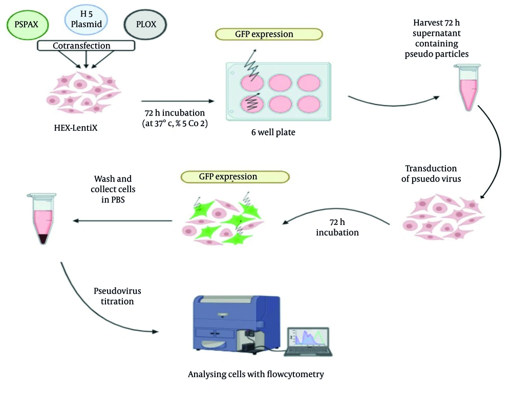
Figure 1.
Schematic presentation of pseudovirus production and titration
To optimize the transfection, different proportions of the three HA:pLOX:psPAX plasmids were used, with or without the NA-encoding plasmid, as well as the addition of sodium butyrate to increase cell permeability. The NA plasmid was tested at a 1:5 ratio to HA. The DNA mixture was added to the Lipofectamine solution at a volume ratio of 1:1. For the positive control, the plasmid containing the VSV glycoprotein gene (pMD2G) was used instead of the HA plasmid. At 24 h following transfection, 1 unit of exogenous neuraminidase from Vibrio cholerae (Sigma-Aldrich, #N7885) type III enzyme was added to each well of the 6-well plate to improve the release of pseudoviruses from the cells. The effect of sodium butyrate (Sigma-Aldrich #156-54-7) on transfection efficiency was also investigated, and a 5 mM concentration was added to the cell medium 24 h post-transfection (9). At 48 and 72 h post-transfection, GFP expression was monitored using a fluorescence microscope, and the supernatant containing the pseudovirus was harvested and stored at -80°C.
Pseudovirus production was also confirmed by measurement of P24 protein using an ELISA kit (PishtazTeb, PT-HIV 1,2 AG-AB-96, Iran). P24 is one of the structural proteins of human immunodeficiency virus (HIV), which is expressed by the primary gag gene in the pseudovirus packaging plasmid (psPAX). Detection of this protein in the supernatant of transfected cells confirms the formation of pseudovirus. The light absorbance of the controls and samples was measured using an ELISA reader at a wavelength of 450 nm. The cut-off was calculated as 0.101 based on the kit information. The “Sample OD / Cut-off” value was used to determine the positive and negative responses (S/Co Index). According to this formula, samples with S/Co of 1 or more were considered positive, while values less than 1 were considered negative. All experiments were performed in 3 independent duplicates to confirm the reproducibility of the results.
3.4. Transduction and Pseudovirus Titration
The infectivity efficiency of the pseudovirus was investigated in HEK293T cell lines. Cells were seeded in 24-well plates (2 × 10⁵ cells/well) for 24 h. Transduction was performed with different dilutions of the harvested pseudovirus. Targeted cells were expected to express GFP as a result of successful pseudovirus entry and infectivity. After 72 h, the level of pseudovirus infectivity was assessed using a fluorescence microscope, and the level of GFP expression was measured by flow cytometry analyzer (Partec, Germany), using WindowsTM FloMax® software. The pseudovirus titer was determined according to the amount of GFP expression detected by flow cytometry using the following formula:
N = Percentage of GFP-expressing cells in flow cytometry; Cn = Number of cells at the time of transduction; DF = Dilution factor; V = Volume of pseudovirus inoculated at the time of transduction (mL); pseudovirus titer = Transduction units per mL (TU/mL).
To optimize transduction, cells were inoculated with different multiplicities of infection (MOIs) and incubation times (4 h and 24 h inoculation times). Additionally, polybrene (Sigma-Aldrich TR-1003) was used to investigate its effect on pseudovirus entry and induction.
3.5. Neutralization Assay
Serum samples from vaccinated chickens, as well as positive and negative control sera, were used to investigate the feasibility of the pseudovirus system for neutralization assays. The control sera were evaluated and confirmed by hemagglutination inhibition (HI) test at Tehran University of Veterinary Medicine (10). The serum samples were heat-inactivated at 56°C for 30 min. Two-fold serial dilutions of sera (starting with 1:16) were prepared in DMEM. An equal amount of pseudovirus (2 × 10⁵ TU/mL) was added to each dilution (ratio 1:1) and incubated for 1 h at room temperature. The mixture of serum and pseudovirus was added to HEK293T cells in a 24-well plate seeded the day before and incubated for 24 h at 37°C in a CO2 incubator. Then, culture medium containing 2% FBS was added to the cells and maintained in the incubator for up to 72 h. To verify GFP expression, the plate was observed under a fluorescence microscope. Subsequently, cells were detached, washed twice with PBS, and the percentage of GFP-expressing cells and pseudovirus titer were measured by flow cytometry.
Additionally, the cross-reactivity of chicken serum containing anti-H5 antibodies was investigated with H1N1 and H3N2 human influenza viruses in a neutralization assay. Two-fold serial dilutions of human and chicken sera were mixed with equal volumes of H1N1 or H3N2 (100 TCID50) and incubated for 1 h at room temperature. The mixture was then added to MDCK cells in a 96-well plate (3 × 10⁴ cells/well) and placed in a CO2 incubator for 48 h. The cytopathic effects caused by H1 and H3 viruses were monitored.
3.6. Statistical Analysis
GraphPad Prism Version 10.2.2 was used for the statistical analysis. The pseudovirus titer and other measured parameters at all stages of the study were analyzed by t-test. A P-value less than 0.05 was considered significant.
4. Results
4.1. Cloning and Cell Transfection
The full lengths of the HA and NA genes were subcloned into the pcDNA3.1 plasmid and confirmed by colony PCR and enzymatic digestion (data not shown). The accuracy of gene cloning was also confirmed by sequencing of the pcDNA-HA and pcDNA-NA plasmids. Single plasmid transfection of MDCK cells with pcDNA encoding the HA sequence was performed, and the transfected cells were exposed to a 0.3% RBC suspension after 48 h. The HA function and expression on the transfected cells were confirmed by hemadsorption test. As shown in Figure 2, the transfected cells were surrounded by RBCs. The cells were exposed to a 0.3% RBC suspension 48 h post-transfection to observe hemadsorption. Binding of RBCs to the surface of the transfected cells (B) was clearly visible compared to the negative control cells (A, magnification: 40×).
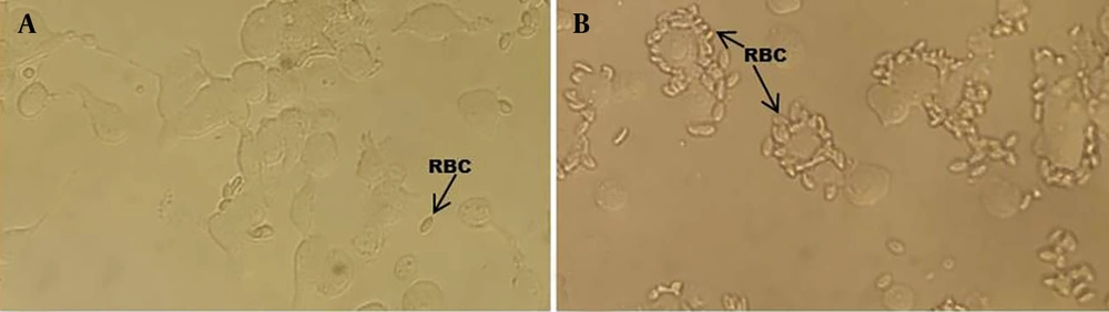
Figure 2.
Hemadsorption assay on HEK cells transfected with pcDNA-H5
4.2. Optimization and Evaluation of H5 Influenza Pseudovirus
Co-transfection of three plasmids into HEK293T cells was carried out using Lipofectamine 2000 reagent. Transfection steps were optimized as described earlier, and successful transfection was confirmed by observation of the GFP reporter gene. Evaluation of different ratios of the three plasmids in co-transfection determined that the greatest efficiency (highest GFP expression) was achieved at a total of 3200 ng of DNA plasmids per well of a 6-well plate, with a ratio of 1.5:1.5:1 of HA:pLOX:psPAX, respectively. To determine the optimal timing for harvesting pseudovirus at 48 or 72 h post-transfection, the supernatant of transfected cells was harvested and P24 expression was assessed using the ELISA test (Figure 3). The “Sample OD / Cut-off” value of pseudoviruses harvested at different times showed a significant increase at 72 h compared to 48 h (P < 0.05).
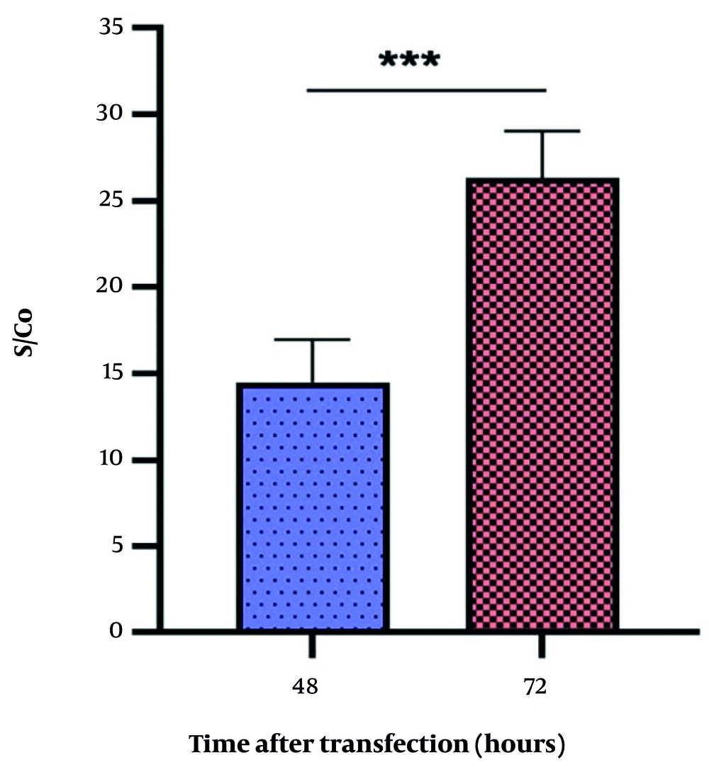
Figure 3.
P24 expression: The chart shows the P24 expression level in cells at 48 and 72 h post-transfection (*** highly significant, P < 0.001).
To optimize the transfection method, 10 μL of 1 M sodium butyrate stock was directly added to each well at 24 h after transfection to achieve a final concentration of 5 mM, to increase the permeability of cell membranes (9). No significant difference was observed between cells with and without sodium butyrate by flow cytometry at 72 h after transfection (data not shown). Since this substance had no effect on increasing GFP expression, it was omitted from the transfection protocol.
The infectivity of pseudovirus was investigated in HEK293T cell lines. Cells were inoculated with different dilutions (1:1, 1:10, 1:100) of the harvested pseudovirus. Transduced cells were confirmed by GFP expression. The pseudovirus titer was determined according to the GFP expression level detected by flow cytometry (Figure 4 and Appendix 2 found in Supplementary File). According to the formula mentioned previously, the titer of H5 pseudovirus was quantified as (2.1 ± 0.13) × 105 TU/mL.
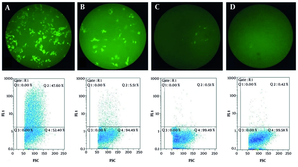
Figure 4.
Transduction results: The images show fluorescence microscope images (top panel, magnification: 10×) and flow cytometry results (bottom panel) of HEK293T cells transduced with different dilutions of H5 pseudovirus; A - C, 1:1, 1:10, and 1:100 dilutions of H5 pseudovirus, respectively; D, negative control. The numerical value of Q2 indicates the quantitative amount of fluorescent emission. The images are representative of 3 independent experiments.
Additionally, during the transfection, the effect of neuraminidase was investigated to increase pseudovirus budding into the supernatant. Microscopic observation and examination of the transduced cells using the flow cytometry analyzer revealed that adding neuraminidase enzyme 24 h after co-transfection of HA:pLOX:psPAX and the NA encoding plasmid resulted in a significantly (P < 0.05) higher titer of pseudovirus (6.1 × 10⁵ TU/mL) than transfection without exogenous enzyme (4.9 × 10⁴ TU/mL) or with neither NA plasmid nor exogenous enzyme (2.8 × 10⁴ TU/mL).
To investigate the effect of initial incubation time, cells were inoculated with pseudovirus and allowed for virus absorption and penetration. Following 4 or 24 h incubation, the medium was replaced and the cells were incubated up to 72 h and examined for GFP expression and flow cytometry analysis. It was revealed that 24 h incubation led to 2-fold higher GFP expression in targeted cells. Assessing the effect of polybrene as a cationic polymer, which increases cell permeability, showed that it can mildly increase the number of GFP-positive cells at an 8 μg/mL concentration to 7.3 × 10⁴ TU/mL compared to the control (6.1 × 10⁴ TU/mL), which was not statistically significant (P > 0.05).
4.3. Pseudovirus Neutralization Test
To set up the neutralization test, heat-inactivated negative and positive chicken control sera (both verified by the HI test) were serially diluted, mixed with the H5 pseudovirus or VSV pseudovirus, and incubated for 1 h at room temperature. The HEK293T cells were inoculated with the mixture and, after 72 h, pseudovirus titer was measured by flow cytometry. The IC50 neutralizing titer was defined as the serum dilution that was capable of a 50% reduction in pseudovirus titer (6). The mixture of pseudovirus and an equal volume of culture medium was also inoculated to determine the pseudovirus titer as the virus control. As presented in Table 1, which shows low variations and highlights three independent repeats of experiments, the 1:32 and 1:64 dilutions of the positive control serum neutralized the virus. In parallel, VSV pseudovirus was used as a control and showed no significant titer reduction when mixed with diluted positive control serum (Appendix 1 can be found in Supplementary File). As expected, negative control serum did not neutralize H5 pseudovirus (data not shown).
Table 1.
Neutralization Assay of Control Chicken Sera Using H5 and Vesicular Stomatitis Virus Pseudoviruses
| Variables | Transduction Unit per mL (TU/mL) | |||||
|---|---|---|---|---|---|---|
| Serum Sample Dilution | Virus Control (No Serum) | 50% of Virus Control | ||||
| 1:32 | 1:64 | 1:128 | 1:256 | |||
| H5 pseudovirus | 5.40 × 10³ | 1.54 × 10⁴ | 2.46 × 10⁴ | 2.93 × 10⁴ | 3.28 × 10⁴ | 1.66 × 10⁴ |
| 6.68 × 10³ | 1.10 × 10⁴ | 2.12 × 10⁴ | 3.10 × 10⁴ | 3.35 × 10⁴ | ||
| 7.80 × 10³ | 1.64 × 10⁴ | 2.36 × 10⁴ | 2.84 × 10⁴ | 3.36 × 10⁴ | ||
| VSV pseudovirus | 2.84 × 10⁴ | 2.93 × 10⁴ | 2.95 × 10⁴ | 3.16 × 10⁴ | 3.19 × 10⁴ | 1.60 × 10⁴ |
| 2.70 × 10⁴ | 2.82 × 10⁴ | 2.84 × 10⁴ | 3.08 × 10⁴ | 3.23 × 10⁴ | ||
| 2.69 × 10⁴ | 2.83 × 10⁴ | 2.89 × 10⁴ | 3.11 × 10⁴ | 3.19 × 10⁴ | ||
Abbreviation: VSV, vesicular stomatitis virus.
To investigate the feasibility of the pseudovirus system for neutralization assays, the titer of neutralizing antibody in twelve chicken sera from a farm vaccinated with the commercial H5 vaccine (Poulvac FluFend) was evaluated in a VNT using H5 pseudovirus. A serum dilution that could reduce the pseudovirus titer by 50% was considered the IC50 neutralizing titer for each serum. Sera with neutralizing titers are shown in Table 2. Given the positive control unit transduction (3.35 × 10⁴), dilutions of serum in which the virus titer was equal to or less than 1.67 × 10⁴ were considered as neutralizing titers. The experiment was performed twice.
Table 2.
Neutralizing Anti-H5 Antibody Titer of 12 Chicken Sera Using H5 Pseudovirus a
| Serum | Transduction Unit per mL (TU/mL) | |||
|---|---|---|---|---|
| 1:64 | 1:128 | 1:256 | 1:512 | |
| 1 | (5.53 ± 0.13) × 103 | (1.57 ± 0.26) × 104 | (2.45 ± 0.21) × 104 | (2.87 ± 0.66) × 104 |
| 2 | (3.32 ± 0.32) × 103 | (1.07 ± 0.43) × 104 | (1.49 ± 0.28) × 104 | (2.46 ± 0.33) × 104 |
| 3 | (5.75 ± 0.25) × 103 | (1.18 ± 0.05) × 104 | (2.23 ± 0.37) × 104 | (3.43 ± 0.26) × 104 |
| 4 | (8.40 ± 0.16) × 103 | (1.22 ± 0.27) × 104 | (2.59 ± 0.88) × 104 | (3.77 ± 0.73) × 104 |
| 5 | (1.13 ± 0.21) × 104 | (1.55 ± 0.30) × 104 | (2.34 ± 0.47) × 104 | (2.60 ± 0.81) × 104 |
| 6 | (3.28 ± 0.05) × 103 | (5.70 ± 0.30) × 103 | (1.46 ± 0.35) × 104 | (2.32 ± 0.32) × 104 |
| 7 | (5.32 ± 0.20) × 103 | (8.15 ± 0.07) × 103 | (1.72 ± 0.09) × 104 | (2.77 ± 0.65) × 104 |
| 8 | (7.09 ± 0.41) × 103 | (1.17 ± 0.68) × 104 | (2.16 ± 0.40) × 104 | (2.99 ± 0.96) × 104 |
| 9 | (1.06 ± 0.55) × 104 | (1.07 ± 0.32) × 104 | (1.47 ± 0.34) × 104 | (2.12 ± 0.18) × 104 |
| 10 | (1.64 ± 0.06) × 104 | (1.61 ± 0.57) × 104 | (1.82 ± 0.44) × 104 | (2.12 ± 0.24) × 104 |
| 11 | (1.48 ± 0.44) × 104 | (1.40 ± 0.23) × 104 | (1.90 ± 0.05) × 104 | (2.65 ± 0.52) × 104 |
| 12 | (7.52 ± 0.28) × 103 | (1.63 ± 0.13) × 104 | (2.33 ± 0.33) × 104 | (2.75 ± 0.97) × 104 |
| Negative | (2.85 ± 0.71) × 104 | (2.81 ± 0.41) × 104 | (3.22 ± 0.43) × 104 | (3.29 ± 0.88) × 104 |
a Values are expressed as mean ± SD.
The titer of neutralizing antibody for each serum sample detected by pseudovirus-based virus neutralization test (pVNT) was compared with anti-HA antibody titer obtained by HI test, as shown in Table 3.
Table 3.
Comparison of Serological Assays, Pseudovirus-Based Virus Neutralization Test and Hemagglutination Inhibition, for Detection of Anti-H5 Antibodies in Chicken Sera a
| Serum Titer | Serum ID | ||||||||||||
|---|---|---|---|---|---|---|---|---|---|---|---|---|---|
| 1 | 2 | 3 | 4 | 5 | 6 | 7 | 8 | 9 | 10 | 11 | 12 | Positive Serum | |
| pVNT | 7 | 8 | 7 | 7 | 7 | 8 | 7 | 7 | 8 | 7 | 7 | 7 | 6 |
| HI | 10 | 9 | 9 | 10 | 10 | 10 | 10 | 10 | 10 | 9 | 9 | 9 | 9 |
Abbreviations: pVNT, pseudovirus-based virus neutralization test; HI, hemagglutination inhibition.
a The pVNT and HI titers are presented as log 2 of reciprocal serum dilution.
In the HI test, the dilution factor of the last well in which the RBCs are not agglutinated indicates the anti-HA titer. In the pVNT, a dilution of the sample that reduced the pseudovirus titer by more than 50% is reported as a neutralizing titer. The results showed a similar titer pattern for serum samples in both evaluation methods. The criterion for neutralization was the positive control serum, which was reported to have a dilution factor of 512 (log2 = 9) in the HI test and 64 (log2 = 6) in the pVNT.
4.4. H5 Pseudovirus Specificity in Cross-reaction
To investigate the specificity of the H5 pseudovirus reaction to antibodies against H5N1, a neutralization test was performed using human sera containing anti-H1 and anti-H3 antibodies, which were provided by the Pasteur Institute of Iran. Compared to the control sera, as shown in Figure 5, human sera containing neutralizing titers of anti-H1 and anti-H3 antibodies could not neutralize the H5 pseudovirus. The H5 pseudovirus was only inhibited by the anti-H5 chicken serum (positive chicken control serum).
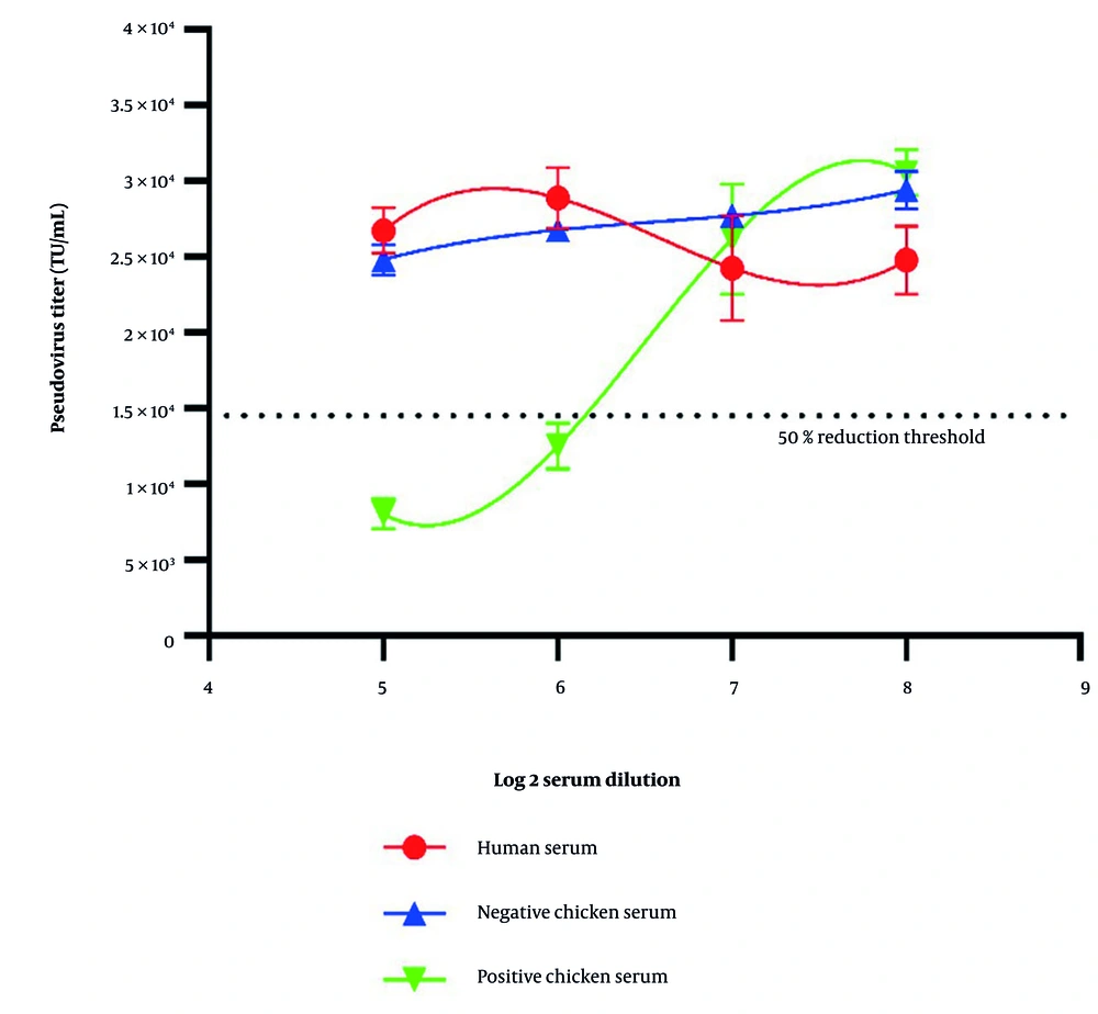
Figure 5.
Cross-neutralization of H5 pseudovirus with human sera
The results showed no neutralization by human serum. Negative (non-vaccinated chicken) and positive (containing anti-H5 antibody) chicken sera were tested in parallel. Additionally, a neutralization test was performed using human influenza viruses. Thus, 2-fold dilutions of positive chicken serum (containing anti-H5 antibody) and negative chicken serum (non-vaccinated chicken, no detectable anti-H5 antibody) were incubated with H1N1 and H3N2 human influenza viruses. Previously confirmed human serum containing anti-H1 and anti-H3 antibodies was used as a positive control and human serum with HI titer less than 32 was used as a negative control. As presented in Table 4, positive chicken serum could not neutralize the H3N2 influenza virus subtype, compared to the human serum. It is noteworthy that the chicken serum neutralized H1N1 virus at a 1:64 dilution. In negative sera, as expected, no non-specific binding was observed, and human serum containing antibody against human influenza viruses neutralized both H1 and H3 viruses up to a 1:512 dilution.
Table 4.
Cross-neutralization of Human Influenza Viruses (A/H1N1 and A/H3N2) and Chicken Sera
| Serum Type | Neutralizing Antibody Titer | |
|---|---|---|
| Influenza Virus A/H1N1 | Influenza Virus A/H3N2 | |
| Negative chicken serum | < 32 a | < 32 |
| H5 positive chicken serum | 64 | < 32 |
| Negative human serum | < 32 | < 32 |
| Positive human serum | 512 | 512 |
a The reciprocal of the last serum dilution that inhibited virus propagation are presented as neutralizing antibody titer.
5. Discussion
In this study, an influenza pseudovirus containing H5 HA was produced using retroviral vectors to evaluate its suitability for assessing virus-neutralizing antibodies in vaccinated chickens. Numerous studies have been conducted to produce inactivated or live attenuated highly pathogenic viruses to enable their study in biosafety level 2 (BSL-2) laboratories (11). One of the recently utilized solutions is the production of pseudoviruses. Pseudotype viruses or pseudotype particles are chimeric viruses that consist of a viral core surrounded by a lipid membrane containing the glycoprotein of another virus. These pseudoviruses are characterized by replication deficiency. By replacing a reporter gene, viruses such as avian influenza virus can be studied in a safe system (12-14).
For the influenza pseudovirus containing H5 HA produced in this study using retroviral vectors, the genes encoding HA (H5) and neuraminidase (N1) were cloned into the pcDNA3.1 expression vector under the CMV promoter. These vectors, along with psPAX vectors containing gag/pol genes and the pLOX-GFP plasmid as a lentiviral transfer vector (which also contains a reporter gene), were amplified after transformation into the host bacteria on a large scale and were extracted using the Qiagen Plasmid Maxi Kit. To produce the pseudovirus encoding the HA gene, three plasmids were simultaneously transfected at the appropriate ratio into HEK293T cells. The ratio between plasmids, cell density, virus harvest time, and the effects of other substances were optimized through several tests. Finally, the confirmed pseudovirus was successfully used to measure the neutralizing antibody titer in related chicken sera vaccinated with a commercial vaccine. This safe method can be used to measure virus-neutralizing antibodies.
All work was conducted using appropriate personal protective equipment and within certified BSL-2 cabinets. Surfaces and materials were disinfected regularly, and waste was autoclaved before disposal. Personnel involved were trained in BSL-2 procedures and followed strict standard operating protocols.
The first report of influenza pseudovirus production using VSV was published in 1972 (15). This virus-like system has been widely used in studies of highly pathogenic viruses, including highly pathogenic avian influenza (HPAI) virus and SARS-CoV-2. This system increases the availability of neutralization methods against these viruses (3, 6, 14). The VNT, as the main diagnostic test for the detection of neutralizing antibody in serum samples, requires BSL-3 equipment, which creates a barrier for such studies (16-18). Regarding the pseudovirus system, HA of the influenza virus on the surface of the pseudovirus is necessary for entry into the host cell. These viruses undergo a single-cycle replication and do not produce progeny virus, thus preventing the possibility of viral leakage from the laboratory. In addition to the influenza virus surface protein, pseudoviruses also encode a reporter gene. The neutralizing antibody titer is measured by the expression of the reporter protein (19).
The successful production of pseudovirus depends on several variables. Related studies have proposed different incubation times during transfection and transduction (20, 21). In this study, various time points were examined, and 72 h post-transfection was determined as the best harvest time. Several studies have investigated the effect of sodium butyrate on the production of pseudoviruses via transfection. Sodium butyrate, as a histone deacetylase inhibitor, selectively modifies gene transcription by altering chromatin and the structure of proteins involved in transcription. It has been reported that sodium butyrate can increase transfection efficiency, especially in plasmids containing the SV40 promoter (22-24). However, in this study, no specific effect was detected on pseudovirus production, which may be due to the high efficiency of Lipofectamine as a cationic reagent in transfection.
Lentiviral and retroviral vectors have been broadly utilized to produce pseudoviruses. In this study, the second-generation lentiviral packaging vector, psPAX (Addgene), was used to complete the viral structure containing influenza H5 on the surface. The packaging condition has been shown to be an effective factor in pseudovirus yield, and this vector has been successfully used in many studies to produce lentiviruses and pseudoviruses (25, 26).
The pVNT is a method of antibody detection that uses chimeric viruses displaying the surface glycoproteins of the virus of interest (such as influenza HA). H5 pseudoviruses possess the same ability to enter cells via the same receptors as wild-type influenza viruses, but can be safely handled under BSL-2 conditions. In addition, these pseudoviruses contain a reporter gene (such as GFP or luciferase) that is expressed only after cell entry. The greater the number of pseudoviruses entering the cells, the higher the intensity of fluorescence, allowing direct and quantitative measurement of viral particle entry by detecting light emission. Similar to the conventional VNT, the pseudovirus is mixed with serial dilutions of serum containing the target antibody, and light emission is measured using a flow cytometer or a fluorescence microscope. A significant reduction in emitted fluorescence indicates effective neutralization of the surface glycoproteins and blocking of viral entry.
The readout can be initially estimated by fluorescence microscope observation, which reduces assay time and increases throughput. In contrast, VNT using wild-type viruses relies on observation of cytopathic effects or other indicators of viral replication, which may require more time and greater technical expertise (27). Mixing serum and pseudovirus in equal volumes and incubating them with target cells constitutes a simple and standardized protocol. The assay does not require complex procedures such as egg inoculation, animal inoculation, or plaque counting. Moreover, pVNT can be performed with various pseudovirus platforms (such as HIV-1, VSV, or lentivirus) and different reporter genes (such as luciferase, GFP, or RFP), which provide greater flexibility and versatility for different applications.
Comparing pVNT titers with HI results demonstrated that, in addition to being reliable, the pVNT is also as sensitive as HI. The HI method is based on the biophysical interaction of blood cells with receptor binding sites on the wild-type virus. The major limitations of the HI test include the need for fresh RBCs, difficulties in wild virus preparation, and the possibility of non-specific serum inhibitors, such as beta lipoproteins. In contrast, the basis of the VNT method is the formation of the antigen-antibody complex, which makes this method more sensitive compared to HI (28-31).
Antibodies against influenza virus recognize different epitopes. Accordingly, antibodies against one subtype of influenza virus may inhibit other subtypes as well. Depending on the domain targeted by the antibody, there is a possibility of cross-reaction. We demonstrated that detection of neutralizing antibodies using H5 pseudovirus is highly specific. Human H1 and H3 antisera failed to inhibit H5 pseudotypes, and chicken H5 antisera failed to neutralize human H3 virus, but did cross-react with H1 virus up to a 1:64 dilution. H5 and H1 both belong to phylogenetic group 1, while H3 is in a separate group, which explains the observed cross-neutralization. These subtypes share conserved epitopes, particularly in the HA stem region, which is a target for broadly neutralizing antibodies. Sanz-Munoz et al. in 2025 also emphasized that cross-protection is more likely within the same HA group due to shared structural features. This also suggests that seasonal vaccines targeting group 1 HAs may offer partial protection against avian strains such as H5, but not against group 2 strains such as H3 (32). Our results are consistent with studies showing that H5 pseudovirus neutralization is dose-dependent and subtype-specific (33).
The main difference between pVNT and VNT is the use of pseudoviruses instead of wild-type viruses, making the assay safer, faster, and more convenient. However, pVNT may not fully reflect the antigenic properties of wild-type viruses, especially if there are mutations or variations in the surface glycoproteins. Therefore, it is important to validate the correlation of neutralizing activity measured by pVNT with other antibody neutralization methods for each new vaccine or therapeutic candidate (34).
This pseudovirus neutralization assay could be adapted for other influenza subtypes or zoonotic viruses. Its application in neutralization assays enables safe, high-throughput evaluation of antibody responses without handling live pathogenic viruses. This system can be readily adapted to other influenza subtypes — such as H7, H9, or even seasonal strains such as H1 and H3 — by substituting the HA gene in the pseudovirus construct. Moreover, the pseudovirus assay framework is highly transferable to emerging viruses, including coronaviruses and paramyxoviruses, facilitating rapid serological surveillance and vaccine efficacy testing. Such adaptability is crucial for pandemic preparedness and studies of cross-species transmission (32).
While pseudoviruses are valuable tools for medical laboratory diagnostic techniques and antiviral drug research, their stability over time must be considered when designing experiments. The stability of pseudoviruses is affected by several factors, such as storage temperature and duration, the backbone used for lentiviral production, pH of the storage medium, and the presence of proteases (35). It is important to note that the stability of pseudoviruses can be improved by optimizing storage conditions and the production process. Their stability can be enhanced by storing pseudoviruses at very low temperatures, such as -80°C, or by adding protease inhibitors to the storage buffer (36, 37).
5.1. Conclusions
This study demonstrated that VNTs based on pseudoviruses expressing H5 on their surface are reliable and safe alternatives for the detection of neutralizing antibodies to avian influenza virus. All procedures for pVNTs can be performed in routine BSL-2 laboratories, and most laboratories equipped with mammalian cell culture facilities meet this biosafety requirement. These systems can be used for broader applications with no threat or harm to the user, such as field diagnostics and vaccine research applications. They may also have potential for adaptation to other influenza subtypes for expanded clinical applications.