
1. Background
Despite strict drinking water quality controls and licensed water and wastewater treatment systems, waterborne illnesses remain one of the world's most serious public health challenges (1). Protozoa, viruses, and bacteria are widely recognized as the primary causes of waterborne illnesses (2). Rotavirus accounted for about 5% of all deaths in children and a cause-specific mortality rate of 86 deaths per 100,000 population of those below 5 years of age (3, 4). Enteric viruses, such as rotavirus, are significant sources of waterborne infections that can have serious economic and social consequences, particularly in underdeveloped countries (5). Viral gastroenteritis is typically induced by sewage contamination of drinking or recreational water (6). However, because treated wastewater is commonly utilized in agriculture, failing treatment operations may transmit infectious virus particles into agricultural areas, raising the risk of crop contamination (7). The enteric viruses are transmitted through the fecal-oral route; their presence in water sources may indicate fecal contamination, and monitoring these viruses may be an effective tool for monitoring fecal contamination in water systems (8). Rotavirus infection typically causes diarrhea, vomiting, fever, and abdominal pain (9). Currently, there is no specific treatment for rotavirus infection, and in most cases, it is a self-limited illness with spontaneous recovery within three to eight days (10).
Vaccination is the best way to prevent rotavirus infection. Live attenuated rotavirus vaccines that have already been approved by the World Health Organization (WHO) and received global/national licensing include Rotarix (RV1), RotaTeq (RV5), ROTAVAC, and ROTASIIL (11, 12). Although rotavirus vaccines have been introduced for infants in more than 100 countries, rotavirus-related mortality remains high in low-income countries (11, 12). Sewage is known to be one of the most concentrated sources of rotavirus in the environment, and contamination of other aquatic matrices is likely when there is damage to the sanitation network or when raw and/or inadequately treated wastewater is discharged into the environment (13). Information on the occurrence of rotavirus vaccine strains in aquatic and wastewater environments is very limited, and further research is needed to identify new genotypes of this virus from the wild-type RVA virus, originating from patients with gastroenteritis, in aquatic and wastewater environments (14).
Based on the group-specific epitopes localized in an immunodominant site of VP6 between amino acid residues 48 and 75, rotaviruses have been separated into five serological species (A-E) and two additional tentative species (F and G) according to the International Committee on Taxonomy of Viruses (ICTV) (15). Molecular characterization of the outer capsid proteins of rotavirus A has led to the identification of several G and P genotypes (16). Currently, there are 42 G genotypes and 58 P genotypes (17). Bacteriological profiles (such as total coliforms and enterococci) used as indicators of microbial contamination in water and wastewater treatment plants (WWTP) have a poor correlation with the presence of viruses compared to bacteria in determining the water pollution index coefficient, due to the small physical size and greater resistance of viruses to wastewater treatment processes (18, 19). Isolation of viruses from wastewater treatment systems (WWTS) where only bacteria are monitored suggests that virus isolation may be a better indicator for determining fecal contamination (20, 21).
The health risk associated with the presence of rotavirus in aquatic ecosystems is greater in poor socioeconomic countries where rotavirus vaccine coverage is low and opportunities for virus transmission are widespread (22). The data provide information on rotavirus infection and the diversity of circulating rotavirus genotypes in the pre-vaccination period, and the data will be useful for evaluating the effectiveness of rotavirus vaccine implementation combined with virus genotyping surveillance in Iran (6). Although many studies have been conducted to monitor the presence of rotaviruses in raw and treated wastewater worldwide (23-25), there is little information about the genotyping of rotaviruses in water and wastewater samples in Iran (26). The five main genotypes of rotavirus in the United States, Australia, and Europe, including G1P (27), G2P (28), G3P (27), G4P (27), and G9P (27), are considered to be the cause of more than 90% of rotavirus gastrointestinal diseases in these countries. Given that rotaviruses with different genotypes have been reported from several studies in different geographical areas, the elimination and monitoring of rotaviruses according to the genotype type can be performed differently and continuously (29-31).
2. Objectives
The aim of this study was to determine prevalence, assess treatment plant efficiency, and genotype circulating rotavirus strains in water and wastewater treatment plant systems (WWTPS) in Ahvaz.
3. Methods
3.1. Study Area
This study was conducted on two water treatment plant systems (WTPS) and a wastewater treatment plant system (WWTPS) in the southwestern region of Iran, in Ahvaz city (Figure 1). These two water treatment plants (WTPs) cover a total of 100% of the drinking water distribution network and 40% of the population of approximately 1,300,000.

Figure 1.
Location of water and wastewater treatment plants in Iran - Ahvaz (red box wastewater treatment plant, blue box water treatment plant)
3.2. Sample Collection
This study collected 60 samples from two WTPS (including raw water inlet points, filtration outlet, and clean water tank outlet) and 48 samples from the WWTPS (from inlet and outlet) using the grab sampling method over a period of one year (from 28 January 2023 to 28 January 2024). The samples were transferred to the Biotechnology and Molecular Laboratory of Jundishapur University of Medical Sciences in sterile plastic capped bottles.
3.3. Concentration of Water Samples
To recover and absorb the virus from water, Zeta Plus 1MDS (Cartridge) filters from Cuno3M, USA (with specifications of 4.25 cm length and 0.22 μm pore size) were used (21). The steps for concentration of water samples included: 60 samples from WTPs were individually filtered into about 20 liters of water by a vacuum pump. Then the filter was removed from the main chamber and washed again in sterile containers with 400 ml of glycine buffer and 1.5% BE-0.05M glycine-PH9 beef extract using compressed air pressure. Then, polyethylene glycol (PEG6000) (Merck, Germany) (30% at 133.6 g) (v/w), and 16 ml (v/v) of 5 M sodium chloride (Merck, Germany) were added to the contents (32). Then, it was centrifuged for 10 min at 5°C with a rotation speed of 15,000 g. The treated supernatant was separated and transferred to sterile microtubes and stored in a freezer at -70°C (32).
3.4. Concentration of Wastewater Samples
Pellet and two-phase methods were used to concentrate 48 wastewater samples (32). 400 mL of the sample was distributed in eight 50 ml falcons and centrifuged for 10 min at 2000 g in a refrigerated centrifuge at 5°C. 1 mL of chloroform (Merck, Germany) was added to the falcons to treat the samples for bacteria and fungi. To remove chloroform, the samples were centrifuged at 1500 g for 10 min. The supernatant was separated and transferred to sterile microtubes. The samples were stored in a freezer at -70°C (32). Two-phase concentration method: 30% polyethylene glycol (PEG6000) at 133.6 g (w/v), 20% dextran from Leuconostoc mesenteroides (D5376, Sigma, USA) with a molecular weight of 2,000,000 at 20 g (w/v), and 16 ml (v/v) of 5 M sodium chloride were transferred to 400 ml of the supernatant from the first stage of centrifugation in the pellet method. Then, the pH of the solution was adjusted to 7 - 8 with 1 N sodium hydroxide (Merck, Germany). The contents of the Erlenmeyer flask were transferred to a separating funnel and stored overnight at 4°C. After the formation of two separate phases, 5 mL of the final milky sediment layer and the layer formed between the two phases were collected. The samples were placed on a shaker for 20 min at 250 rpm. In the next step, the sample was centrifuged at 5°C for 10 min at 2000 g. The treated liquid was separated and stored in a freezer at -70°C (32).
3.5. Recovery Efficiency
To draw the graph using the GraphPad software and to calculate the efficiency of each of the water and wastewater treatment plants in Ahvaz city in order to completely remove rotaviruses, the following formula was used:
3.6. Molecular Identification of Rotavirus
3.6.1. Nucleic Acid Extraction and Complementary DNA Synthesis
RNA extraction was performed using the QIAamp mini viral RNA kit (Qiagen, Germany). Reverse transcription of RNA was performed using the Jena Bioscience kit and the Fermentas complementary DNA (cDNA) synthesis kit. The final reaction volume of 20 μL consisted of 4 μL of 5x reaction buffer, 2 μL of dNTPs mix (100 mM), 1 μL of Revert Aid M-Mul-V reverse transcriptase (200 U/mL, Fermentas, Germany), 2 μL of random hexamer (Fermentas, Germany), 8 μL of RNA template, and 3 μL of Ribo Lock RNase inhibitor (20 U/mL). The reaction mixture was incubated at 25°C for 10 min, 42°C for 60 min, and then at 70°C for 5 min. The synthesized cDNA was stored at -80°C.
3.6.2. Reverse Transcription Polymerase Chain Reaction
The RT-PCR was performed on cDNA. The final reaction volume was 20 μL containing 10 μL Master mix (Amplicon, Takapou Zist, Iran), 2 μL DNA template, and 100 nM of each primer (CinnaGen, Tehran, Iran). The designed rotavirus primers were RVA-F: 5'- CAAACTGACGAAGCGAATAAATG-3' and RVA-R: 5'-TGTAGCATCAGTTGTCAAGCATC-3' [Tm] at 61°C (21). Polymerase chain reaction was performed on an ABI Thermocycler, consisting of initial denaturation at 94°C for 7 min, 40 cycles (denaturation 94°C for 45 s, annealing 61°C for 30 s, extension 72°C for 30 s), and final extension at 72°C for 7 min (21). Polymerase chain reaction products were then electrophoresed on a 2% agarose gel for 30 min and photographed using a gel doc.
3.6.3. Rotavirus G Genotyping
Multiplex nested reverse transcription polymerase chain reaction (MN-RT-PCR) was used for G genotyping using cDNA products from specific primers designed from the WHO guideline (2009) (Table 1) (33). For genotype determination, specific primers G1 to G4, G8 to G9, G10, and G12 were used using the RT-PCR-nested multiplex method. The PCR reaction mixture contained MgCl2 (50 mM), deoxynucleoside triphosphates (10 mM), primers (10 pmol), 10X PCR buffer, and Taq DNA polymerase (1U). The polymerase chain reaction consisted of 30 cycles (94°C for 1 min, 42°C for 2 min, and 72°C for 2 min) and a final extension (72°C for 5 min). Analysis of the amplicons was performed by 2% agarose gel electrophoresis with ethidium bromide (10 μg/mL) to increase the resolution of the products.
Table 1.
Sequence and Position of Primers for Rotavirus Genotype Determination in the Present Study
| Primers | Sequence (5′→3′) | Nucleotide Position | Type |
|---|---|---|---|
| aBT1 | CAAGTACTCAAATCAATGATGG | 314 - 335 | G1 |
| aCT2 | CAATGATATTAACACATTTTCTGTG | 411 - 435 | G2 |
| aET3 | CGTTTGAAGAAGTTGCAACAG | 689 - 709 | G3 |
| aDT4 | CGTTTCTGGTGAGGAGTTG | 480 - 498 | G4 |
| aAT8 | GTCACACCATTTGTAAATTCG | 178 - 198 | G8 |
| aFT9 | CTAGATGTAACTACAACTAC | 757 - 776 | G9 |
| mG10 | ATGTCAGACTACATATACTGG | 666 - 687 | G10 |
| G12 | CCGATCGACGTAACGTTGTA | 548 - 567 | G12 |
| Beg9 | GGCTTTAAAAGAAATTTCCGTCTGG | 1 - 28 | - |
| End9 | GGTCACATCATACAATTCTAATCTAAG | 1062 - 1036 | - |
| RVG9 | GGTCACATCATACAATTCT | 1062 - 1044 | - |
3.7. Statistical Analysis
Data were analyzed using SPSS statistical software (version 16.0, USA). Statistical differences between the concentrations of enteric viruses in the water samples were determined using one-way ANOVA and the chi-square test. Differences at P < 0.05 were considered significant.
4. Results
4.1. Distribution of Rotavirus in Wastewater Treatment Plant Samples
Distribution of rotavirus in wastewater treatment plant samples using the concentration method: Out of 48 wastewater samples, 14 positive cases (29.17%) of rotavirus were identified using the pellet and two-phase methods. Of these positive cases, 6 cases (42.85%) and 3 cases (21.42%) were identified in the input and output wastewater samples using the pellet concentration method, respectively, while this rate was 4 cases (28.58%) and 1 case (7.15%) using the two-phase concentration method (Table 2). The highest prevalence of rotaviruses in the study with two concentration methods was in spring with 5 cases (35.71%) and summer with 5 cases (35.71%). There was no statistically significant difference between the number of wastewater inlet and outlet samples using the pellet and two-phase methods in different months of the year (P = 0.315).
Table 2.
Frequency of Rotavirus in Wastewater Treatment Plant in Different Seasons with Two Concentration Methods a
| Type of Samples | Influent | Effluent | Total | |||
|---|---|---|---|---|---|---|
| Concentration methods | Pellet | Two-Phase | Pellet | Two-Phase | Pellet | Two-Phase |
| Seasons | ||||||
| Spring | 1 (10.00) | 2 (20.00) | 1 (25.00) | 1 (25.00) | 2 (14.29) | 3 (21.42) |
| Summer | 2 (20.00) | 2 (20.00) | 1 (25.00) | 0 (0.00) | 3 (21.42) | 2 (14.29) |
| Autumn | 2 (20.00) | 0 (0.00) | 0 (0.00) | 0 (0.00) | 2 (14.29) | 0 (0.00) |
| Winter | 1 (20.00) | 0 (0.00) | 1 (25.00) | 0 (0.00) | 2 (14.29) | 0 (0.00) |
| Total | 6 (60.00) | 4 (40.00) | 3 (75.00) | 1 (25.00) | 9 (64.29) | 5 (35.71) |
| P-value | 0.383 | 0.513 | 0.315 | |||
a Values are expressed as No. (%).
4.2. Distribution of Rotavirus in Water and Wastewater Treatment Plant Samples Using Reverse Transcription Polymerase Chain Reaction Method
Out of 108 water and wastewater treatment plant samples (WWWTPs), 45 positive samples (41.67%) of rotavirus were identified by RT-PCR method. Of the 60 water treatment plant samples, the frequency of rotavirus by RT-PCR method was 16 cases (26.67%) in the input samples, 5 cases (8.33%) in the filtration outlet, and 10 cases (16.67%) in the tank outlet, while of the 48 wastewater treatment plant samples, it was 10 cases (20.83%) in the input samples and 4 cases (8.33%) in the output samples. Table 3 shows the frequency of rotavirus in WWWTPs by season using RT-PCR method. The highest prevalence of rotaviruses in WWWTPs was observed in the summer season with 15 cases (33.34%), followed by 12 cases (26.67%) in spring, 10 cases (22.22%) in winter, and 8 cases (17.77%) in autumn. In general, there was no statistically significant difference between the number of samples entering and leaving the water tank in all seasons (P = 0.879). Also, there was no significant difference between the number of samples entering and leaving the wastewater in different seasons (P = 0.626).
Table 3.
Distribution and Frequency of Rotavirus in Wastewater and Water Treatment Plant Systems in Different Seasons by Reverse Transcription Polymerase Chain Reaction a
| Type of Samples/Seasons | Influent | Filtration Output | Tank Outlet | Total |
|---|---|---|---|---|
| Water treatment plant | ||||
| Spring | 4 (12.90) | 1 (3.23) | 2 (6.45) | 7 (22.58) |
| Summer | 4 (12.90) | 2 (6.45) | 4 (12.90) | 10 (32.25) |
| Autumn | 3 (9.68) | 1 (3.23) | 2 (6.45) | 6 (19.36) |
| Winter | 5 (16.93) | 1 (3.23) | 2 (6.45) | 8 (25.81) |
| Total | 16 (51.61) | 5 (16.14) | 10 (32.25) | 31 (100) |
| Wastewater treatment plant | Input | - | Output | Total |
| Spring | 3 (21.35) | - | 2 (14.30) | 5 (35.70) |
| Summer | 4 (28.60) | - | 1 (7.15) | 5 (35.70) |
| Autumn | 2 (14.30) | - | 0 (0.00) | 2 (14.30) |
| Winter | 1 (7.15) | - | 1 (7.15) | 2 (14.30) |
| Total | 10 (71.40) | - | 4 (28.60) | 14 (100) |
a Values are expressed as No. (%).
4.3. Distribution of Rotavirus Genotypes
G-typing was performed on positive rotavirus samples via MN-RT-PCR. The most common genotypes were G9 and G10 types, which were identified in 40 (88.89%) and 5 (11.11%) of 45 positive samples in WWWTPs, respectively (Figure 2). In Table 4, G9 and G10 genotypes of rotaviruses were identified at the highest frequency in the summer season with a distribution of 15 cases (33.34%). A significant relationship was not observed between the distribution of rotavirus genotypes and different seasons of the year in the study by MN-RT-PCR method (P > 0.05).
Table 4.
Distribution of Rotavirus Genotypes in Different Sources of the Water and Wastewater Treatment Plants by Multiplex Nested Reverse Transcription Polymerase Chain Reaction Method a
| Type of Samples/Seasons | Rotavirus Genotypes | |||||||
|---|---|---|---|---|---|---|---|---|
| G1N | G2 | G3 | G4 | G8 | G9 | G10 | G12 | |
| Water treatment plant | ||||||||
| Spring | - | - | - | - | - | 5 (11.11) | 2 (4.44) | - |
| Summer | - | - | - | - | - | 10 (22.22) | - | - |
| Autumn | - | - | - | - | - | 6 (13.34) | - | - |
| Winter | - | - | - | - | - | 8 (17.79) | - | - |
| Wastewater treatment plant | ||||||||
| Spring | - | - | - | - | - | 2 (4.44) | 3 (6.67) | - |
| Summer | - | - | - | - | - | 5 (11.11) | - | - |
| Autumn | - | - | - | - | - | 2 (4.44) | - | - |
| Winter | - | - | - | - | - | 2 (4.44) | - | - |
| Total | - | - | - | - | - | 40 (88.89) | 5 (11.11) | - |
a Values are expressed as No. (%).
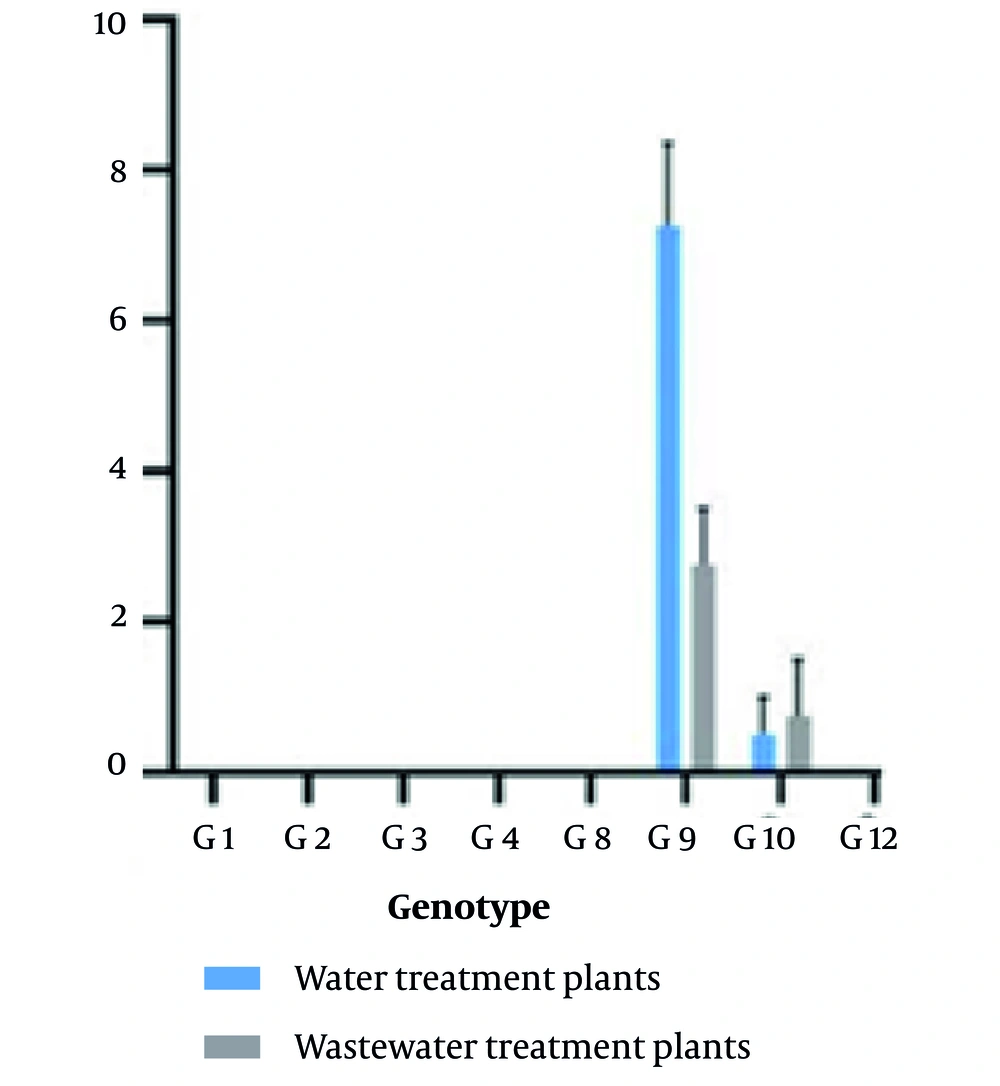
Figure 2.
Frequency of different rotavirus genotypes in water and wastewater treatment plants
4.4. Treatment Plant Efficiency
The efficiency of WWWTPs in removing rotavirus was 75% and 60%, respectively.
5. Discussion
In the present study, we found rotaviruses in 41.66% of the samples analyzed from water and wastewater treatment plant systems. Our findings are comparable to those of prior studies conducted around the world, which have described an incidence of rotaviruses of 11 to 42% in wastewater samples (32, 34-37). Several studies have identified rotaviruses in water and wastewater sources used for various purposes, including domestic, agricultural, and industrial uses, highlighting the need to look more broadly at the global prevalence of the virus in different aquatic environments (7, 25, 38). Co-infection with different rotavirus strains due to high genetic diversity leads to the emergence of new strains that are resistant to rotavirus vaccines, some of which have been identified in aquatic environments (39). The identification of rotaviruses in environmental specimens has been challenging because rotaviruses are fastidious and need more than one week to produce apparent cytopathic effect (CPE) (40). Also, inoculated cell cultures often deteriorate before the presence of characteristic CPE, making the obtainment of reliable and reproducible outcomes problematic (40).
Therefore, molecular monitoring of viruses in water and wastewater has been recommended worldwide by the World Health Organization to monitor vaccination and the efficiency of wastewater treatment plant systems. There are many methods for concentrating wastewater samples to determine the amount of virus, including ultracentrifugation, filtration, ultrafiltration, adsorption, and sedimentation-based methods (pellet and two-phase) (21, 41, 42). The two-phase concentration method was first proposed by Hovi et al. (43). With this method, it is possible to concentrate water and wastewater samples by 50 to 100 times (43). In the two-phase method, the very expensive 20% dextran material is used to increase the resorption and aggregation of rotaviruses, but in the Pellet method proposed by Kargar et al., there is no need to use the expensive dextran and polyethylene glycol materials (44). The results of this study indicate that the type and number of viruses isolated in the two-phase and pellet methods are different, so both methods were used together to concentrate wastewater samples. However, the two-phase concentration method using PEG 6000 and dextran is a very efficient recovery method for water and wastewater viruses (43).
The prevalence of rotavirus in the present study was reported from 108 samples, 45 cases (41.67%) from wastewater treatment plant systems. A similar prevalence of the virus was reported by Arraj et al. in France, who detected rotaviruses in 44.8% of treated wastewater samples and in 37.9% of raw wastewater samples using RT-PCR (45). Redwan and Attar in Saudi Arabia detected rotaviruses in 65% of treated wastewater samples using RT-PCR (46). Myrmel et al. reported rotavirus levels in the inlet and outlet samples of three wastewater treatment plant systems in Norway using nested PCR, in 64.3% of the inlet samples and in 53.3% of the outlet samples (47). The higher prevalence of rotavirus in China was detected in 100% of the wastewater input and also in 90% of the recovered wastewater samples (48). It seems that the higher rate of rotavirus in some studies compared to the present study can be attributed to the method of virus concentration and recovery. In contrast to the present study, previous research has also shown a higher incidence of rotaviruses during the cold months of the year in different types of environmental waters (32, 36, 49, 50).
Rotavirus genotype analysis in this study showed that the G9 and G10 genotypes were the dominant genotypes in water and wastewater samples. The type of genotypes in previous studies conducted on the monitoring of two wastewater treatment plants in the north and south of Isfahan was different, and in their study, the dominant genotypes were G10 and G1. A study conducted by Kargar et al. in Shiraz Hospital showed that the dominant genotypes were G1 and G4 (32). The highest seasonal distribution of genotype G in the present study was in the warm season, while the seasonal distribution of genotype G in previous studies indicated that genotypes G10, G1, and G9 were more frequently reported in the cold seasons (32, 51). In a study by Tavakoli et al., three rotavirus genotypes were reported from Tehran wastewater samples, including G9P[4] (collected in February), G4P[8] (collected in March), and G9P[8] (collected in June) (7). Rodriguez et al. reported two G genotypes, including G1 and G9, from Venezuelan wastewater samples (52). In a study by Azaran et al. in Ahvaz, rotavirus prevalence was reported in 32% of children's stool samples, and G/P genotyping showed that G9P[8] and G2P[4] were dominant (53).
Our genotyping results, which indicated the implications of detecting G9 and G10 strains, should be analyzed in the context of vaccination policy and epidemiology in Iran. The limitations of the present study were the small sample size, sampling frequency, and the lack of genetic correlation between the genotypes identified in municipal wastewater systems, clinical, and hospital isolates of rotavirus. Previous studies have shown that rotavirus strains may be released into the environment and thus contaminate water sources (32, 54). This contamination can facilitate the circulation of genotypes between the environment and the population, as well as the generation of new rotavirus strains through the process of rearrangement. Therefore, they can be considered as a reference for risk assessment. Therefore, continuous monitoring of rotavirus through molecular methods on clinical and environmental samples is essential to better understand the distribution and circulation of rotavirus for any community. While biological assays, such as cell culture, are required as the gold standard to accurately assess the infectious potential (infectivity) of the virus in environmental samples, the molecular method is considered a sensitive tool for monitoring genetic contamination (52).
In previous studies, we used an integrated cell culture reverse transcription polymerase chain reaction (ICC-RT-PCR) method to identify enteroviruses from wastewater samples from Tehran province (55) and recently to identify enteroviruses from wastewater samples from Ahvaz (data not published). For this reason, using the ICC-RT-PCR method in molecular monitoring of water and wastewater can give us deeper insight into the spread of infectious viruses. The difference between the genotypes identified in the present study and other studies could be due to differences in the type of sample, sampling method, number of samples examined, detection methods, and geographical area of study. Since the prevalence of circulating virus in water and wastewater samples of a region can reflect the frequency of the virus in a community, the presence of virus in water and wastewater samples can be a suitable method for monitoring the virus throughout a community (56). Molecular monitoring of wastewater treatment plant systems plays an important role in removing human pathogens from aquatic environments in the world. Therefore, high efficiency of effective wastewater treatment plant systems, along with molecular monitoring of circulating rotaviruses, is essential for all communities.
Although our study showed that rotavirus elimination in wastewater treatment plant systems was not complete, the viral contamination was significant and revealed a critical public health risk. This result is consistent with previous studies that have shown the presence of rotavirus not only in water and wastewater (32, 51, 57, 58). In addition to the implementation protocol for water and wastewater treatment plants, the use of supplementary treatment systems of wastewater treatment plant systems, such as ozone gas, is recommended. Molecular detection and monitoring of rotavirus in municipal wastewater treatment plant systems is widely used worldwide to understand information about circulating genotypes, correlate the level of vaccine protection, and provide a signal of imminent outbreak of the virus through water consumption in certain populations.
5.1. Conclusions
Given the importance of rotavirus transmission via water, it is critical to monitor wastewater treatment plant system activities on a regular basis in order to control these waterborne viruses and other microbes. Importantly, these findings indicate that molecular-based rotavirus surveillance of water and wastewater may be an important epidemiological tool for assessing the actual community structure of rotaviruses from wastewater treatment plant systems in that region. Along with population-based studies in Iran, it is suggested that vaccination against rotavirus genotypes G9 and G10 should be included in national vaccination programs.