Jundishapur J Microbiol

Image Credit:Jundishapur J Microbiol

Effects of Postbiotics Derived from Lactobacillus plantarum and Bifidobacterium bifidum on Biofilm Formation and Virulence Gene Expression of Enterococcus faecalis

Author(s):
Samira SaediSamira SaediSamira Saedi ORCID1, Javad NezhadiJavad NezhadiJavad Nezhadi ORCID2, Roya Abedi SoleimaniRoya Abedi SoleimaniRoya Abedi Soleimani ORCID3, Mahdi Asghari OzmaMahdi Asghari OzmaMahdi Asghari Ozma ORCID1, Hossein Samadi KafilHossein Samadi KafilHossein Samadi Kafil ORCID2, 4,*
1Student Research Committee, Tabriz University of Medical Sciences, Tabriz, Iran
2Department of Clinical Microbiology, Faculty of Medicine, Tabriz University of Medical Sciences, Tabriz, Iran
3Department of Food Science and Technology, Faculty of Nutrition and Food Sciences, Tabriz University of Medical Sciences, Tabriz, Iran
4Drug Applied Research Center, Tabriz University of Medical Sciences, Tabriz, Iran

Jundishapur Journal of Microbiology:Vol. 18, issue 10; e165694
Published online:Nov 04, 2025
Article type:Research Article
Received:Aug 23, 2025
Accepted:Oct 24, 2025
How to Cite:Saedi S, Nezhadi J, Abedi Soleimani R, Asghari Ozma M, Samadi Kafil H. Effects of Postbiotics Derived from Lactobacillus plantarum and Bifidobacterium bifidum on Biofilm Formation and Virulence Gene Expression of Enterococcus faecalis. Jundishapur J Microbiol. 2025;18(10):e165694. doi: https://doi.org/10.5812/jjm-165694

Abstract

Background:

Enterococcus faecalis is an opportunistic pathogen capable of forming biofilms and developing antibiotic resistance, which complicates infection treatment.

Objectives:

This study investigates the effect of postbiotics derived from Lactobacillus plantarum and Bifidobacterium bifidum on biofilm formation and virulence gene expression in E. faecalis.

Methods:

In the present study, standard strains of L. plantarum ATCC 8014, B. bifidum ATCC 15696, and E. faecalis ATCC 29212 were used. Postbiotics/cell-free supernatants (CFSs) were prepared from probiotics and added to E. faecalis. The contents of the obtained postbiotics were evaluated by GC-MS. Biofilm formation was examined using the microtiter plate method, and the expression of endocarditis- and biofilm-associated pilus A (ebpA), enterococcal fibronectin-binding antigen A (efaA), aggregation substance (asa), and adhesin to collagen of Enterococcus (ace) genes was assessed by real-time (RT)-PCR.

Results:

The CFSs significantly reduced biofilm formation in a dose-dependent manner. Bifidobacterium bifidum CFSs at 20, 10, and 5 mg/mL significantly decreased biofilm formation. Similarly, L. plantarum CFSs at 20 and 10 mg/mL showed a significant inhibitory effect. The qRT-PCR analysis revealed that L. plantarum CFSs downregulated efaA, asa, and ace genes but had no effect on the ebpA gene. Conversely, B. bifidum CFSs reduced ebpA and ace gene expression but did not significantly alter efaA and asa genes.

Conclusions:

These findings suggest that postbiotics may help reduce the pathogenicity of E. faecalis, particularly in preventing infections caused by E. faecalis.

1. Background

Enterococcus faecalis is a Gram-positive bacterium commonly found in the large intestine, where it aids digestion and vitamin production. However, it can act as an opportunistic pathogen, especially in hospitalized or immunocompromised patients (1, 2). Its ability, similar to that of Escherichia coli, to acquire antibiotic resistance genes increases the risk of severe hospital-acquired infections, particularly during prolonged antibiotic therapy (3, 4). Enterococcus faecalis can cause urinary tract infections (UTIs), bacteremia, meningitis, endocarditis, wound infections, and gastrointestinal infections, especially after complex surgeries or organ transplants (5). Another key virulence factor of E. faecalis is its strong biofilm-forming ability. Biofilms are organized communities of microorganisms embedded in a self-produced extracellular polymeric substance (EPS) and attached to living or non-living surfaces. This structure protects microorganisms from adverse environmental conditions, disinfectants, and host immune responses.
Once E. faecalis forms a biofilm, treatment becomes extremely difficult, as antimicrobials cannot effectively penetrate its depths. Consequently, the infection may become chronic and easily spread through the bloodstream or other tissues (6, 7). In addition to biofilm formation, E. faecalis expresses various virulence genes that significantly contribute to its pathogenicity. By combining biofilm formation, virulence factor expression, and the acquisition of antibiotic-resistance genes, this bacterium poses serious clinical challenges (8). Given its role in life-threatening infections — particularly in immunocompromised patients — there is an increasing focus on developing new therapeutic approaches to combat E. faecalis (9).
One of the most promising new therapeutic methods that has attracted the attention of researchers in recent years is the use of probiotics and postbiotics to treat diseases (10, 11). Probiotics are live microorganisms that provide health benefits to the host, while postbiotics — their metabolic products — support immune function, inhibit pathogens, and reduce inflammation (12). Among the common microorganisms used in the production of postbiotics are Lactobacillus plantarum and Bifidobacterium bifidum, which have gained much attention due to their unique properties in producing antibacterial compounds and regulating the immune system (13, 14). As previously mentioned, postbiotics refer to compounds or products obtained from the metabolic activity of probiotics, which can have beneficial effects on host health (15). These compounds include proteins, peptides, fatty acids, enzymes, and various metabolites that can directly or indirectly enhance antibacterial, anti-inflammatory, and immunogenic activities in the body (16).

2. Objectives

This study investigated the effects of L. plantarum and B. bifidum postbiotics on E. faecalis virulence genes — endocarditis- and biofilm-associated pilus A (ebpA), enterococcal fibronectin-binding antigen A (efaA), aggregation substance (asa), and adhesin to collagen of Enterococcus (ace) — as well as on biofilm formation. The findings provide insights into the potential use of postbiotics as therapeutic or preventive agents against E. faecalis and related pathogens. By reducing biofilm formation and downregulating virulence genes, these postbiotics demonstrate preliminary in vitro promise as adjunct strategies to decrease bacterial pathogenicity, potentially leading to alternative treatments with fewer side effects than conventional antibiotics.

3. Methods

3.1. Bacterial Strains

All experiments were performed on standard laboratory strains. To investigate the effects of postbiotics derived from probiotics on the expression of virulence genes and biofilm-forming ability in the E. faecalis ATCC 29212 strain, the L. plantarum ATCC 8014 and B. bifidum ATCC 15696 strains were used.

3.2. Preparation of Cell-Free Supernatants

To prepare CFSs, L. plantarum ATCC 8014 and B. bifidum ATCC 15696 were grown on De Man-Rogosa-Sharpe (MRS) agar at 37°C for 18 - 24 h. Colonies were transferred into MRS broth (Merck, Germany) supplemented with 0.6% yeast extract (Sigma-Aldrich) and incubated overnight at 37°C and 150 rpm. Cultures were centrifuged (6000 × g, 10 min, 4°C), and the supernatants were filtered (0.22 μm, Millipore), aliquoted, and stored at -20°C.

3.3. Determination of Cell-Free Supernatant Concentration

To determine the CFS concentration, supernatants were obtained by centrifugation and sterile filtration, then freeze-dried or low-temperature dried to yield a powder. Dry weight was measured, and the powder was reconstituted in sterile medium to the desired concentration, expressed as mg dry matter/mL. This procedure ensured accurate application of CFS bioactive components and improved experimental reproducibility.

3.4. GC-MS Postbiotic Metabolite Identification

GC-MS analysis of postbiotics was performed using a Shimadzu QP-5050 apparatus, a Gas Chromatograph GC-17A with an HP-5 capillary column (phenylmethyl siloxane, 30 m, 0.25 mm internal diameter), and a mass spectrometer with a mass range of 50 - 600 m/z. Helium served as the carrier gas at 1 mL/min with a 1:30 split ratio. The injector and detector were calibrated at 250°C and 280°C, respectively. The column temperature was programmed to rise linearly at 5°C per minute from 60°C to 250°C and held at 250°C for 10 minutes. Retention indices were determined based on the retention times of injected n-alkanes under identical chromatographic conditions prior to sample injection. By comparing mass spectra with Willey (n17) and Adams libraries, GC-MS identified and characterized the compounds (17).

3.5. Preparation and pH Adjustment of Cell-Free Supernatants for Biofilm Formation Assay

The CFSs from L. plantarum and B. bifidum were collected under standard sterile laboratory conditions, including controlled temperature, pH, and incubation time. Their naturally acidic pH was neutralized to approximately 7 with NaOH to avoid acidity-related effects on biofilm formation. The neutralized CFSs were then applied in assays evaluating their impact on E. faecalis biofilm formation (18).

3.6. Biofilm Formation Assay

The effect of CFSs on E. faecalis ATCC 29212 biofilm formation was evaluated using a 96-well microtiter plate assay. A bacterial suspension (0.5 McFarland, ~107 CFU/mL, OD600 = 0.08 - 0.1) in trypticase soy broth (TSB) containing 0.5% glucose was prepared, and 10 μL were added to 90 μL of medium per well. Then, 100 μL of CFSs were added, and the plates were incubated at 37°C for 24 h, with untreated wells serving as controls. After incubation, wells were washed with saline, fixed with methanol, stained with 0.1% crystal violet, washed, air-dried, and destained with 33% acetic acid. Biofilm biomass was quantified at OD570.

3.7. Quantitative Real-time PCR Assay

Enterococcus faecalis ATCC 29212 was grown on blood agar (BA) at 37°C under aerobic conditions until reaching the mid-logarithmic phase (OD600 ≈ 0.5). The samples were then treated with CFSs derived from L. plantarum and B. bifidum at a concentration of 20 mg/mL. Enterococcus faecalis ATCC 29212 was exposed to the CFSs for 4 hours, allowing sufficient time for the transcriptional response while avoiding late-stage stress effects.
A q-real-time (RT)-PCR assay was performed using the SYBR Premix EX Taq II, Tli RNase H+ (Takara Bio Inc.), and a RT-PCR machine (Applied Biosystems StepOnePlus™). In the present study, the universal gene (16S rRNA) was used as the reference gene (19). DNA-free RNA was used for cDNA synthesis according to the manufacturer’s instructions (Yekta Tajhiz Azma, Tehran, Iran; Cat. No. YT4500). RNA extraction was performed using the Favorgen kit (Favorgen, Pingtung, Taiwan).
The quality and purity of the extracted RNA were assessed using a NanoDrop spectrophotometer (Thermo Scientific, USA) by measuring the absorbance ratio at 260/280 nm. RNA samples with an A260/A280 ratio between 1.8 and 2.1 were considered acceptable. To eliminate potential genomic DNA contamination, all RNA samples were treated with DNase I (Acell Teb Rad Med) prior to reverse transcription. The cDNA synthesis was performed using the Parstous cDNA Synthesis Kit (Parstous Co., Iran) following the manufacturer’s instructions. As a negative control, a no-RT control (without reverse transcriptase enzyme) was included for each sample to confirm the absence of DNA contamination in downstream qPCR reactions. The reaction protocol consisted of an initial temperature of 95°C for 15 minutes, followed by 40 cycles of 30 s at 95°C, 45 s at 60°C, and 45 s at 72°C.

3.8. Statistical Analysis

GraphPad Prism (v.9.4.1) and the Statistical Package for the Social Sciences (SPSS) v.20.0 (SPSS, Inc., Chicago, IL, USA) were employed for statistical analysis. The two groups were compared using Student’s t-test. Data normality was assessed using the Shapiro-Wilk test, and since the data were normally distributed, one-way ANOVA was applied to compare the means between independent groups and stages. A P-value of less than 0.05 was considered statistically significant. Furthermore, all experiments were conducted using three independent biological replicates.

4. Results

4.1. Metabolite Profiling of Postbiotics by GC-MS

Table 1 shows the bioactive compounds identified in the postbiotic samples, as revealed by GC-MS analysis. By comparing the metabolite profiles of B. bifidum and L. plantarum, two postbiotic sources, it is evident that they possess distinct metabolic capabilities.
Table 1.Postbiotic GC-MS-Identified Major Chemicals
No.Bifidobacterium bifidumLactobacillus plantarum
1Lactic acidLactic acid
2Glycolic acidAcetic acid
33-Hydroxybutyric acidSuccinic acid
4Succinic acidButyric acid
53-Hydroxypropionic acid2,3-Butanediol
6Stearic acidDiacetyl
7Heptadecanoic acidPhenyllactic acid

4.2. Biofilm Formation

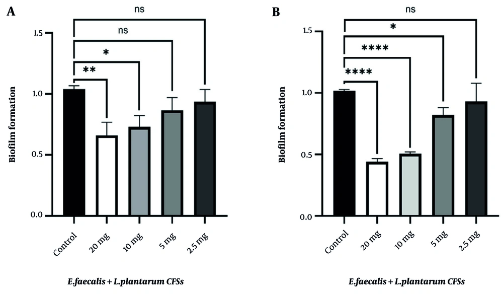
The biofilm formation assay confirmed the inherent ability of E. faecalis to form biofilms, which play a crucial role in its pathogenicity and resistance to antimicrobial treatments. Treatment with CFSs derived from L. plantarum and B. bifidum resulted in a concentration-dependent reduction in biofilm formation, as indicated by decreasing optical density (OD) values. Notably, CFSs from B. bifidum exhibited stronger inhibitory effects than those from L. plantarum (Figure 1S in Supplementary File).
Furthermore, it was determined that postbiotics derived from B. bifidum exhibited a greater capacity to inhibit the formation of E. faecalis biofilms than those derived from L. plantarum. Statistical analyses revealed that concentrations of 20 (P < 0.0001), 10 (P < 0.0001), and 5 mg (P = 0.04) of B. bifidum CFSs significantly decreased biofilm formation compared to the control group. Likewise, concentrations of 20 (P = 0.003) and 10 mg (P = 0.01) of L. plantarum CFSs significantly reduced biofilm formation relative to the control group (Figure 1, Table 2).
Statistical analysis revealed that <i>Lactobacillus plantarum</i> and <i>Bifidobacterium bifidum</i> cell-free supernatants (CFSs) significantly reduced <i>Enterococcus faecalis</i> biofilm formation in a dose-dependent manner, with <i>B. bifidum</i> showing a stronger inhibitory effect at equivalent concentrations. All experiments were performed in triplicate to ensure result accuracy and reliability (* P-value = 0.01, ** P-value = 0.001, **** P-value = 0.0001).
Figure 1.

Statistical analysis revealed that Lactobacillus plantarum and Bifidobacterium bifidum cell-free supernatants (CFSs) significantly reduced Enterococcus faecalis biofilm formation in a dose-dependent manner, with B. bifidum showing a stronger inhibitory effect at equivalent concentrations. All experiments were performed in triplicate to ensure result accuracy and reliability (* P-value = 0.01, ** P-value = 0.001, **** P-value = 0.0001).

Table 2.Effect of Different Cell-Free Supernatant Concentrations on Biofilm Formation Reduction in Enterococcus faecalisa
CFSs Concentration (mg/mL)Biofilm Reduction of Enterococcus faecalis (%)
Lactobacillus plantarum
2044
1027
514
2.57
Bifidobacteriumbifidum
2056
1050
518
2.57

Abbreviation: CFS, cell-free supernatant.

a Evaluated with all experiments performed in triplicate to ensure reproducibility.

4.3. q-Real-time PCR

4.3.1. The Ability of Lactobacillus plantarum Cell-Free Supernatants to Reduce the Expression of Virulence Genes

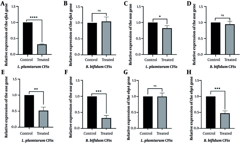
The analysis of E. faecalis isolates revealed a significant reduction in the expression of key virulence genes — efaA, asa, and ace — following exposure to CFSs produced by L. plantarum, as compared to the control group. The downregulation of efaA (P < 0.0001), asa (P = 0.01), and ace (P = 0.001) suggests that L. plantarum CFSs may interfere with mechanisms associated with bacterial adhesion, invasion, and biofilm formation, all of which are critical for E. faecalis pathogenicity. These findings align with the hypothesis that probiotic-derived postbiotics can mitigate bacterial virulence factors, thus offering potential therapeutic strategies for controlling E. faecalis-associated infections.
However, no significant difference was observed in the relative expression of the ebpA gene (P > 0.05) in the treated group compared to the control. This may indicate that L. plantarum CFSs selectively target certain virulence factors more effectively than others, or that the ebpA gene — associated with biofilm formation — may require higher concentrations or longer exposure to fully modulate its expression. It is also possible that different bacterial strains respond variably to postbiotic treatments. Overall, these results suggest that while L. plantarum CFSs can significantly reduce the expression of several critical virulence genes, factors such as exposure time, dosage, and strain variability may influence the full extent of their effects (Figure 2A, C, E, and G).
A, C, E, G, the effect of cell-free supernatants (CFSs)-derived from <i>Lactobacillus plantarum</i> on the expression of virulence genes including enterococcal fibronectin-binding antigen A (<i>efaA</i>), aggregation substance (<i>asa</i>), adhesin to collagen of <i>Enterococcus</i> (<i>ace</i>), and endocarditis- and biofilm-associated pilus A (<i>ebpA</i>); B, D, F, H, the effect of CFSs-derived from <i>Bifidobacterium bifidum</i> on the expression of virulence genes including <i>efaA</i>, <i>asa</i>, <i>ace</i>, and <i>ebpA</i> (the student's <i>t</i>-test was used to compare the groups; * P-value = 0.01, ** P-value = 0.001, *** P-value = 0.0001 - 0.0003, and **** P-value &lt; 0.0001; Abbreviation: ns, not significant).
Figure 2.

A, C, E, G, the effect of cell-free supernatants (CFSs)-derived from Lactobacillus plantarum on the expression of virulence genes including enterococcal fibronectin-binding antigen A (efaA), aggregation substance (asa), adhesin to collagen of Enterococcus (ace), and endocarditis- and biofilm-associated pilus A (ebpA); B, D, F, H, the effect of CFSs-derived from Bifidobacterium bifidum on the expression of virulence genes including efaA, asa, ace, and ebpA (the student's t-test was used to compare the groups; * P-value = 0.01, ** P-value = 0.001, *** P-value = 0.0001 - 0.0003, and **** P-value < 0.0001; Abbreviation: ns, not significant).

4.3.2. The Ability of Bifidobacterium bifidum Cell-Free Supernatants to Reduce the Expression of Virulence Genes

The gene expression analysis of E. faecalis isolates showed that exposure to CFSs derived from B. bifidum led to a significant reduction in the expression of the ebpA (P = 0.0003) and ace (P = 0.0001) genes compared to the control group. These findings suggest that B. bifidum CFSs may interfere with key virulence mechanisms of E. faecalis, including biofilm formation and adhesion, which are crucial for bacterial persistence and pathogenicity.
On the other hand, no significant difference was observed in the relative expression of the efaA and asa genes (P > 0.05) in the treated group compared to the control. This could imply that the postbiotic compounds derived from B. bifidum selectively target specific virulence factors, while others — such as those involved in stress response or other regulatory pathways — may be less sensitive to this treatment. It is also possible that higher concentrations or prolonged exposure may be required to significantly modulate the expression of these genes.
Overall, these results indicate that B. bifidum CFSs exhibit selective anti-virulence activity, particularly affecting genes related to biofilm formation and adhesion. These findings suggest a potential strategy for managing E. faecalis-associated infections (Figure 2B, D, F, and H).

5. Discussion

The observed inhibitory effects on biofilm formation and virulence gene expression suggest that postbiotics interfere with bacterial communication and metabolic pathways. Postbiotics contain various bioactive compounds, such as short-chain fatty acids (SCFAs), peptides, and bacteriocins, which can influence bacterial physiology (20). Studies have shown that postbiotic compounds can disrupt quorum sensing (QS), a key regulator of biofilm formation, thereby reducing bacterial adhesion and aggregation. Bacteriocins, organic acids (e.g., lactic and acetic acids), and SCFAs can interfere with QS in E. faecalis by blocking QS receptors, disrupting cell membranes, altering intracellular pH, or modulating global regulatory systems. Some Lactobacillus-derived metabolites inhibit the FSR QS system, which controls virulence factors such as gelatinase (gelE) and serine protease (sprE). Differences in gene expression between L. plantarum and B. bifidum CFSs may reflect variations in their bioactive compound profiles and interactions with pathogen regulatory pathways (21). Additionally, specific metabolites from B. bifidum and L. plantarum may modulate bacterial gene expression, leading to a decrease in virulence factor production (13).
This study examined the effects of L. plantarum and B. bifidum CFSs on the expression of key E. faecalis virulence genes (efaA, asa, ace, and ebpA), which are essential for adhesion, biofilm formation, and survival in hostile environments, while also analyzing the CFS composition using GC-MS. The efaA gene encodes a surface protein that mediates adhesion to host cells and supports stable biofilm formation, an essential step in infection. Since adhesion is central to many virulence processes, targeting efaA can reveal how postbiotics may inhibit this mechanism (3, 22). The asa gene encodes an adhesin that enables E. faecalis to attach to host cells and promote biofilm formation. Adhesins play critical roles in chronic infections, as biofilms enhance bacterial resistance to treatment and immune evasion. Studying this gene can clarify how postbiotics may modulate or inhibit adhesion and biofilm formation, offering potential for new therapeutic strategies (19, 23).
Biofilms not only confer antibiotic resistance but also protect bacteria from host immune responses. Therefore, studying the impact of postbiotics on this gene can help identify new ways to interfere with biofilm formation and improve the treatment of resistant infections (22). Finally, the ebpA gene is involved in the production of proteins that enable E. faecalis to bind to host cells. These proteins directly contribute to adhesion and biofilm formation processes. Examining the effects of postbiotics on this gene can help us better understand the molecular pathways involved in virulence and could lead to the development of targeted therapies (24). The selected genes play crucial roles in virulence processes such as host cell adhesion, biofilm formation, and treatment resistance. Our results showed that postbiotics reduce the expression of some of these genes and inhibit biofilm formation, highlighting their potential as biological agents to control E. faecalis infections. Notably, B. bifidum postbiotics had a stronger effect on biofilm reduction than those from L. plantarum.
Specifically, L. plantarum postbiotics decreased efaA, asa, and ace expression but did not affect ebpA, whereas B. bifidum postbiotics reduced ebpA and ace expression without significantly impacting efaA and asa. These differences likely stem from variations in their composition and metabolic activities. Our findings on L. plantarum postbiotics align with Kim et al., who reported that lactic acid bacteria postbiotics suppress biofilm formation in mastitis-associated bacteria, including E. faecalis (25). Similarly, Nezhadi and Ahmadi found that postbiotics derived from L. plantarum can prevent biofilm formation in nosocomial bacteria, including E. faecalis and Pseudomonas aeruginosa (1). Furthermore, Knysh et al. demonstrated that postbiotics derived from B. bifidum effectively prevented biofilm formation in pathogenic bacteria, including E. coli and P. aeruginosa, and significantly reduced the biofilm formation rate compared to the control group (26). Additionally, a study by Asghari Ozma et al. showed that postbiotics derived from lactic acid bacteria such as B. bifidum can play a significant role in inhibiting biofilm formation and serve as novel therapeutic agents for treating infections caused by Clostridium difficile (27).
This study represents the first investigation into the effects of postbiotics derived from B. bifidum and L. plantarum on the expression of virulence genes, including efaA, ebpA, asa, and ace. Nevertheless, several previous studies have demonstrated that L. plantarum can influence the expression of other virulence genes. For example, in a study conducted by Oumaima et al., the effect of L. plantarum was investigated on P. aeruginosa, revealing that L. plantarum can reduce the activity of the MexXY-OprM efflux pump in this bacterium, thereby aiding in overcoming antibiotic resistance (28). Furthermore, a study by Zabolyova et al. showed that postbiotics (enterocins) can positively influence the treatment of methicillin-resistant S. aureus strains and contribute to overcoming antibiotic resistance (29). Additionally, Ishikawa et al. reported that postbiotics produced by lactobacilli modify the transcription of virulence genes in Aggregatibacter actinomycetemcomitans, thereby reducing its pathogenicity and antibiotic resistance (30).
Our results indicate that postbiotics from B. bifidum were more effective than those from L. plantarum in reducing E. faecalis biofilm formation. The strains exerted distinct effects on virulence gene expression, likely reflecting differences in their metabolic profiles. Postbiotics from L. plantarum reduced efaA, asa, and ace, but not ebpA, whereas B. bifidum reduced ebpA and ace without altering efaA and asa. A limitation of this study is that it did not assess the synergistic effects of postbiotics with antibiotics or other drugs; this will be addressed in future research.

5.1. Conclusions

This study provides new insights into the potential of postbiotics from L. plantarum and B. bifidum to reduce the pathogenicity of E. faecalis by inhibiting biofilm formation and suppressing the expression of key virulence genes. These findings support the growing interest in postbiotics as a novel and sustainable alternative to combat bacterial infections. Further research, particularly clinical studies, is required to confirm these results and explore the practical applications of postbiotics in future healthcare.

Acknowledgments

Footnotes

References

  • 1.
    Nezhadi J, Ahmadi A. Assessing the efficacy of postbiotics derived from Lactobacillus plantarum on antibiotic resistance genes in nosocomial pathogens such as Enterococcus faecalis and Pseudomonas aeruginosa. Lett Appl Microbiol. 2024;77(12). [PubMed ID: 39657994]. https://doi.org/10.1093/lambio/ovae127.
  • 2.
    Kafil HS, Mobarez AM. Assessment of biofilm formation by enterococci isolates from urinary tract infections with different virulence profiles. J King Saud Univ Sci. 2015;27(4):312-7. https://doi.org/10.1016/j.jksus.2014.12.007.
  • 3.
    Kafil HS, Mobarez AM, Moghadam MF, Hashemi ZS, Yousefi M. Gentamicin induces efaA expression and biofilm formation in Enterococcus faecalis. Microb Pathog. 2016;92:30-5. [PubMed ID: 26724739]. https://doi.org/10.1016/j.micpath.2015.12.008.
  • 4.
    Derakhshan S, Saedi S, Ahmadi A, Hedayati MA. Virulence genes, phylogenetic analysis, and antimicrobial resistance of Escherichia coli isolated from urinary tract infection in hospitalized patients and outpatients. J Appl Genet. 2022;63(4):805-13. [PubMed ID: 35972677]. https://doi.org/10.1007/s13353-022-00718-8.
  • 5.
    Codelia-Anjum A, Lerner LB, Elterman D, Zorn KC, Bhojani N, Chughtai B. Enterococcal urinary tract infections: A review of the pathogenicity, epidemiology, and treatment. Antibiotics (Basel). 2023;12(4). [PubMed ID: 37107140]. [PubMed Central ID: PMC10135011]. https://doi.org/10.3390/antibiotics12040778.
  • 6.
    Yang S, Meng X, Zhen Y, Baima Q, Wang Y, Jiang X, et al. Strategies and mechanisms targeting Enterococcus faecalis biofilms associated with endodontic infections: A comprehensive review. Front Cell Infect Microbiol. 2024;14:1433313. [PubMed ID: 39091674]. [PubMed Central ID: PMC11291369]. https://doi.org/10.3389/fcimb.2024.1433313.
  • 7.
    Akbari Aghdam M, Soroush Barhaghi MH, Aghazadeh M, Jafari F, Beomide Hagh M, Haghdoost M, et al. Virulence genes in biofilm producer Enterococcus faecalis isolates from root canal infections. Cell Mol Biol (Noisy-le-grand). 2017;63(5):55-9. [PubMed ID: 28719346]. https://doi.org/10.14715/cmb/2017.63.5.11.
  • 8.
    Golob M, Pate M, Kusar D, Dermota U, Avbersek J, Papic B, et al. Antimicrobial resistance and virulence genes in Enterococcus faecium and Enterococcus faecalis from humans and retail red meat. Biomed Res Int. 2019;2019:2815279. [PubMed ID: 31211134]. [PubMed Central ID: PMC6532320]. https://doi.org/10.1155/2019/2815279.
  • 9.
    Gong J, Bai T, Zhang L, Qian W, Song J, Hou X. Inhibition effect of Bifidobacterium longum, Lactobacillus acidophilus, Streptococcus thermophilus and Enterococcus faecalis and their related products on human colonic smooth muscle in vitro. PLoS One. 2017;12(12). e0189257. [PubMed ID: 29216305]. [PubMed Central ID: PMC5720742]. https://doi.org/10.1371/journal.pone.0189257.
  • 10.
    Saedi S, Derakhshan S, Hasani A, Khoshbaten M, Poortahmasebi V, Milani PG, et al. Recent advances in gut microbiome modulation: Effect of probiotics, prebiotics, synbiotics, and postbiotics in inflammatory bowel disease prevention and treatment. Curr Microbiol. 2024;82(1):12. [PubMed ID: 39589525]. https://doi.org/10.1007/s00284-024-03997-y.
  • 11.
    Saedi S, Derakhshan S, Sadeghi J, Hasani A, Khoshbaten M, Poortahmasebi V, et al. A study on gut microbiota and short-chain fatty acids in patients with inflammatory bowel disease from northwest Iran. Lett Appl Microbiol. 2025;78(8). [PubMed ID: 40802486]. https://doi.org/10.1093/lambio/ovaf111.
  • 12.
    Zaib S, Hayat A, Khan I. Probiotics and their beneficial health effects. Mini Rev Med Chem. 2024;24(1):110-25. [PubMed ID: 37291788]. https://doi.org/10.2174/1389557523666230608163823.
  • 13.
    Li S, Huang R, Shah NP, Tao X, Xiong Y, Wei H. Antioxidant and antibacterial activities of exopolysaccharides from Bifidobacterium bifidum WBIN03 and Lactobacillus plantarum R315. J Dairy Sci. 2014;97(12):7334-43. [PubMed ID: 25282420]. https://doi.org/10.3168/jds.2014-7912.
  • 14.
    Lalezadeh A, Fadaee M, Saedi S, Nezhadi J, Ozma MA, Ahmadi S, et al. A critical review on the potential of inactivated bacteria in counteracting human pathogens. Curr Microbiol. 2025;82(7):295. [PubMed ID: 40394322]. https://doi.org/10.1007/s00284-025-04282-2.
  • 15.
    Vinderola G, Sanders ME, Salminen S. The concept of postbiotics. Foods. 2022;11(8). [PubMed ID: 35454664]. [PubMed Central ID: PMC9027423]. https://doi.org/10.3390/foods11081077.
  • 16.
    Wang P, Wang S, Wang D, Li Y, Yip RCS, Chen H. Postbiotics-peptidoglycan, lipoteichoic acid, exopolysaccharides, surface layer protein and pili proteins-structure, activity in wounds and their delivery systems. Int J Biol Macromol. 2024;274(Pt 1):133195. [PubMed ID: 38885869]. https://doi.org/10.1016/j.ijbiomac.2024.133195.
  • 17.
    Chang HM, Foo HL, Loh TC, Lim ETC, Abdul Mutalib NE. Comparative studies of inhibitory and antioxidant activities, and organic acids compositions of postbiotics produced by probiotic Lactiplantibacillus plantarum strains isolated from Malaysian foods. Front Vet Sci. 2020;7:602280. [PubMed ID: 33575277]. [PubMed Central ID: PMC7870707]. https://doi.org/10.3389/fvets.2020.602280.
  • 18.
    Alshanta OA, Albashaireh K, McKloud E, Delaney C, Kean R, McLean W, et al. Candida albicans and Enterococcus faecalis biofilm frenemies: When the relationship sours. Biofilm. 2022;4:100072. [PubMed ID: 35313556]. [PubMed Central ID: PMC8933684]. https://doi.org/10.1016/j.bioflm.2022.100072.
  • 19.
    Kafil HS, Mobarez AM, Moghadam MF. Adhesion and virulence factor properties of Enterococci isolated from clinical samples in Iran. Indian J Pathol Microbiol. 2013;56(3):238-42. [PubMed ID: 24152500]. https://doi.org/10.4103/0377-4929.120375.
  • 20.
    Gurunathan S, Thangaraj P, Kim JH. Postbiotics: Functional food materials and therapeutic agents for cancer, diabetes, and inflammatory diseases. Foods. 2023;13(1). [PubMed ID: 38201117]. [PubMed Central ID: PMC10778838]. https://doi.org/10.3390/foods13010089.
  • 21.
    Machado MG, Sencio V, Trottein F. Short-chain fatty acids as a potential treatment for infections: A closer look at the lungs. Infect Immun. 2021;89(9). e0018821. [PubMed ID: 34097474]. [PubMed Central ID: PMC8370681]. https://doi.org/10.1128/IAI.00188-21.
  • 22.
    Ghazvinian M, Asgharzadeh Marghmalek S, Gholami M, Amir Gholami S, Amiri E, Goli HR. Antimicrobial resistance patterns, virulence genes, and biofilm formation in enterococci strains collected from different sources. BMC Infect Dis. 2024;24(1):274. [PubMed ID: 38438983]. [PubMed Central ID: PMC10910731]. https://doi.org/10.1186/s12879-024-09117-2.
  • 23.
    Khalil MA, Alorabi JA, Al-Otaibi LM, Ali SS, Elsilk SE. Antibiotic resistance and biofilm formation in Enterococcus spp. isolated from urinary tract infections. Pathogens. 2022;12(1). [PubMed ID: 36678381]. [PubMed Central ID: PMC9863506]. https://doi.org/10.3390/pathogens12010034.
  • 24.
    Ho FK, Lam LN, Matysik A, Watts TD, Wong JJ, Chong KKL, et al. Role of sortase-assembled Ebp pili in Enterococcus faecalis adhesion to iron oxides and its impact on extracellular electron transfer. Microbiol Spectr. 2025;13(3). e0233724. [PubMed ID: 39902984]. [PubMed Central ID: PMC11878085]. https://doi.org/10.1128/spectrum.02337-24.
  • 25.
    Kim H, Youn H, Moon J, Kim H, Seo K. Comparative anti-microbial and anti-biofilm activities of postbiotics derived from kefir and normal raw milk lactic acid bacteria against bovine mastitis pathogens. Lwt. 2024;191. https://doi.org/10.1016/j.lwt.2023.115699.
  • 26.
    Knysh OV, Isayenko OY, Voyda YV, Kizimenko OO, Babych YM. Influence of cell-free extracts of Bifidobacterium bifidum and Lactobacillus reuteri on proliferation and biofilm formation by Escherichia coli and Pseudomonas aeruginosa. Regul Mech Biosyst. 2019;10(2):251-6. https://doi.org/10.15421/021938.
  • 27.
    Asghari Ozma M, Mahmoodzadeh Hosseini H, Ataee MH, Mirhosseini SA. Evaluating the antibacterial, antibiofilm, and anti-toxigenic effects of postbiotics from lactic acid bacteria on Clostridium difficile. Iran J Microbiol. 2024;16(4):497-508. [PubMed ID: 39267941]. [PubMed Central ID: PMC11389761]. https://doi.org/10.18502/ijm.v16i4.16309.
  • 28.
    Oumaima A, Molavi F, Tehranipoor M. [Synergistic effect of silver oxide nanoparticles and probiotic Lactobacillus plantarum on gene expression of MexX component of pump efflux system in drug-resistant Pseudomonas aeruginosa strains]. J Microb World. 2020;14(3):47-58. FA.
  • 29.
    Zabolyova N, Laukova A, Pogany Simonova M. Susceptibility to postbiotics - enterocins of methicillin-resistant Staphylococcus aureus strains isolated from rabbits. Vet Res Commun. 2024;48(3):1449-57. [PubMed ID: 38324077]. [PubMed Central ID: PMC11147817]. https://doi.org/10.1007/s11259-024-10323-1.
  • 30.
    Ishikawa KH, Bueno MR, Kawamoto D, Simionato MRL, Mayer MPA. Lactobacilli postbiotics reduce biofilm formation and alter transcription of virulence genes of Aggregatibacter actinomycetemcomitans. Mol Oral Microbiol. 2021;36(1):92-102. [PubMed ID: 33372378]. https://doi.org/10.1111/omi.12330.

Crossmark
Crossmark
Checking
Share on
Cited by
Metrics

Purchasing Reprints

  • Copyright Clearance Center (CCC) handles bulk orders for article reprints for Brieflands. To place an order for reprints, please click here (   https://www.copyright.com/landing/reprintsinquiryform/ ). Clicking this link will bring you to a CCC request form where you can provide the details of your order. Once complete, please click the ‘Submit Request’ button and CCC’s Reprints Services team will generate a quote for your review.
Search Relations

Author(s):

Related Articles