
1. Background
Infective endocarditis (IE) is a mysterious disease that continues to puzzle scientists. After penetrating the bloodstream, bacteria are moved via the heart at a speed of nearly 60 km/h (1, 2). However, because of this whirling stream, some bacteria connect to one of the valves of the heart. There, they conscript fibrin and platelets in a growing endocarditis lesion called vegetation. This vegetation, which is at its core an infected blood clump dangling off one of the heart valves, protects the pathogens from the immune system's continuous attacks and allows them to grow uncontrolled and achieve higher bacterial densities than those in skin abscesses (1). If left untreated, the mortality rate of IE reaches almost 100%, and even with optimal therapy, one in three patients dies (3). Even though the high mortality rate of IE compared to other infectious and cardiovascular diseases has not changed in the past decades, and even though IE remains a rather rare illness, affecting yearly between 2 - 12 × 105 persons, it is estimated that the cause of death is above 105 persons around the world per year (4). The most common type of IE is mitral or aortic valve endocarditis (5, 6).
Different types of Staphylococcus are the most commonly involved bacteria (7, 8) and are correlated with high rates of morbidity and mortality because of their strong avidity for endothelial tissue, their ability to create an endovascular infection, and their aggressive nature (9-11). New treatments and prophylactic methods for this devastating disease are urgently required. Unfortunately, the IE pathogenesis is so complex and strange that up to now it is not clear how and why some bacteria like Staphylococcus aureus, Streptococcus spp., and S. epidermidis, flourish in places such as the cardiac valves. In this study, we explore the hospital microorganisms that lead to IE.
2. Objectives
This study aims to identify, monitor, and characterize nosocomial pathogens causing IE to enhance therapeutic strategies.
3. Methods
3.1. Study Subjects and Sampling
For this study, 20 IE patients (55% male and 45% female) over 18 months from September 2018 to February 2020 were selected from hospitalized patients in Namazi, Shahid Faqihi, and Qalb al-Zahra hospitals in Shiraz, Iran. All included cases were selected from Iranian adults (18 - 80 years old) who had the initial symptoms of IE. Written informed consent was obtained from all study participants. In addition, all protocols used complied with the medical guidelines of the Declaration of Helsinki. The sample size of 20 was specified by the number of confirmed IE cases presenting at the selected hospitals during the study period that met the inclusion criteria, reflecting the preliminary and descriptive nature of this study. Blood sampling of all participants after obtaining written consent was done in a final volume of 20 mL in a blood culture tube.
3.2. Blood Culture
The blood culture was performed in blood culture tubes. In the first step, the caps of blood culture tubes were sterilized, and then blood was added to the tubes. The blood culture tubes were incubated in a 37°C, CO₂ incubator for 48 hours. All of them were assessed periodically; hence, when the growth of bacteria was clear, the amount of blood was sub-cultured on an efficient culture medium such as blood agar, chocolate agar, and Eosin Methylene Blue agar (EMB) for a variable time between 7 - 21 days. After colony growth, to identify the gram-negative bacteria, the lysine decarboxylase, ornithine decarboxylase, phenylalanine decarboxylase, urease, Triple Sugar-Iron (TSI), Voges-Proskauer (VP), methyl red (MR), citrate, and sulfide indole motility (SIM) tests were performed. Also, to identify the gram-positive bacteria, standard biochemical tests were used.
3.3. Antibiotic Susceptibility Evaluation by Disk Diffusion Method
After bacteria isolation, an amount of each bacterial colony was added to sterile normal saline, and the concentration was confirmed compared to 0.5 McFarland solution. In the next step, the solution was cultured on a Mueller Hinton agar medium. In the third step, appropriate antibiogram discs for the type of studied bacteria were placed in a circular pattern with a 12 mm distance on the cultured medium. All discs were selected based on the Clinical and Laboratory Standards Institute (CLSI 2021) standards (12). After that, all plates were incubated in a 37°C, CO₂ incubator for 24 hours. At the end of 24 hours, zones of lack of growth were assessed in plates and compared to the company instruction (Padtanteb company, Iran) (https://padtanteb.ir/), and results were published in three different statuses: Sensitive, Intermediate, and Resistant. In this study, 20 antibiotics from 12 different antibiotic classes were used (Table 1).
Table 1.
Antibiotic Classes and Agents Used in Microorganism Resistance Evaluation
| No. | Antibiotic Classification | Antibiotic (μg) |
|---|---|---|
| 1 | Aminoglycoside | Gentamicin (10), amikacin (30) |
| 2 | Carbapenem | Imipenem (10) |
| 3 | First and second Cephalosporin | Cefalexin (30) |
| 4 | Third and fourth Cephalosporin | Cefotaxime (30), ceftazidime (30), cefixime (5) |
| 5 | Macrolide | Erythromycin (15), azithromycin (15), clindamycin (2), lincomycin (2) |
| 6 | Glycopeptide | Vancomycin (30) |
| 7 | Sulfonamide | Co-trimoxazole (10) |
| 8 | Fluroquinolone | Ciprofloxacin (5), ofloxacin (5) |
| 9 | Penicillin | Ampicillin (10), cloxacillin (10) |
| 10 | Phenicol | Chloramphenicol (30) |
| 11 | Polymyxin | Colistin (10) |
| 12 | Tetracycline | Tetracycline (30) |
3.4. Molecular Tests
3.4.1. Bacterial DNA Extraction
For specific bacterial DNA extraction, the studied individual’s erythrocytes should be removed, so based on the company protocol, the MR buffer was used. Then, for gram-positive and gram-negative bacteria DNA extraction, all steps were done according to the DNA extraction kit company instruction (Bacteria DNA Extraction kit, Poya Gene azma, Iran). Extracted DNA samples' purity and concentration were determined using a Nanodrop spectrophotometer (Thermo Fisher Scientific, USA) by measuring the optical density at 260/280 nm.
3.4.2. Molecular Identification
To improve extracted DNA copies, a polymerase chain reaction (PCR) was performed by the listed primers in Table 2 (13). The amount of each required agent and the condition of PCR are shown in the following table. After the end of PCR, to evaluate the PCR products, electrophoresis was done. To investigate the final results, after the electrophoresis duration, the agarose gel was placed in an ultraviolet transillumination device, and the final picture appeared. All PCR products were sent to Macrogen, South Korea for sequencing, and the sequencing method was Sanger sequencing. The phylogenetic tree of 16S rRNA gene sequences was constructed using the neighbor-joining method with 100 bootstrap replicates in MEGA 10.
Table 2.
The Primer Sequences and Polymerase Chain Reaction Conditions
| Genes | Primer Sequence 5´to 3´ | PCR Condition | Reaction Mixture |
|---|---|---|---|
| 16SF(a) | GCTCAGATTGAACGCTGG | 1 cycle: 94◦C /5 min 35 cycles: Denaturation: 95◦C/60 s Annealing: 58◦C/60 s Extension: 72◦C/60 s Followed by 1 cycle: 72◦C/7min | 5 μL of PCR buffer, 1.5 μL of Mgcl2 solution, 0.5 μL of dNTP mix,1 μL of each forward primer a and b, and reverse primer (10 pm), 0.5 μL of Taq DNA polymerase, and 3μL of extracted DNA, Add water up to a total volume of 50 μL |
| 16SF(b) | GCTCAGGACGAACGCTGG | ||
| 16SR | TACTGCTGCCTCCCGTA |
Abbreviation: PCR, polymerase chain reaction.
3.5. Statistical Analysis
GraphPad Prism 9.0 was used for statistical analysis (GraphPad Software, Inc., San Diego, CA, USA). The chi-square and Fisher tests evaluated the correlation among the investigated variables. P-values less than 0.05 were considered statistically significant.
4. Results
The most frequent persons with IE belonged to ages 41 - 60 years old (6 females and 5 males), and the lowest frequency belonged to ages 61 - 80 years old (2 males) (For more information see Table 3).
Table 3.
Demographic and Descriptive Characteristics in Study Groups a
| Factors | IE Cases |
|---|---|
| Number of participants | 20 |
| Gender of participants | |
| Female | 9 (45) |
| Male | 11 (55) |
| Age of participants (y) | |
| 1 - 20 | 3 (15) |
| 21 - 40 | 4 (20) |
| 41 - 60 | 11 (55) |
| 61 - 80 | 2 (10) |
| Underline disease | |
| Cardiac rheumatism | 6 (30) |
| Congenital heart disease | 4 (20) |
| Coronary heart disease | 7 (35) |
| Other diseases | 3 (15) |
| History of surgery | |
| Valve replacement or repair surgery | 6 (30) |
| Gingiva surgery | 4 (20) |
| Pacemaker implantation surgery | 3 (15) |
| Other surgery | 2 (10) |
| Without surgery | 5 (25) |
Abbreviation: IE, infective endocarditis
a Values are expressed as No. (%).
4.1. Antibiotic Susceptibility
In an investigation of S. aureus antibiotic susceptibility, it was found that the highest resistance of this microorganism is to clindamycin, erythromycin, and cloxacillin, which are respectively located in the macrolide and penicillin groups of antibiotics (Table 1). This microorganism's susceptibility to different antibiotics is listed in Table 4, which shows the pattern of S. aureus sensitivity. Following this, in the evaluation of S. epidermidis, we found that it is highly sensitive to vancomycin (glycopeptide group) and also showed a low-level resistance to lincomycin (macrolide group) and chloramphenicol from the phenicol group of antibiotics.
In the same investigation on Streptococcus spp., it was revealed that it has a high resistance to a member of the penicillin group, called ampicillin, and it has a high sensitivity to co-trimoxazole, lincomycin, and imipenem, which respectively belong to the sulfonamide, macrolide, and carbapenem groups of antibiotic classification. For Enterococcus spp., our results showed high sensitivity to ampicillin from the penicillin family. Also, the same tests for Escherichia coli showed high sensitivity to amikacin from the aminoglycoside family and colistin from the polymyxin family. In Pseudomonas spp., susceptibility tests revealed a high resistance against cefotaxime and chloramphenicol, which belong to the phenicol and cephalosporin families.
Table 4.
Percentage of Resistant Antibiotic among Clinical Isolates from Infective Endocarditis Patients
| Antibiotics | Susceptibility | Staphylococcus aureus (N = 5) | S. epidermidis (N = 3) | Streptococcus sp. (N = 4) | Enterococcus sp. (N = 2) | Escherichia coli (N = 2) | Pseudomonas sp. (N = 1) |
|---|---|---|---|---|---|---|---|
| Vancomycin | Resistant | 1 (20) | 0 | 1 (25) | 2 (100) | - | - |
| Chloramphenicol | Resistant | 1 (20) | 1 (33.33) | 2 (50) | 2 (100) | 1 (50) | 1 (100) |
| Gentamicin | Resistant | 2 (40) | 3 (100) | - | - | 1 (50) | 0 |
| Lincomycin | Resistant | 1 (20) | 1 (33.33) | 0 | 2 (100) | - | - |
| Co-trimoxazole | Resistant | 1 (20) | 2 (66.67) | 0 | 2 (100) | 1 (50) | 0 |
| Ciprofloxacin | Resistant | 1 (20) | 3 (100) | 1 (25) | 2 (100) | 2 (100) | 1 (100) |
| Cloxacillin | Resistant | 2 (40) | 3 (100) | 1 (25) | 2 (100) | - | - |
| Cefalexin | Resistant | 1 (20) | 2 (66.67) | 1 (25) | 2 (100) | 2 (100) | 1 (100) |
| Erythromycin | Resistant | 2 (40) | 2 (66.67) | - | 2 (100) | - | - |
| Clindamycin | Resistant | 2 (40) | 2 (66.67) | - | 2 (100) | - | - |
| Ampicillin | Resistant | - | - | 3 (75) | 0 | - | - |
| Imipenem | Resistant | - | - | 0 | 2 (100) | 2 (100) | 0 |
| Ofloxacin | Resistant | - | - | 1 (25) | 2 (100) | - | - |
| Azithromycin | Resistant | - | - | - | 2 (100) | - | - |
| Amikacin | Resistant | - | - | - | - | 0 | 0 |
| Colistin | Resistant | - | - | - | - | 0 | 0 |
| Tetracycline | Resistant | - | - | - | - | 2 (100) | 1 (100) |
| Cefotaxime | Resistant | - | - | - | - | 2 (100) | 1 (100) |
| Cefixime | Resistant | - | - | - | - | 2 (100) | 0 |
| Ceftazidime | Resistant | - | - | - | - | 2 (100) | 0 |
a Values are expressed as No. (%).
4.2. Antimicrobial Susceptibility Patterns of Identified Pathogens
After the investigation of microorganisms’ resistance to antibiotics, our studied microorganisms were classified into three different groups: Multi drug resistant (MDR), pandrug resistant (PDR), and extensively drug-resistant (XDR). Three (60%) of 5 cases of S. aureus were resistant to different antibiotics, and the remaining 2 patients were sensitive. So, 4 (80%) of those infected with S. aureus were non-MDR and 1 (20%) person was MDR. All infected cases with S. epidermidis [3 cases, (2, 66.67% MDR and 1, 33.33% PDR)], Streptococcus spp. [4 cases (3, 75% non-MDR and 1, 25% MDR)], Enterococcus spp. (2 cases were PDR), E. coli [2 cases (1, 50% XDR and 1, 50% MDR)], and Pseudomonas spp. (1 person MDR) were resistant to different types of antibiotics, and their classification is shown in Table 5.
Table 5.
Resistance Pattern of Isolated Bacteria from Infective Endocarditis Patients
| Antibiotic Resistance Pattern | Staphylococcusaureus (N = 5) | S.epidermidis (N = 3) | Streptococcus spp. (N = 4) | Enterococcus spp. (N = 2) | Escherichia coli (N = 2) | Pseudomonas spp. (N = 1) |
|---|---|---|---|---|---|---|
| MDR | 0 | 2 | 1 | 0 | 1 | 1 |
| XDR | 0 | 0 | 0 | 0 | 1 | 0 |
| PDR | 1 | 1 | 0 | 2 | 0 | 0 |
| Non-MDR | 4 | 0 | 3 | 0 | 0 | 0 |
Abbreviation: IE, infective endocarditis; MDR, multi drug resistant; PDR, pandrug resistant; XDR, extensively drug-resistant.
4.3. Blood Culture Test
Based on blood culture, it was found that 17 [11 males (55%) and 6 females (30%)] IE cases were positive and 3 [all of them were female (15%)] were negative (Figure 1A). So, a significant relation was detected between the most positive (N = 11) and negative (N = 0) blood culture in the male group (P < 0.001). The microorganisms of negative blood culture cases were sequenced and it was discovered that the genomes belonged to S. sanguinis, S. mitis, and Brucella melitensis. In the frequency distribution investigation of blood culture tests in different age groups, the analysis showed that the most positive blood culture frequency belonged to ages 41 - 60 (Figure 1B). Also, the most frequent microorganism in blood culture positive was S. aureus in males, with a percentage of 17.65% (Table 6).
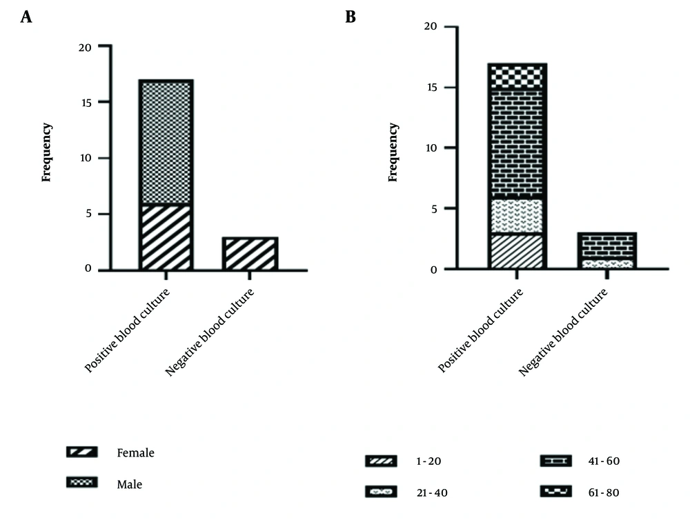
Figure 1.
A, distribution of infective endocarditis (IE) patients by blood culture results stratified by gender; B, distribution of IE patients by blood culture results across different age groups
Table 6.
The Frequency Distribution of the Study Population Based on the Genus and Bacteria a
| Microorganism | Female | Male |
|---|---|---|
| Pseudomonas sp. | 0 | 1 (5.88) |
| Escherichiacoli | 1 (5.88) | 1(5.88) |
| Enterococcus sp. | 0 | 2 (11.76) |
| Streptococcus sp. | 2 (11.76) | 2 (11.76) |
| Staphylococcusepidermidis | 1 (5.88) | 2 (11.76) |
| S.aureus | 2 (11.76) | 3 (17.65) |
a Values are expressed as No. (%).
4.4. Phylogenetic Trees of Blood Culture Negative Microorganisms
Phylogenetic trees of S. sanguinis, S. mitis, and B. melitensis are shown, respectively, in Figure 2.
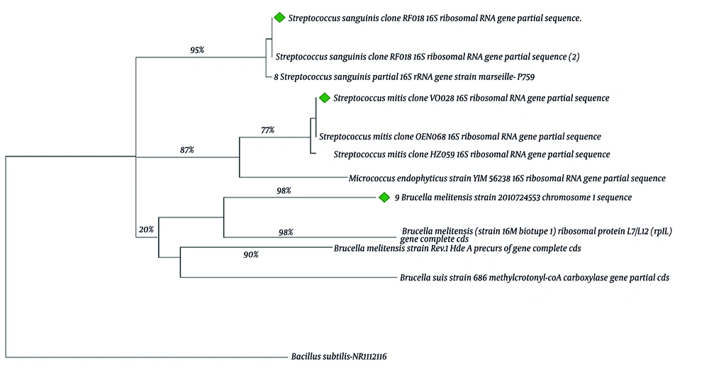
Figure 2.
Phylogenetic tree using the neighbor-joining method with 100 bootstrap replicates. Bacillus subtilis has been selected as outgroup. Isolates are shown.
5. Discussion
Infective endocarditis remains a significant clinical challenge and is currently recognized as the fourth leading cause of life-threatening infectious syndromes worldwide. Despite advances in antimicrobial therapy, diagnostic imaging, particularly echocardiography, and surgical interventions, the disease continues to carry considerable morbidity and mortality rates (14). The implementation of updated clinical diagnostic criteria, such as those outlined in the modified Duke criteria, alongside an emphasis on early and accurate echocardiographic assessment, has markedly improved diagnostic precision. Nonetheless, the evolving landscape of microbial resistance and shifts in patient demographics necessitate continual reassessment of treatment protocols and prevention strategies (15).
This study underscores the critical importance of pathogen identification and antimicrobial susceptibility profiling in the management of IE. The increasing prevalence of IE in many regions, including Iran, highlights the urgent need to revisit and reinforce effective treatment strategies. Particularly concerning is the growing incidence of IE among men aged 41 - 60, a demographic that appears disproportionately affected, consistent with previous studies that have identified male sex and older age as risk factors for IE (16).
Antibiotic resistance is a pivotal matter in the infectious disease treatment process, and in recent years it has become a world concern; as a result, antibiotic resistance is a crisis. This resistance can increase the rate of disease transmission, disease duration (which may raise the costs of treatment), and mortality. Chirillo et al. reported that 73% of IE patients in their cohort were male, with a predominant age range of 34 - 60 years (17), findings echoed across multiple epidemiological investigations. In several studies, it was confirmed that most IE patients are men and their ages are higher than 35 years (18-21).
Comorbidities and predisposing factors continue to play a vital role in the pathogenesis of IE. In our study, the most prevalent underlying conditions among IE patients were coronary heart disease (35%) and cardiac rheumatism (30%). These findings align with prior reports indicating that structural heart diseases and previous cardiac surgeries are major contributors to IE susceptibility. For instance, Day et al. observed that among 1,588 IE patients, 662 had cardiac comorbidities, including congenital heart disease (81%), valve replacement or repair (80%), and cardiac rheumatism (5%) (22). Additional studies have reinforced the link between IE and prior cardiac surgery, gingival procedures, and rheumatic heart disease (17-20).
Microbiological evaluation remains a cornerstone in the diagnosis and management of IE. In our cohort, blood cultures were positive in 85% of patients. The predominant pathogens identified included S. aureus (25%), Streptococcus spp. (20%), S. epidermidis (15%), Enterococcus spp. (10%), E. coli (10%), and Pseudomonas spp. (5%). These findings are consistent with global data indicating that Staphylococcus, Streptococcus, and Enterococcus species are the leading causative agents in IE (17, 19, 20). Notably, among patients with negative blood cultures (15%), the most frequently identified organisms were Streptococcus spp. (66.66%) and Brucella spp. (33.33%). This result reflects recent reports suggesting that fastidious organisms, such as Bartonella spp. and Brucella spp., are increasingly implicated in culture-negative endocarditis cases (18, 23, 24).
The challenge of antimicrobial resistance cannot be overstated. The widespread emergence of resistant strains significantly complicates IE management and contributes to prolonged disease courses, increased healthcare costs, and elevated mortality rates. The findings from Shiraz, Iran, are consistent with a broader national scoping review which highlighted antibiotic resistance as a major public health threat in Iran, underscoring the need for local antimicrobial stewardship programs (25). The resistance patterns observed in our study support the growing concern surrounding the efficacy of standard antibiotic regimens and underscore the necessity of tailoring therapy based on local microbiological data and resistance trends. These findings further reinforce the global call for antimicrobial stewardship and the rational use of antibiotics in clinical practice. Given the complexity of IE and the evolving epidemiological and microbiological profiles, adherence to updated international guidelines is essential.
Recommendations from authoritative bodies such as the European Society of Cardiology (ESC) and the American Heart Association (AHA) provide crucial direction for clinicians in diagnosing, managing, and preventing IE (26-28). These guidelines emphasize individualized patient assessment, early surgical intervention when indicated, and rigorous follow-up, all of which are pivotal in improving clinical outcomes.
5.1. Conclusions
This study highlights the epidemiological and microbiological characteristics of IE in our patient cohort, with a marked predominance among males aged 41 - 60. A significant proportion of IE cases (74%) with positive blood cultures were associated with Staphylococcus species, reaffirming their central role as primary pathogens in IE. Furthermore, antimicrobial susceptibility testing revealed a concerning level of resistance among the isolated microorganisms, underscoring the growing threat of antibiotic resistance in managing IE. These findings emphasize the urgent need for ongoing surveillance, routine antimicrobial resistance profiling, and adherence to updated clinical guidelines to optimize therapeutic outcomes. Future studies with larger sample sizes and molecular diagnostic approaches are recommended to further investigate resistance mechanisms and refine targeted treatment strategies.