
1. Background
Coronavirus disease-19 (COVID-19), first identified in late 2019, rapidly evolved into a global pandemic, causing substantial morbidity and mortality worldwide (1). Before vaccines became available, preventive strategies such as mask use, hand hygiene, and social distancing were implemented; yet achieving long-term control required herd immunity and the rapid development of safe and effective vaccines (2). Vaccines based on different platforms, including inactivated virus and novel messenger RNA (mRNA) technologies, were promptly authorized for emergency use. In Türkiye, the national program began with CoronaVac in January 2021, followed by BNT162b2 in April 2021 (3).
The immune system generates protection either through natural infection or vaccination; while both induce antibodies (4), vaccine-induced immunity — particularly from mRNA vaccines — has shown superior efficacy in preventing severe disease, especially in vulnerable populations (5). Antibody responses have been widely assessed using commercial serological assays such as enzyme-linked immunosorbent assay (ELISA), chemiluminescence immunoassay (CLIA), lateral flow assay (LFA), and enzyme-linked fluorescent assay (ELFA), targeting spike (S) or nucleocapsid (N) proteins as reliable indicators of humoral immunity (6, 7).
Although the duration of vaccine-mediated protection remains uncertain due to the decline in neutralizing antibodies over time, mRNA vaccines generally elicit stronger and more persistent responses than inactivated vaccines (8-10). Despite these findings, comparative data on the long-term persistence of humoral immunity across vaccine platforms remain limited, particularly in the Turkish population. Therefore, this study aimed to longitudinally assess antibody durability over one year following BNT162b2 or CoronaVac vaccination, addressing an important gap in understanding vaccine-induced immunity.
2. Objectives
The present study evaluated neutralizing antibody responses in individuals with a documented negative history of COVID-19 during the six months before the first sampling (TP-I, December 2022) who had received at least three homologous doses of either an inactivated or mRNA vaccine. The COVID-19-negative status was confirmed by reverse transcription polymerase chain reaction (RT-PCR) at TP-I and, at later time points (TP-II and TP-III), through systematic symptom screening, medical record review, and negative BinaxNOW antigen test results. Differences in antibody levels between vaccine types and their relation to vaccine preference were analyzed. Serial samples from the same individuals were also used to evaluate temporal changes in antibody responses and the duration of detectable serological protection.
3. Methods
This prospective observational study was conducted between December 2022 and December 2023. A total of 100 participants were included, determined through a priori power analysis ensuring 80% statistical power at a 0.05 significance level. Adult volunteers who had received at least three homologous doses of either CoronaVac or BNT162b2 and had no confirmed COVID-19 diagnosis within six months before December 2022 were enrolled. Participants were recruited voluntarily from the general community, including students in health-related fields, physicians, nurses, and academic staff. Inclusion criteria required a negative RT-PCR test and age ≥ 18 years. Individuals were excluded if they were pregnant, underage, or had immunodeficiency, autoimmune disease, immunosuppressive therapy, transplantation, or organ failure (Figure 1).
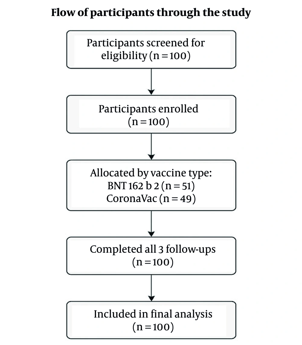
Figure 1.
Flow of participants through the study
Serum samples were collected at three predefined time points: TP-I (December 2022), TP-II (June 2023), and TP-III (December 2023). All samples were obtained within one week of each time point. The same participants were followed longitudinally, and antibody changes were analyzed across three intervals: PI (TP-I to TP-II), PII (TP-II to TP-III), and PIII (TP-I to TP-III). At each sampling, participants were screened for COVID-19. At TP-I, prior infection was excluded using RT-PCR. At TP-II and TP-III, symptomatic illness, healthcare visits, and RT-PCR results were reviewed, and BinaxNOW™ COVID-19 Antigen Self Test (Abbott Laboratories, USA) were used to confirm negative status. These verification procedures were consistent across all time points and are also referenced in the objectives section for clarity.
Blood samples (≥ 5 mL) were collected in serum separator tubes, centrifuged at 4000 rpm for 5 minutes, and stored at -80°C. Anti-severe acute respiratory syndrome coronavirus 2 (SARS-CoV-2) immunoglobulin G (IgG) levels were measured using the VIDAS Anti-SARS-CoV-2 IgG assay (BioMerieux, France), a two-step ELFA targeting the protein S. Analyses were performed on the VIDAS analyzer with 100 µL of serum per test, and results were interpreted according to the manufacturer’s criteria [Cutoff Index (COI) < 1.0 = negative; COI ≥ 1.0 = positive].
3.1. Statistical Analysis
All analyses were performed using SPSS v25.0 (IBM Corp., USA). Continuous variables were expressed as mean ± SD or median (min-max), and categorical variables as No. (%). As variables were non-normally distributed (Kolmogorov-Smirnov P < 0.001), non-parametric tests were applied. Potential confounding factors, including age and sex, were examined using Spearman’s rank correlation coefficient and non-parametric between-group tests (Mann-Whitney U and Kruskal-Wallis), allowing evaluation of their influence on antibody levels without assuming data normality. Differences among three time points were assessed with the Friedman test, followed by Wilcoxon signed-rank tests with Bonferroni correction (P < 0.0167). Between-group comparisons used Mann-Whitney U or Kruskal-Wallis tests, and correlations were analyzed with Spearman’s coefficient. Subgroup analyses were conducted by vaccine type, age, and sex to assess potential interactions in antibody kinetics. A P-value < 0.05 was considered statistically significant.
4. Results
4.1. Demographic Characteristics of Participants and Vaccine Preferences
A total of 100 participants were included, with a mean age of 34.9 ± 12.8 years (range: 20 - 59); 62% (n = 62) were female and 38% (n = 38) male. Vaccine distribution was nearly equal: 51% (n = 51) received BNT162b2 and 49% (n = 49) CoronaVac. In the BNT162b2 group, mean ages were 41.8 ± 10.6 years for females and 30.8 ± 11.5 for males; in the CoronaVac group, 37.8 ± 14.0 and 30.9 ± 12.1 years, respectively. A significant association was observed between vaccine type and gender (χ2 = 3.968, P = 0.046), with more females receiving CoronaVac and more males BNT162b2. Age distribution did not differ significantly between vaccine groups (U = 1217.000, P = 0.822). Detailed demographic data are shown in Table 1.
Table 1.
Demographic Characteristics of Participants by Vaccine Type a
| Demographic Characteristics | CoronaVac (n = 49) | BNT162b2 (n = 51) | P-Value |
|---|---|---|---|
| Gender [No. (%)] | 0.046 | ||
| Female | 28 (57.1) | 19 (37.3) | |
| Male | 21 (42.9) | 32 (62.7) | |
| Age (M ± SD) | 34.1 ± 13.6 | 34.9 ± 12.3 | 0.822 |
| Age range (y) | 20 - 59 | 20 - 58 | - |
a All participants were healthy at baseline, without reported comorbidities or immuno-compromising conditions. The relationship between vaccine type and gender was evaluated with the chi-square test; age comparisons were conducted using the Mann-Whitney U test.
4.2. Immunoglobulin G Antibody Titers by Vaccine Type
All participants remained IgG-positive (COI ≥ 1) at all three time points. Median IgG titers were consistently higher in the BNT162b2 group than in the CoronaVac group. Mann-Whitney U tests showed significant differences at all time points: TP-I: U = 335.5, P < 0.001; TP-II: U = 198.0, P < 0.001; TP-III: U = 150.0, P < 0.001. These differences remained significant after Bonferroni correction (P < 0.0167). Median values and ranges are presented in Table 2, and the decline trend is illustrated in Figure 2.
Table 2.
Median Immunoglobulin G Levels by Vaccine Type and Time Points (BAU/mL) a
| Study Time and Vaccine Type | Median (BAU/mL) | Min-Max (BAU/mL) | Q1-Q3 (BAU/mL) b |
|---|---|---|---|
| TP-I | |||
| BNT162b2 | 51.6 | 17.71 - 61.20 | 44.84 - 57.26 |
| CoronaVac | 23.2 | 15.66 - 56.83 | 20.08 - 41.67 |
| TP-II | |||
| BNT162b2 | 34.5 | 19.09 - 49.76 | 29.97 - 39.71 |
| CoronaVac | 15.4 | 6.07 - 35.89 | 10.41 - 20.06 |
| TP-III | |||
| BNT162b2 | 29.4 | 10.47 - 41.23 | 22.90 - 32.84 |
| CoronaVac | 8.23 | 1.02 - 34.30 | 1.40 - 11.17 |
a Immunoglobulin G (IgG) values represent median concentrations measured at three time points (TP-I: December 2022, TP-II: June 2023, TP-III: December 2023).
b Q1 - Q3: interquartile range.
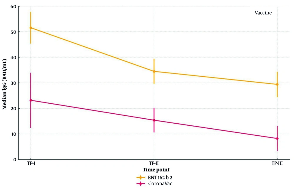
Figure 2.
Change in severe acute respiratory syndrome coronavirus 2 (SARS-CoV-2) immunoglobulin G (IgG) levels over time by vaccine type
Median IgG concentrations (BAU/mL) measured at three time points (TP-I, TP-II, TP-III) in individuals vaccinated with either BNT162b2 or CoronaVac. Error bars represent the interquartile range (Q1 - Q3). No participant experienced symptomatic COVID-19 or tested antigen-positive during the study period. Minor fluctuations, including the lowest value at TP-II, likely reflect biological or analytical variation rather than breakthrough infection.
4.3. Temporal Changes in Immunoglobulin G Levels
Within-group comparisons using the Wilcoxon signed-rank test showed a significant decline in IgG levels over time for both vaccine types (Table 3). Reductions were observed in all intervals — TP-I to TP-II (PI), TP-II to TP-III (PII), and TP-I to TP-III (PIII) — each with P < 0.001.
Table 3.
Statistical Comparison of Within-Group Immunoglobulin G Titer Decline by Vaccine Type Across Three Time Points a
| Vaccine Type and Period | Median IgG Start (BAU/mL) | Median IgG End (BAU/mL) | Z | P-Value |
|---|---|---|---|---|
| BNT162b2 | ||||
| PI | 51.60 | 34.50 | -6.215 | < 0.001 |
| PII | 34.50 | 29.40 | -6.158 | < 0.001 |
| PIII | 51.60 | 29.40 | -6.215 | < 0.001 |
| CoronaVac | ||||
| PI | 23.20 | 15.40 | -6.093 | < 0.001 |
| PII | 15.40 | 8.23 | -6.093 | < 0.001 |
| PIII | 23.20 | 8.23 | -6.093 | < 0.001 |
a Immunoglobulin G levels were compared across three time points using the Wilcoxon signed-rank test. Statistically significant differences were observed at each time point within both vaccine groups (P < 0.001 for all comparisons).
4.4. Percent Decline Analysis of Immunoglobulin G Levels
A time-dependent decrease in IgG concentrations was observed in all intervals based on median values. In the CoronaVac group, antibody levels fell by 33.6% from TP-I to TP-II and 46.6% from TP-II to TP-III, totaling a 64.5% annual decline. The BNT162b2 group showed a 33.1% reduction in the first six months and 14.8% thereafter, with a total one-year decline of 43.0%. Although both groups exhibited marked reductions, BNT162b2 responses remained higher throughout follow-up (Figure 3).The graph illustrates the relative percent reduction in median IgG concentrations (BAU/mL) between TP-I and TP-II (period I), TP-II and TP-III (period II), and TP-I and TP-III (period III), stratified by vaccine group.
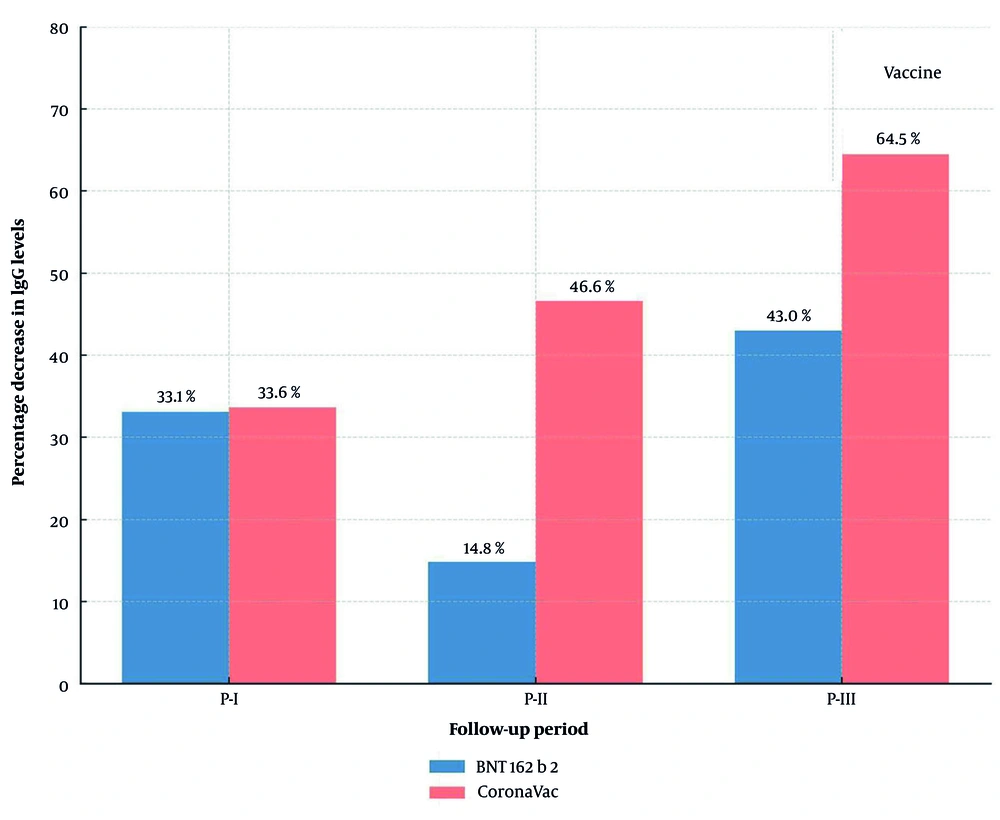
Figure 3.
Percentage decrease in immunoglobulin G (IG) antibody levels by vaccine type across follow-up periods
To examine the time effect on antibody kinetics, a Friedman test across TP-I, TP-II, and TP-III revealed a significant temporal trend (χ2 = 198.020; P < 0.001). Post-hoc Wilcoxon tests confirmed significant differences between all time points (P < 0.001; Bonferroni-corrected P < 0.0167), supporting the robustness of the observed decline. An overall estimation by averaging median IgG values at TP-I and TP-III across both vaccine groups indicated an approximate 49.7% reduction over one year. These results demonstrate a substantial and statistically significant decline in IgG levels, independent of vaccine type.
4.5. Association Between Age, Sex, and Decline in Immunoglobulin G Antibody Levels
Spearman’s rank correlation showed no significant relationship between participants’ age and IgG levels at any time point (TP-I, TP-II, or TP-III) (P > 0.05). Similarly, IgG levels did not differ significantly between males and females at any point (P > 0.05). Analysis of annual percent changes revealed no correlation with age (rs = 0.145; P = 0.149) and no significant difference between sexes (P > 0.05). These results indicate that the decline in antibody levels occurred independently of demographic factors such as age and sex.
5. Discussion
The COVID-19 pandemic has brought the understanding of immune responses against SARS-CoV-2 — and their long-term persistence — to the forefront of global research. Comparative evaluation of vaccine platforms is essential for guiding public health strategies and developing next-generation vaccines. Neutralizing antibody dynamics consistently confirm the efficacy of vaccine-induced adaptive immunity in preventing infection; numerous studies have shown that vaccination substantially reduces the risk of severe illness and mortality, particularly in symptomatic and comorbid individuals (8, 11-13). In countries where multiple vaccines are in use, analyzing the specific immune responses elicited by each is critical for assessing durability and informing optimized vaccination strategies. Although vaccines were deployed rapidly, ongoing follow-up studies remain vital to understanding the persistence of immunity and preparing for future outbreaks.
Aligned with this study’s aim, we longitudinally evaluated antibody kinetics induced by two widely used COVID-19 vaccines in Türkiye — BNT162b2 (mRNA-based) and CoronaVac (inactivated) — by measuring IgG levels at three time points to assess response magnitude, durability, and the influence of age and sex. Participants vaccinated with BNT162b2 showed significantly higher IgG levels at all three time points than those receiving CoronaVac (P < 0.001 for all). This finding supports the superior ability of mRNA vaccines to elicit strong antibody responses and aligns with previous studies reporting more durable immunity after BNT162b2 (14, 15). Similar findings were reported by Panahi et al., who evaluated antibody responses among healthcare professionals vaccinated with various COVID-19 platforms and demonstrated that vaccine type substantially influenced humoral immunity levels (16). The enhanced immunogenicity of mRNA vaccines likely stems from their focused protein S targeting, whereas inactivated vaccines, despite presenting the full virus, elicit weaker responses due to limited antigen presentation (17).
Over one year, IgG levels declined significantly in both groups, decreasing gradually among BNT162b2 recipients but more sharply in the CoronaVac group. Six-month interval analyses confirmed a time-dependent decline, with time identified as the main determinant regardless of vaccine type or individual factors. Overall, antibody levels fell by about 50% within twelve months. Comparable findings were also reported by Saeed et al. from Pakistan, who demonstrated significantly higher SARS-CoV-2 S antibody titers following Sputnik V compared to Sinopharm and CoronaVac after only the first dose, supporting the superior immunogenicity of viral-vector and mRNA-based vaccines over inactivated platforms (18). BNT162b2 not only produced higher initial IgG titers but also showed a slower decline than CoronaVac; the 64.5% reduction at twelve months among CoronaVac recipients indicates a shorter-lived immune response. This pattern aligns with reports that mRNA-induced antibodies wane more slowly than those from inactivated vaccines (19, 20). Although based on a limited sample, these temporal trends underscore the need for sustained serological surveillance at the population level.
Demographic analysis revealed a significant association between vaccine type and sex, with more women receiving CoronaVac and more men BNT162b2. However, IgG levels and annual declines did not differ significantly by sex or age, nor was age associated with vaccine preference. These findings are consistent with studies suggesting that antibody responses are not independently influenced by demographic factors. While some reported stronger responses in women and age-related declines (21, 22), others found no significant associations (23, 24). Our results align with the latter, though interpretation should consider sample size, age homogeneity, and unmeasured factors such as hormonal or genetic influences.
All participants remained IgG-positive throughout follow-up. Despite declining titers, no seronegativity or symptomatic infection occurred, indicating continued protection likely mediated by immunological memory and other adaptive mechanisms. Hybrid immunity, arising from vaccination and incidental exposure, also contributes to protection against variants (25). During the study, Omicron and its subvariants predominated, suggesting possible asymptomatic exposures. Such exposures could have reinforced vaccine-induced immunity, sustaining IgG levels and preventing symptomatic infection. This interplay underscores the evolving nature of vaccine-induced and hybrid immunity amid variant circulation.
Several limitations should be acknowledged. The sample size was limited to 100 participants, restricting generalizability. Asymptomatic infections and individual factors such as genetic, hormonal, or environmental variables were not controlled. Although participants with immunosuppressive or major chronic conditions were excluded, some variability likely remained. Only IgG levels were measured, and functional neutralization assays were not performed; therefore, the neutralizing capacity of the detected antibodies could not be directly confirmed. The follow-up period was limited to one year. In addition to these limitations, several potential sources of bias and confounding should be considered. Because participants were voluntarily recruited from healthcare-related fields, selection bias may exist, as these individuals might exhibit greater health awareness and vaccine adherence than the general population. Furthermore, undetected asymptomatic infections could have acted as confounders affecting antibody persistence despite regular screening at each time point. Finally, as only humoral immunity was assessed, measurement bias cannot be entirely excluded.
Despite these limitations, the study provides meaningful comparative data on humoral immune responses over twelve months after two vaccine types and offers insight into how vaccine platform, time, and demographics shape antibody dynamics. Although antibody levels declined significantly, all participants remained seropositive and free of symptomatic infection, suggesting sustained clinical protection potentially supported by hybrid immunity. Overall, the results should be interpreted in light of the study population’s characteristics and sample size. While they provide valuable insight into antibody persistence within this cohort, further research involving larger and more diverse populations would strengthen generalizability.
5.1. Conclusions
In conclusion, this study demonstrates that while both vaccines maintained seropositivity over one year, BNT162b2 induced stronger and more persistent IgG responses than CoronaVac. These findings highlight the need for continued immune monitoring and larger, long-term studies to inform future vaccination strategies.