1. Introduction
Severe fever with thrombocytopenia syndrome (SFTS) is currently classified within the Phenuiviridae family, Bandavirus genus, and possesses a tripartite genome comprising L, M, and S segments. It is a spherical, enveloped, and segmented negative-strand RNA virus (1). The incubation period of severe fever with thrombocytopenia syndrome virus (SFTSV) is generally five to fourteen days, and transmission occurs primarily through arthropods such as ticks, with a reported mortality rate of up to 30% (2). Patients with SFTSV infection accompanied by central nervous system involvement often experience rapid symptom progression, including headaches, nausea, vomiting, limb tremors, and disturbances of cognition and consciousness. Studies indicate that SFTSV infection increases the risk of opportunistic fungal infections, particularly Aspergillus, which can lead to multi-organ system failure and higher mortality rates (3).
Among opportunistic fungi, both Aspergillus and Rhizopus are notable for causing extensive vascular invasion, resulting in vascular thrombosis and significant tissue infarction. Pulmonary infections caused by Rhizopus are frequently associated with respiratory distress and hemoptysis, with a mortality rate of 70% to 100% if left untreated (4).
Here we report a rare case of co-infection with SFTSV and mold fungi in eastern China. To the best of our knowledge, no previous reports have documented simultaneous pulmonary infections caused by both Aspergillus and Rhizopus in the context of SFTS. Therefore, this case represents the first documented instance of dual fungal co-infection associated with SFTS, highlighting a novel and clinically important manifestation of this emerging viral disease. Based on the detection results of metagenomic next-generation sequencing (mNGS), we rapidly identified pathogenic evidence, thereby facilitating prompt and precise treatment for the patient.
2. Case Presentation
The patient consented to research authorization for record review, and the study received approval from the institutional review board. A forty-five-year-old male, resident of Xiangshan County, Zhejiang province — a hilly region in southeast China — was bitten by ticks on the inner side of his right ankle while mowing grass on April 6, 2024. He developed generalized malaise, including chills, fever, and diarrhea, and presented to a local hospital on April 18. He was initially prescribed levofloxacin and returned home. On April 23, due to worsening symptoms, he was admitted to the Infection Department of Ningbo Yinzhou No. 2 Hospital.
The patient was previously healthy, with no history of hypertension, diabetes, or hematologic/oncologic disease. Upon admission, routine microbiological and virological examinations were performed. Results for common respiratory pathogens, including influenza viruses A and B, parainfluenza virus, respiratory syncytial virus, adenovirus, cytomegalovirus, Epstein-Barr virus, and bacteria such as Mycobacterium tuberculosis, were all negative. Blood and sputum cultures showed no evidence of bacterial or fungal growth, and serologic tests for parasites were negative.
On admission, the patient was alert with a body temperature of 37.9°C (ear temperature), heart rate of 80 beats per minute, respiratory rate of eighteen breaths per minute, blood pressure of 117/66 mmHg, and oxygen saturation (SpO2) of 99% on room air. A dark red, round papular scab was observed on the medial aspect of the right ankle (Figure 1). The remainder of the skin was intact, and the neurologic examination was unremarkable.
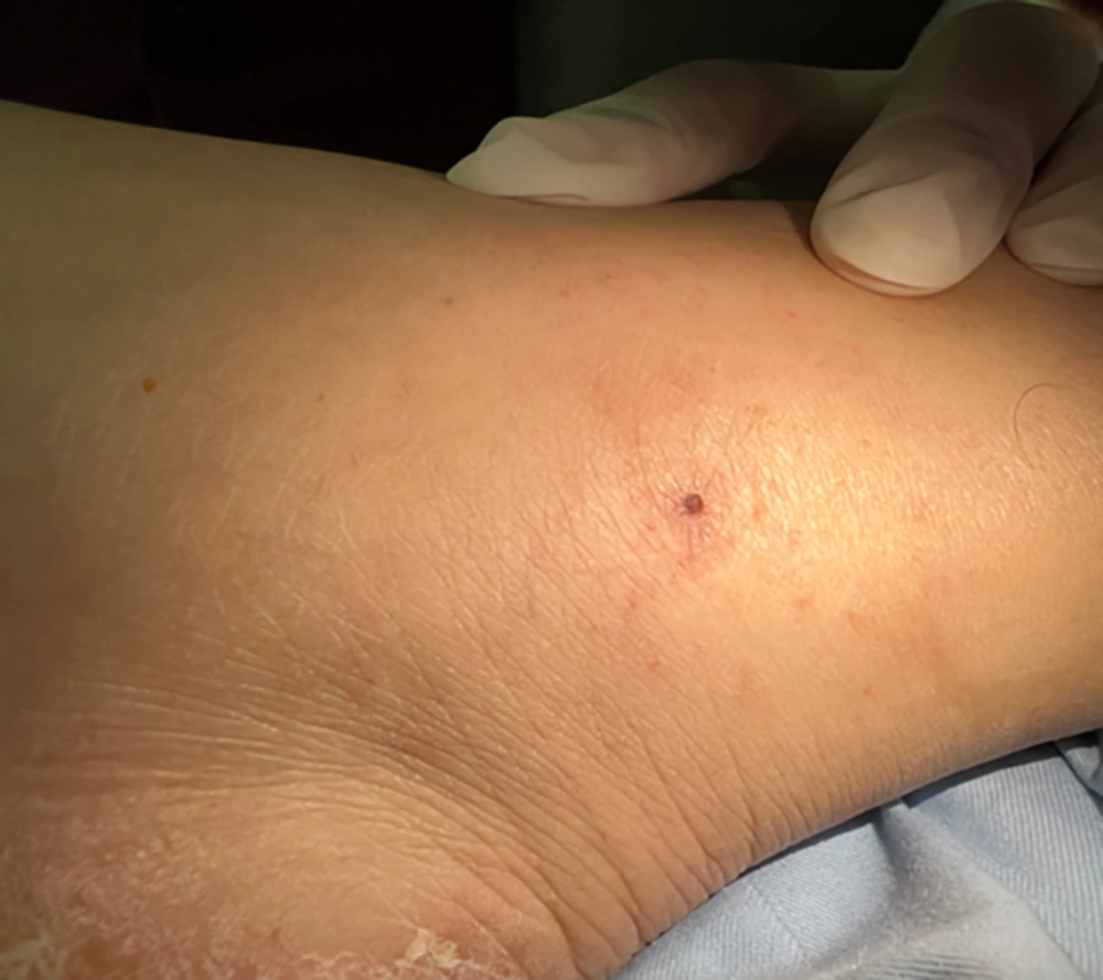
Figure 1.
Bite mark on the medial ankle
Initial laboratory studies revealed leukocytopenia (white blood cell count: 2,700 cells/mm3), thrombocytopenia (platelet count: 82,000 cells/mm3), elevated C-reactive protein (CRP) level (11.2 mg/L), elevated aspartate aminotransferase (AST) level (407 U/L), elevated alanine aminotransferase (ALT) level (136 U/L), elevated creatine kinase (CK) level (14,897 U/L), elevated lactate dehydrogenase (LDH) level (1,168 U/L), acute renal insufficiency (serum creatinine: 192 μmol/L), elevated cardiac troponin I (0.072 ng/mL), and significantly increased ferritin (> 1,500 ng/mL). Results for the Widal test, cytomegalovirus-DNA, Epstein-Barr virus-DNA, Mycoplasma pneumoniae-DNA, influenza A-RNA, influenza B-RNA, respiratory syncytial virus-RNA, rhinovirus-RNA, adenovirus-DNA, and COVID-19-RNA were negative. Hemorrhagic fever with renal syndrome antibodies and dengue fever antibodies were also negative. No Plasmodium was found. Chest computed tomography (CT) revealed flocculent high-density shadows in both lungs (Figure 2). Electrocardiogram and echocardiographic findings were normal.
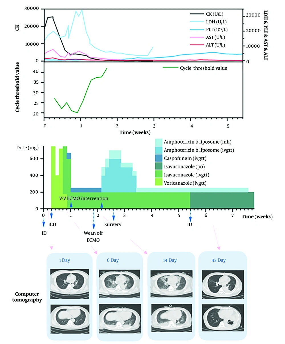
Figure 2.
Antifungal treatment regimen administered during the patient’s intensive care unit (ICU) admission
On April 25, the patient developed hand tremor, followed by a generalized tonic-clonic seizure and trismus. He was transferred to the intensive care unit (ICU) after endotracheal intubation. Due to these clinical manifestations, serum samples were sent to the Infectious Disease Diagnosis Laboratory of Ningbo City Center for Disease Control and Prevention for SFTSV analysis. Polymerase chain reaction (PCR) testing of cerebrospinal fluid (CSF) for SFTSV was weakly positive, and serum reverse transcription Polymerase chain reaction (RT-PCR) for SFTSV was positive, with a cycle threshold (Ct) value of 27 (Figure 2). The CSF examination revealed CSF pressure of 18 cm H2O, leukocyte count of 0/mm3, erythrocyte count of 0/mm3, protein concentration of 351 mg/L, LDH of 15 U/L, and glucose concentration of 4.82 mmol/L. Serum mNGS detected 3,244 RNA sequence reads with a relative abundance of 58.61% corresponding to SFTSV. The patient was thus diagnosed with SFTSV-associated encephalopathy.
Management included antiepileptic drugs, methylprednisolone (40 mg/day for three days), immunoglobulin (400 mg/kg/day for six days, totaling 180 g) to modulate the immune response, favipiravir as antiviral therapy, and doxycycline to address potential co-infection (given the possibility of Rickettsia and Borrelia burgdorferi sensu lato from ticks) (5). Additionally, three sessions of therapeutic plasma exchange were performed. Platelets, LDH, CK, Ct values, AST, and ALT were monitored throughout the course and are depicted as curves in Figure 2. Notably, LDH, AST, and ALT levels fluctuated on April 25, 26, and 27, coinciding with plasma exchange procedures.
On day three of admission, fiberoptic bronchoscopy revealed multiple white patches in the main bronchus, congested and edematous bronchial mucosa, and a small amount of adherent white mucous sputum. Sputum culture smear showed septate hyphae, suggestive of Aspergillus. The patient was started on voriconazole antifungal therapy.
On day seven of admission, the patient's oxygenation and circulation worsened and septic shock was suspected, accompanied by further respiratory decline. Veno-venous extracorporeal membrane oxygenation (V-V ECMO) was initiated. The same day, mNGS from serum and bronchoalveolar lavage fluid (BALF) confirmed Aspergillus fumigatus infection. The antifungal regimen was adjusted to caspofungin in combination with isavuconazole. After six days, the patient was successfully weaned from V-V ECMO, with a decline in plasma viral load to undetectable levels (Figure 2).
Following modification of the antifungal regimen, the patient’s respiratory status initially improved, with reduced oxygen requirements and gradual resolution of pulmonary infiltrates on follow-up CT performed on May 2. Serum inflammatory markers, including CRP and procalcitonin, declined, and both platelet and leukocyte counts normalized. However, around May 7, clinical deterioration recurred with renewed fever (38.5°C), increased oxygen demand, and rising CRP and LDH levels. Repeat imaging revealed new consolidations and cavitation in the left lower lobe, inconsistent with isolated aspergillosis. This prompted re-evaluation of the antifungal strategy and repeat mNGS, which subsequently identified Rhizopus microsporus.
Bronchoscopy was repeated, and on May 8, mNGS of BALF identified both A. fumigatus and R. microsporus (sequence count: 4,151; relative abundance: 3.07%). Initial bronchoscopic specimens and smears had shown only septate hyphae consistent with Aspergillus species; direct microscopy and culture for Mucorales were not initially performed due to predominance of Aspergillus on mNGS and culture, and rapid clinical deterioration. After detection of R. microsporus by mNGS, repeated bronchoscopy and BALF culture targeting Mucorales were performed, resulting in isolation and confirmation of R. microsporus, consistent with mNGS findings.
Later that evening, the patient developed a dark bloody clot in the airway after choking. Bedside bronchoscopy showed active bleeding in the basal segment of the left lower lobe and cast thrombosis of the main bronchial airway. Bronchial angiography revealed hyperplasia and tortuosity of the left lower lung bronchial artery, with a few abnormal staining foci. After bronchoscopic hemostasis failed, bilateral bronchial artery embolization (BAE) was performed. Liposomal amphotericin B was administered at 3 mg/kg/day, increasing to 5 mg/kg/day (Figure 2). On May 11, emergency left lower lobectomy was performed for severe hemoptysis (Figure 1 in Supplementary File). Simultaneously, R. microsporus was detected in BALF culture (Figure 2 in Supplementary File), confirming previous mNGS results.
The patient’s consciousness gradually returned to normal, and a head MRI scan was completed (Figure 3 in Supplementary File). He was subsequently transferred to the Infection Unit for rehabilitation. The patient received five weeks of amphotericin B liposome followed by oral isavuconazole (200 mg once daily) until complete radiological resolution was achieved on follow-up chest CT. The clinical progression and treatments are summarized in Figure 2. Surgical pathology revealed two types of fungal hyphae: Narrow, septate, acute-angle branching hyphae consistent with Aspergillus species, and broad, ribbon-like, sparsely septate hyphae with right-angle branching characteristic of Rhizopus species. These features confirmed coexistence of Aspergillus and Mucorales infection, as annotated in Figure 4 in Supplementary File.
On day ten of admission, despite combined antifungal therapy targeting Aspergillus (caspofungin plus isavuconazole), fever persisted and oxygenation worsened. Follow-up CT showed progressive left lower lobe consolidation and new cavitation inconsistent with typical invasive aspergillosis alone. Laboratory assessment revealed rising serum β-D-glucan and persistently elevated inflammatory markers, while galactomannan did not further increase. These atypical findings, in combination with worsening hemoptysis and lack of improvement on anti-Aspergillus therapy, raised suspicion for mucormycosis. Consequently, repeat mNGS of BALF confirmed R. microsporus sequences. A detailed timeline of the patient’s clinical course, diagnostics, and interventions is presented in Figure 3.
![Timeline summarizing the patient’s clinical course, diagnostic findings, and treatments – the figure illustrates the sequence of key clinical events, including the onset of severe fever with thrombocytopenia syndrome (SFTS), respiratory deterioration requiring extracorporeal membrane oxygenation (ECMO), detection of <i>Aspergillus fumigatus</i> and <i>Rhizopus microsporus</i>, antifungal therapy adjustments, and surgical interventions [bronchial artery embolization (BAE) and lobectomy].](https://brieflands.com/journals/jjm/articles/166706/figures/jjm-18-11-166706-i002-preview.webp "Timeline summarizing the patient’s clinical course, diagnostic findings, and treatments – the figure illustrates the sequence of key clinical events, including the onset of severe fever with thrombocytopenia syndrome (SFTS), respiratory deterioration requiring extracorporeal membrane oxygenation (ECMO), detection of <i>Aspergillus fumigatus</i> and <i>Rhizopus microsporus</i>, antifungal therapy adjustments, and surgical interventions [bronchial artery embolization (BAE) and lobectomy].")
Figure 3.
Timeline summarizing the patient’s clinical course, diagnostic findings, and treatments – the figure illustrates the sequence of key clinical events, including the onset of severe fever with thrombocytopenia syndrome (SFTS), respiratory deterioration requiring extracorporeal membrane oxygenation (ECMO), detection of Aspergillus fumigatus and Rhizopus microsporus, antifungal therapy adjustments, and surgical interventions [bronchial artery embolization (BAE) and lobectomy].
3. Discussion
This case highlights the diagnostic and therapeutic complexity of managing SFTS-associated encephalopathy complicated by dual pulmonary fungal infections. The patient presented with progressive respiratory failure and septic shock, despite standard antiviral and antibacterial therapy. Early bronchoscopy identified septate hyphae, leading to a diagnosis of invasive aspergillosis and initiation of targeted antifungal therapy (6). One of the main clinical challenges was the atypical disease progression despite adequate anti-Aspergillus therapy. Although the patient initially improved, with decreasing oxygen requirements and inflammatory markers, he soon deteriorated, developing new pulmonary cavities and hemoptysis. These findings prompted reconsideration of the original diagnosis and repeat mNGS testing, which revealed R. microsporus co-infection. This diagnostic turnaround underscores the importance of maintaining suspicion for mucormycosis in immunocompromised or critically ill patients, even when another fungal pathogen has already been identified (7, 8).
Previous reports from China and South Korea have primarily described SFTS patients with single fungal infections, such as invasive aspergillosis or candidiasis, but not dual mold co-infections (9-12). In those cases, the fungal infection typically occurred during the late phase of SFTS, often associated with severe immunosuppression or corticosteroid exposure. In contrast, our case demonstrates that simultaneous infection with both Aspergillus and Rhizopus can occur early in the disease course, emphasizing the need for early fungal surveillance and broad diagnostic testing in critically ill SFTS patients.
Another major challenge involved balancing aggressive antifungal treatment with the patient’s hepatic and renal dysfunction secondary to SFTS. Initial antifungal therapy with voriconazole was effective against Aspergillus but provided limited coverage for Mucorales. Therefore, the regimen was switched to liposomal amphotericin B and isavuconazole to broaden antifungal coverage and ensure activity against both Aspergillus and Rhizopus species, while minimizing toxicity, consistent with current treatment guidelines (13). Therapeutic drug monitoring guided dosing adjustments.
The patient also experienced life-threatening hemoptysis, necessitating timely multidisciplinary intervention — including BAE and eventual left lower lobectomy (14). These steps were crucial for achieving hemostasis and source control. In this case, BAE was performed immediately after failure of bronchoscopic hemostasis, when active bleeding from the left lower lobe resulted in airway obstruction and oxygen desaturation. Although temporary hemostasis was achieved, recurrent massive hemoptysis occurred three days later, and emergency lobectomy was performed. The timing of surgery was based on persistent bleeding, localized lesions observed on imaging, and stabilization of vital signs following antifungal therapy and supportive care. Early surgical intervention after initial hemostatic control, once systemic condition permits, is critical to preventing fatal airway obstruction and achieving complete removal of necrotic foci colonized by Mucorales. This experience suggests that prompt but well-timed surgery, guided by clinical stability and lesion localization, can substantially improve outcomes in patients with invasive fungal co-infection.
Similarly, recent multicenter evidence demonstrates that early, multidisciplinary intervention significantly improves survival among patients with invasive mucormycosis. For example, a large retrospective study from Iran involving ninety-seven patients with rhino-orbito-cerebral mucormycosis reported that timely combined antifungal therapy and surgical management markedly reduced mortality rates (15). These findings further support our clinical observation that prompt diagnosis and well-timed surgical resection are critical to improving outcomes in patients with mixed mold infections.
Several risk factors may have contributed to the development of dual fungal infection in this case. The SFTS-related immune dysregulation and corticosteroid therapy can suppress host immunity and facilitate fungal proliferation (16). Specifically, SFTSV infection induces marked endothelial injury and a cytokine storm characterized by elevated levels of interleukin 6, tumor necrosis factor alpha, and interferon gamma, which compromise vascular integrity and tissue oxygenation. Increased vascular permeability and endothelial apoptosis not only contribute to multi-organ failure but also create a microenvironment favorable for fungal invasion. Furthermore, SFTS can transiently increase blood–brain barrier permeability and disrupt innate immune cell function — including neutrophil chemotaxis and macrophage phagocytic activity — thereby facilitating deeper tissue invasion by both viral and fungal pathogens. In addition, iron overload and previous exposure to voriconazole may predispose to mucormycosis by promoting fungal resistance (17-19).
The key lessons from this case include: (1) Early mNGS testing can rapidly detect mixed fungal infections that conventional microscopy and culture may miss; (2) dynamic reassessment of antifungal efficacy is essential when clinical deterioration occurs; and (3) combined medical and surgical management significantly improves outcomes in invasive fungal co-infections. In summary, this case demonstrates that prompt recognition of mixed mold infections and individualized, multidisciplinary management are critical to improving survival in patients with SFTS complicated by invasive fungal disease.