
1. Background
Mycoplasma pneumoniae is a common respiratory pathogen with a high incidence among children, adolescents, and the elderly, posing a significant burden on public health (1-5). The clinical manifestations of M. pneumoniae infection are often nonspecific and overlap with those of other respiratory pathogens, making accurate laboratory diagnosis essential for guiding appropriate treatment decisions. Current detection methods for M. pneumoniae include culture, serological tests, and molecular assays. Culture methods are complex, require special media, and take several weeks to obtain results (6). Serological tests are limited by the window period in early infection and cross-reactivity with other pathogens (7-9). Molecular assays, particularly fluorescent PCR, have become the preferred method due to their high sensitivity and specificity (10-12). However, most existing fluorescent PCR methods require nucleic acid extraction, which increases operational complexity, reagent costs, and detection time to over 1 hour (13, 14).
To address these challenges, we developed an extraction-free fluorescent PCR for M. pneumoniae detection. To the best of our knowledge, this is the first report of applying extraction-free PCR technology to M. pneumoniae. While extraction-free PCR approaches have been developed for other pathogens such as SARS-CoV-2 (15), their application to M. pneumoniae has not been explored. By employing an optimized reaction system containing Triton X-100 and an inhibitor-tolerant polymerase, combined with a novel two-stage denaturation strategy based on amplicon melting temperature analysis, this method enables direct sample processing and detection within 30 minutes, providing a rapid, reliable, and user-friendly diagnostic solution for clinical settings.
2. Objectives
Our study introduces a novel extraction-free fluorescent PCR assay that eliminates nucleic acid purification steps, enabling direct sample-to-result detection within 30 minutes.
3. Methods
3.1. Clinical Samples and Mycoplasma pneumoniae Positive Control
A total of 322 throat swab samples were collected from patients at the first hospital of Jiaxing in 2024, including 32 samples positive for M. pneumoniae, 22 positive for influenza virus, 13 positive for SARS-CoV-2, 9 positive for respiratory adenovirus, 11 positive for rhinovirus, 13 positive for parainfluenza virus, 6 positive for coronavirus NL63, 3 positive for coronavirus OC43, 1 positive for respiratory syncytial virus, 33 positive for Haemophilus influenzae, 18 positive for Klebsiella pneumoniae, 15 positive for Streptococcus pneumoniae, 8 positive for S. pyogenes, 2 positive for Pneumocystis, 3 positive for Aspergillus, 2 positive for Chlamydiapsittaci, and 131 samples negative for all the aforementioned pathogens. The M. pneumoniae positive control with a concentration of 10,000 copies/mL was purchased from Fantasia Biopharma Co., Ltd.
3.2. Primers and Probe Design
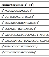
The P1 gene sequences of M. pneumoniae were downloaded from GenBank and subjected to multiple sequence alignment using Clustal X software. Following the comparative analysis, a highly conserved region was selected to design primers and probe using Primer Express 3.0 software. The specificity of the designed primers and probe was verified using NCBI BLAST analysis against the nucleotide database. The sequences exhibited high identity exclusively with M. pneumoniae P1 gene sequences, with no significant homology to other Mycoplasma species, common respiratory pathogens, or the human genome. The sequences of the primers and probe are presented in Table 1.
Table 1.
The Sequences of the Primers and Probe
| Variables | Sequence | Length (bp) |
|---|---|---|
| Primer F | GGCAGTCAACAAACCACGTATG | 22 |
| Primer R | GGTGGTTGATGCGGTCAAA | 19 |
| Probe | FAM-TCCCACCCGAACCGAAGCGG-BHQ1 | 20 |
| Amplicon | - | 63 |
3.3. Analysis of Amplicon Melting Temperature
To determine the melting temperature of the amplicons for designing the two-stage denaturation strategy, melting curve analysis was performed using a conventional PCR protocol. A standard primer concentration of 0.2 µM was used. The detailed reaction components and cycling conditions are summarized in Table 2. After PCR amplification, a melting temperature curve analysis was performed. The PCR products were cooled to 75°C and then heated to 90°C at a rate of 0.1°C per second. Fluorescence signals were continuously monitored during the whole process. All reactions were performed using a Bio-Rad CFX 96™ fluorescent PCR system.
Table 2.
Summary of PCR Conditions Used in Different Experiments
| Parameters | Melting Curve Analysis (Section 2.3) | Commercial Kit (Section 2.7) | Extraction-free Fluorescent PCR (Section 3.2) |
|---|---|---|---|
| Template | Clinical sample (5 µL) | Extracted nucleic acid (5 µL) | Clinical sample (5 µL) |
| Detection chemistry | EvaGreen dye (1 µL) | TaqMan probe | TaqMan probe |
| Primer concentration | 0.2 µM | Not disclosed by manufacturer | 1 µM |
| Probe concentration | N/A (no probe required for EvaGreen dye-based detection) | Not disclosed by manufacturer | 0.6 µM |
| Triton X-100 | No | No | Yes (0.5%) |
| Initial denaturation | 95°C, 2 min | 95°C, 2 min | 95°C, 3 min |
| Cycling conditions | 40 cycles: 95°C 5s, 60°C 20s; fluorescence monitored continuously during melting curve analysis | 40 cycles: 95°C 5s, 60°C 30s, fluorescence collected once per cycle post-annealing/extension | Stage 1: 10 cycles (95°C 1s, 65°C 10s, no fluorescence collection); Stage 2: 30 cycles (86°C 1s, 60°C 5s, fluorescence collected once per cycle post-annealing/extension) |
| Total reaction volume | 20 µL | 25 µL | 20 µL |
3.4. Optimization of Fluorescent PCR
A one-factor-at-a-time optimization strategy was employed to optimize the extraction-free fluorescent PCR assay. The key parameters evaluated included primer pair concentration (0.2 - 1.6 µM), probe concentration (0.1 - 0.8 µM), annealing/extension temperature (55 - 65°C), and annealing/extension time (5 - 20 s). Each parameter was varied individually while all other parameters were kept constant to identify the optimal reaction conditions.
3.5. Specificity and Sensitivity of Fluorescent PCR
The specificity of fluorescent PCR was evaluated by amplifying the 322 throat swab samples mentioned above. For sensitivity, clinical samples negative for Mycoplasma pneumoniae were used for diluting Mycoplasma pneumoniae positive control to achieve final concentrations of 2500, 1000, 500, 250, and 100 copies/mL, respectively, and tested by fluorescent PCR. Each concentration was tested in 16 replicates. The limit of detection was calculated by probit analysis with 95% probability.
3.6. Reproducibility of Fluorescent PCR
For the reproducibility evaluation of the fluorescent PCR, different concentrations of M. pneumoniae positive control were tested repeatedly. Each concentration was tested in triplicate in the same run to evaluate intra-assay variability, and each concentration was tested in three different runs to evaluate inter-assay variability. The intra- and inter-assay variabilities were calculated as coefficient of variation based on quantification cycle (Cq) values of each test (16).
3.7. Comparison of Newly Developed Fluorescent PCR with Commercial Fluorescent PCR
Thirty-two M. pneumoniae-positive samples were diluted 10-fold with clinical samples negative for M. pneumoniae. Both methods were tested using the same set of diluted samples to enable a direct head-to-head comparison. The newly developed fluorescent PCR utilized the optimized PCR solution and amplification conditions as described in Table 2. For the commercial fluorescent PCR, nucleic acids were first extracted using a magnetic bead-based DNA/RNA extraction kit, followed by the preparation of PCR solution and amplification conditions according to the manufacturer's instructions. The detailed PCR conditions are summarized in Table 2.
4. Results
4.1. Analysis of Amplicon Melting Temperature
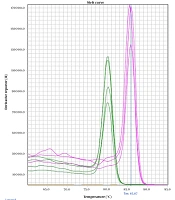
Melting curve analysis after PCR amplification revealed that by 86°C, nearly all amplicons had completely denatured (Figure 1). The melting curve showed a single transition, confirming the amplification of only one specific product without primer-dimers or nonspecific amplification. Based on this finding, we determined to set the denaturation temperature for 30 cycles in the second stage to 86°C.
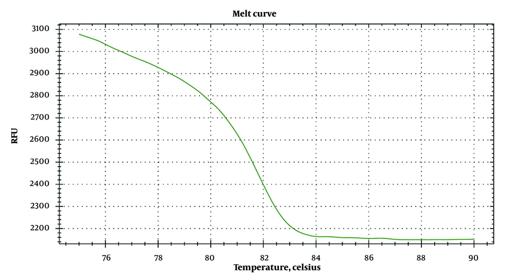
Figure 1.
The melting curve of amplification products
4.2. Optimization of Fluorescent PCR
To determine the optimum primer concentration, probe concentration, and annealing/extension temperature for the newly developed fluorescent PCR, optimization was performed. Based on the optimized results, the optimal primer combination was 1 µM, the optimal probe concentration was 0.6 µM, the optimal annealing/extension temperature and time for the first stage were 65°C and 10 s, and the optimal annealing/extension temperature and time for the second stage were 60°C and 5 s. The complete optimized reaction conditions, including the two-stage denaturation protocol, are summarized in Table 2.
4.3. Specificity of Fluorescent PCR
The specificity of the fluorescent PCR was evaluated using 322 clinical samples, including samples positive for 16 different respiratory pathogens (8 viruses, 4 bacteria, 2 fungi, 1 Mycoplasma species, and 1 Chlamydia species). Clinical samples positive for M. pneumoniae showed positive amplification, whereas no positive fluorescent signal was observed in other clinical samples. All 32 M. pneumoniae-positive samples showed positive amplification, while no false-positive signals were observed in samples containing other pathogens or in pathogen-negative samples, demonstrating 100% specificity. No cross-reaction with other pathogens or false positives was observed (Figure 2).
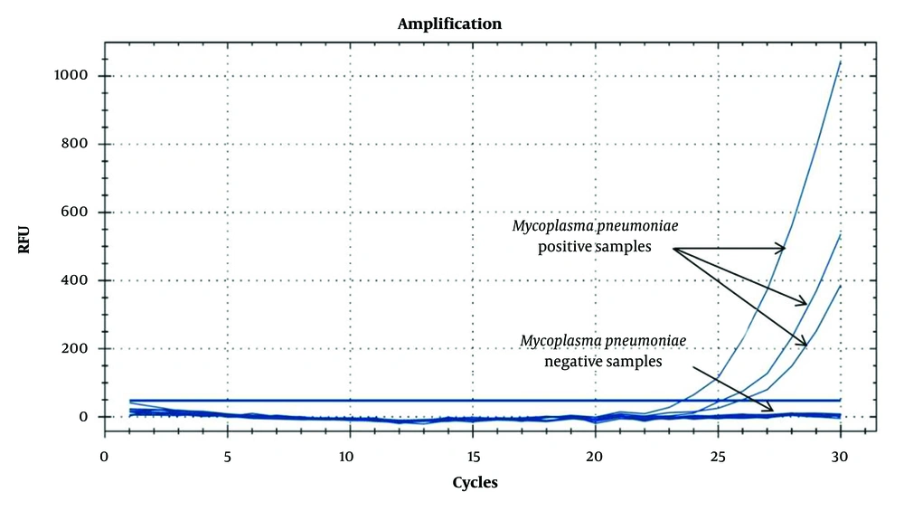
Figure 2.
Specificity of fluorescent PCR
4.4. Sensitivity of Fluorescent PCR
To identify the sensitivity of this method, various concentrations of M. pneumoniae positive control were tested in 16 replicates at each concentration level. The probit analysis showed that the limit of detection of the fluorescent PCR in 95% of cases was 553.26 copies/mL (95% CI: 341.32 - 2063.47 copies/mL) (Figure 3). The raw data are provided in Supplementary Table S1.
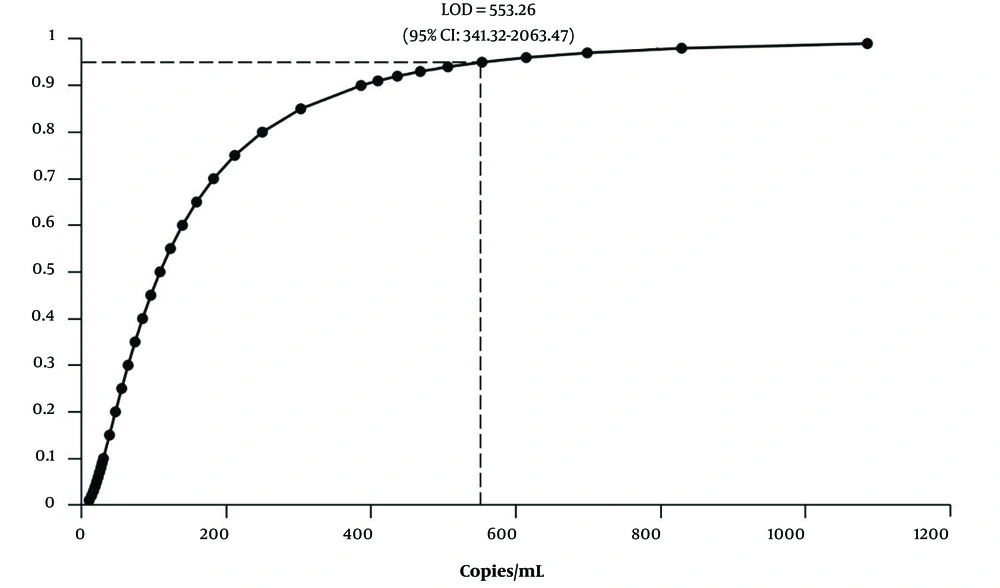
Figure 3.
Probit analysis of fluorescent PCR
4.5. Reproducibility of Fluorescent PCR
To test the reproducibility of this fluorescent PCR, assessment based on intra- and inter-assay variabilities was performed. The intra-assay variability was 1.86% and 3.17% at positive control concentrations of 2500 and 1000 copies/mL, respectively, while the inter-assay variability was 3.73% and 4.70% at these concentrations. These results indicated that the fluorescent PCR method demonstrated good repeatability.
4.6. Comparison of Newly Developed Fluorescent PCR with Commercial Fluorescent PCR
To enable a direct head-to-head comparison, both the newly developed fluorescent PCR and the commercial fluorescent PCR were used to test the same set of 32 M. pneumoniae-positive samples that had been diluted 10-fold. The newly developed fluorescent PCR detected 20 samples as positive for M. pneumoniae (62.5%), while the commercial fluorescent PCR detected 17 samples as positive (53.1%). These results demonstrate that the extraction-free method achieved superior detection performance for low-concentration samples compared with the commercial extraction-based kit evaluated in this study (Table 3).
Table 3.
Head-to-Head Comparison Between the Newly Developed Method and Commercial Fluorescent PCR Kit
| Parameter | Newly Developed Method | Commercial Kit |
|---|---|---|
| Nucleic acid extraction | Not required | Required |
| Total turnaround time | < 30 min | > 1 h |
| Detection of 10-fold diluted positive samples a | 20/32 (62.5%) | 17/32 (53.1%) |
| Hands-on steps | 1 | Multiple steps |
| Equipment required | PCR instrument only | PCR instrument + extraction system |
| Technical expertise | Minimal | Moderate |
a Both methods were tested using the same set of 10-fold diluted M. pneumoniae positive clinical samples.
5. Discussion
To the best of our knowledge, this is the first report of an extraction-free fluorescent PCR method for M. pneumoniae detection. While extraction-free PCR approaches have been reported for other pathogens, particularly SARS-CoV-2 (15), their application to M. pneumoniae has not been previously described. The key innovation of our method is the two-stage denaturation strategy. Conventional PCR typically uses 95°C denaturation to ensure strand separation of complex genomic DNA. However, based on melting curve analysis, we found that the specific amplicons had completely denatured by 86°C. We therefore designed a protocol using 95°C for the first 10 cycles to effectively denature the genomic DNA template, followed by 86°C for the subsequent 30 cycles to amplify the short amplicons. This approach significantly reduces thermal cycling time while maintaining detection sensitivity.
To enable a fair comparison of detection performance, both the newly developed method and the commercial kit were evaluated using the same set of 10-fold diluted M. pneumoniae-positive clinical samples (Table 3). The total turnaround time was reduced from over 1 hour to less than 30 minutes. Notably, the extraction-free method demonstrated a higher detection rate (62.5%, 20/32) compared with the commercial extraction-based kit (53.1%, 17/32), indicating that the extraction-free approach does not impair detection sensitivity for clinical samples. A common concern regarding extraction-free PCR methods is the potential reduction in sensitivity compared with extraction-based approaches, primarily due to the presence of PCR inhibitors in crude clinical samples (17, 18). However, our results demonstrated the opposite. This finding can be explained by nucleic acid loss during the extraction process. Studies have reported that DNA loss of up to 83% can occur during extraction (19). For samples with low pathogen loads, this loss may outweigh the theoretical benefit of concentration. Our extraction-free approach retains all the nucleic acid present in the sample. Additionally, the use of an inhibitor-tolerant polymerase effectively mitigates the impact of PCR inhibitors (20), reducing the main advantage of nucleic acid purification.
Several limitations of this study should be acknowledged. First, although an optimized reaction system was used, extremely high concentrations of PCR inhibitors in certain clinical samples may still affect amplification efficiency. Second, the direct sample input volume is limited to 5 µL to prevent inhibition. While our results demonstrated superior detection for the clinical samples tested, extraction-based methods theoretically allow for template concentration from larger volumes. This capability might be advantageous for samples with extremely low pathogen loads falling below the limit of detection of our assay. Third, this method has been validated only for throat swab samples; additional validation is required for other respiratory specimen types such as nasopharyngeal swabs, sputum, or bronchoalveolar lavage fluid (21). Fourth, the sample size of M. pneumoniae-positive cases (n = 32) is relatively small, and larger-scale multi-center clinical validation studies will be considered in the future.
Potential sources of error in this method include: (1) Variability in sample collection techniques, which may affect the amount of target pathogen captured; (2) differences in sample matrix composition among patients, potentially leading to variable PCR inhibition; (3) temperature accuracy and ramping speed of different PCR instruments, which may affect the performance of the two-stage denaturation protocol; and (4) sample degradation during transport or storage. Despite these limitations, the extraction-free approach offers significant advantages for specific clinical scenarios. The rapid turnaround time (< 30 minutes) makes this method particularly suitable for outpatient settings where prompt diagnosis is essential for guiding antibiotic treatment decisions. The simplified workflow reduces the need for specialized training and expensive extraction equipment, potentially enabling M. pneumoniae testing in resource-limited settings or point-of-care environments (22, 23). Future work will focus on validating this method with other respiratory specimen types and integrating it into automated or portable platforms to expand its applicability to point-of-care testing.
5.1. Conclusions
We developed a rapid, simple, and reliable extraction-free fluorescent PCR assay for M. pneumoniae detection. By eliminating nucleic acid extraction and integrating a two-stage denaturation strategy, the assay shortens total turnaround time to < 30 minutes while maintaining excellent specificity, sensitivity, and reproducibility, outperforming commercial extraction-based kits for low-concentration samples. This method shows great promise for clinical application, particularly in outpatient settings requiring prompt diagnosis.