
1. Background
The skin is the most exposed organ of the human body and is therefore vulnerable to various injuries, including burns, scrapes, and cuts. Damage to epithelial structures compromises the body’s ability to protect itself from external threats (1). Burns caused by excessive heat or caustic chemicals disrupt the skin barrier and increase the risk of infection. Infection is one of the most common and serious complications of wound healing, particularly in chronic wounds, and may pose considerable risks during the acute phase after burn injury (2). With the increasing frequency of burn infections and antibiotic resistance, innovative wound-care treatments are urgently needed. Although cotton gauze is commonly used for burn care, it has important drawbacks, including pain during removal and potential delays in healing (3, 4).
According to wet wound-healing theory, maintaining a moist environment is essential for promoting granulation tissue growth and skin cell division, thereby facilitating complete wound healing (5). An ideal dressing should maintain high humidity at the wound site while absorbing excess exudate. It should also be hypoallergenic, comfortable, affordable, capable of oxygen and water-vapor exchange, and protective against microbial invasion (6). Hydrogels formed by combining different polymers typically show improved wound-dressing properties compared with those of their individual components. By supporting moist wound healing and adequate fluid absorption, hydrogels can also facilitate monitoring of the healing process because of their transparency. They are among the most promising materials for wound care because they meet essential dressing requirements, including maintaining a moist wound environment, absorbing excess exudate, covering sensitive tissue without adhesion, and reducing pain through cooling effects while actively contributing to healing. However, because hydrogels alone do not eliminate pathogenic microbes, infection in burn wounds remains a major challenge (7).
Recent advances in wound care have focused on developing active dressings in which hydrogels are combined with antimicrobial components to create an optimal healing environment. In this context, new strategies are being developed to use antimicrobial hydrogels as burn wound dressings. Natural polymers such as alginate (ALG) and gelatin (GEL) are commonly used in wound-healing applications. Alginate, derived from brown seaweed, is a biocompatible and cost-effective biomaterial suitable for various biomedical applications, including wound healing, bone repair, and nerve regeneration. Alginate dressings maintain a moist microenvironment, minimize bacterial infection, and promote healing (8). The calcium ions released from alginate dressings play a crucial physiological role in the clotting mechanism during the initial stages of wound healing (9). Conversely, GEL, obtained from the hydrolysis of the collagen triple-helix structure, is favored in biomedical applications because of its hemostatic properties, low antigenicity, physicochemical stability, biocompatibility, and biodegradability (10).
According to current guidelines, 1% (w/w) silver sulfadiazine (SSD) cream is widely used to treat burns and skin infections (11). Silver sulfadiazine is effective against a broad spectrum of microorganisms, including gram-negative and gram-positive bacteria and fungi such as Candida albicans. This cream is used to prevent and treat infections in burns, for the short-term management of leg ulcers and pressure ulcers, and to prevent infections in skin grafts. However, frequent reapplication of SSD can interfere with healing by exposing patients to infectious agents and causing pain during dressing removal because the cream is not biodegradable (12). Pandey et al prepared SSD-containing hydrogel sponges and evaluated their effectiveness in wound healing using animal models. Their results showed that the antimicrobial activity and wound-healing rate of this dressing were increased compared with those of conventional commercially available creams (13). In our previous work, we used a composite of nanofibers and tragacanth hydrogel containing Aloe vera extract and SSD as a wound dressing. The results showed that wound healing occurred because of SSD disintegration and the release of silver ions in the wound bed. Silver sulfadiazine damages the bacterial cell membrane and cell wall, thereby inhibiting bacterial growth without affecting healthy skin cells. In addition, the angiogenic properties of Aloe vera showed favorable synergistic results (14).
2. Objectives
Given the growing importance of hydrogel formulations, this study aimed to develop and evaluate an SSD-loaded GEL/ALG hydrogel for topical antimicrobial applications.
3. Methods
3.1. Chemical Materials
Sodium alginate (Mv = 1.2 × 10⁵, μ = 280 mPa·s) and gelatin were obtained from Shanghai Chemical Reagent Co. (Shanghai, China). Phosphate-buffered saline (PBS) and ethanol (C2H5OH, 98% purity) were obtained from Merck (Germany). Calcium chloride (CaCl2; CAS No. 10043 - 52 - 4) and SSD were purchased from Sigma-Aldrich Co. (USA). All other chemicals were commercially available, of analytical grade, and used as received.
3.2. Biological Materials
Mouse fibroblast cells (L-929), Staphylococcus aureus ATCC 25923, Acinetobacter baumannii ATCC 19606, Pseudomonas aeruginosa ATCC 27853, and Candida albicans ATCC 10231 were obtained from the Pasteur Institute, Iran.
3.3. Preparation of Gelatin/Alginate
First, 2% (w/v) solutions of GEL and sodium ALG were prepared separately. The GEL and sodium ALG solutions were then mixed at an 80:20 ratio and magnetically stirred for 24 hours to obtain a uniform solution of the two polymers.
3.4. Synthesis of Hydrogels
A 1% (w/w) SSD solution was added to the polymer mixture and stirred at room temperature for 60 minutes until complete dissolution. The hydrogel solution was then transferred to a Petri dish (6 cm) and dried at room temperature (25°C, 30% - 40% relative humidity). After drying, the material was immersed in a 1% CaCl2 solution for 15 minutes to crosslink the sodium ALG. Thickness was measured at 5 random points per film using a digital micrometer; the target thickness was 0.18 - 0.22 mm. The mass of each hydrogel was recorded, and acceptable batch variation was ≤ 5%. Each experimental condition was prepared in triplicate independent batches (n = 3), with each batch cast separately.
3.5. Hydrogel Characterization
3.5.1. Swelling
A swelling test was conducted to evaluate the adsorption and swelling properties of the GEL/ALG hydrogel containing SSD. The initial weight of the sample was recorded, and the hydrogel film was immersed in PBS at 37°C for a specified duration. The swelling rate was determined at 30, 60, 90, 120, 150, 180, and 210 minutes. Once equilibrium was reached, the sample was reweighed, and water absorption (swelling rate) was calculated using Equation 1, where W0 represents the weight of the dry sample and W1 represents the weight of the sample after water absorption.
Equation 1:
3.5.2. Biodegradability
Degradation tests were conducted by submerging hydrogel samples (1 × 1 cm) in 10 mL of PBS at pH 7.4. The hydrogel samples were weighed and then placed in Falcon tubes containing PBS, which were maintained in an incubator at 37°C. At intervals of 1, 3, 5, 7, and 14 days, the samples were removed from PBS, dried in an oven for 2 hours, and weighed again. Hydrogel weight loss was determined under sterile conditions using Equation 2, where Wi represents the initial weight of the hydrogel and Wf represents the final weight after degradation.
Equation 2:
3.5.3. Water Vapor Transmission Rate
Samples of GEL/ALG hydrogel (control) and GEL/ALG/SSD hydrogel, each with a diameter of 20 mm, were secured in separate beakers containing 25 mL of double-distilled water. A control was also established using a tube filled with 25 mL of double-distilled water without a cap. The initial weights of the test tubes, including the water and respective samples, were recorded. The samples were then incubated for 24 hours at 37°C and 40% humidity (15). After incubation, the samples were weighed again, and the water vapor transmission rate was determined using Equation 3, where Wi is the initial weight of the system, including the test tube, water, and hydrogel; Wf is the final weight of the system after reduction; and A represents the area of the bottle opening.
Equation 3:
3.5.4. Porosity
To determine hydrogel porosity, samples were submerged in ethanol until saturation. Porosity was calculated as follows: A hydrogel sample with weight W was placed in a graduated cylinder containing ethanol. The initial volume of ethanol (V1) and the combined volume of the hydrogel and ethanol (V2) were recorded. After 1 hour, the samples were removed, and the remaining ethanol volume in the cylinder was measured as V3. Porosity was calculated using Equation 4.
Equation 4:
3.5.5. Mechanical Study
Samples were prepared according to ASTM D00882, with dimensions of 1 × 6 cm. The hydrogels were subjected to tensile strain at 50 mm/min, with the distance between the two jaws of the device set at approximately 3 cm. Elongation percentage, elastic modulus, and tensile strength were determined from the stress-strain curve (15). Strength was calculated using Equation 5.
Equation 5:
3.5.6. Scanning Electron Microscopy
The surface morphology of the prepared hydrogels was examined using scanning electron microscopy (SEM; TESCAN-Vega3, Czech Republic). The hydrogels were frozen in liquid nitrogen and then fractured with tweezers. A gold layer was applied to the cross-sections of the fractured samples.
3.5.7. Fourier-Transform Infrared Spectroscopy
The functional groups and bonds present in the samples were analyzed using Fourier-transform infrared (FT-IR) spectroscopy. The samples were processed into thin tablets. FT-IR absorption peaks were detected in the wavelength range of 400 - 4000 cm-1, and bond types were identified by analyzing the absorption bands.
3.5.8. Hemocompatibility
The hemocompatibility of the GEL/ALG and GEL/ALG/SSD hydrogels was assessed using a hemolysis assay. Fresh anticoagulated blood from human volunteers (2.50 mL) was treated with heparin, diluted with 5.00 mL of normal saline, and centrifuged at 9000 rpm for 5 minutes to isolate red blood cells (RBCs). The RBCs were washed 3 times with normal saline and then diluted to a final volume of 20 mL with normal saline. Subsequently, 1 mL of RBC suspension was exposed to different hydrogel concentrations (100, 200, 300, 400, 500, and 600 μg/mL). The samples were incubated at 37°C for 3 hours and centrifuged again at 9000 rpm for 5 minutes. A 100-μL aliquot of supernatant from each sample was transferred to a 96-well plate, and absorbance was measured using a microplate reader at 577 nm (16). The degree of hemolysis was calculated using Equation 6, where ODs represents the absorbance of the sample, ODnc represents the absorbance of the negative control (normal saline), and ODpc represents the absorbance of the positive control (deionized water).
Equation 6:
3.5.9. Antimicrobial Activity
For the assay, microbial suspensions of A. baumannii and P. aeruginosa (gram-negative bacteria), S. aureus (gram-positive bacterium), and C. albicans were prepared at a concentration of 108 colony-forming units/mL. A 100-μL volume of each suspension was evenly spread on the surface of Mueller-Hinton agar plates (Merck, Germany) to ensure uniform microorganism distribution for accurate measurement of antimicrobial effects. Hydrogel samples were prepared by cutting the hydrogels into circular disks with a diameter of 1 cm. The disks were then placed on the surface of the inoculated agar plates, allowing the active components of the hydrogels to diffuse into the agar over time and potentially create an inhibition zone around each disk.
The plates were incubated at 37°C for 24 hours to allow microbial growth and interaction with the hydrogel samples. After incubation, inhibition zones, defined as areas around the hydrogel disks with no microbial growth, were measured using a Vernier caliper and recorded in millimeters. Inhibition-zone size indicated the antimicrobial effectiveness of the hydrogel. To ensure reliability, gentamicin disks (20 μg/disk; Mast, England) were included as positive controls in the disk diffusion test. Comparing the inhibition zones of the hydrogels with those of the gentamicin control provided insight into the antimicrobial potency of the hydrogels. These measurements helped determine the potential of the hydrogels as antibacterial agents for wound care.
3.6. Statistical Analysis
Cytotoxicity analysis was performed using GraphPad Prism version 8.0 (GraphPad Software Inc., San Diego, CA). A P value ≤ 0.05 was considered statistically significant.
4. Results
4.1. Morphological Observation
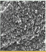
Scanning electron microscopy evaluation of the morphology, porosity, and structural integrity of the prepared hydrogels revealed a distinct three-dimensional porous structure (Figure 1). This porosity is essential for facilitating drug release and enhancing the absorption of biological fluids. Pore size and distribution can substantially affect hydrogel performance in applications such as wound dressings and can influence drug-release kinetics (17). Incorporation of SSD into the hydrogel matrix altered the smooth, uniform morphology of the GEL/ALG hydrogel. It increased pore size and affected the crosslinking density and network formation of the hydrogel. The results also indicated good compatibility between the matrix and the drug (18).
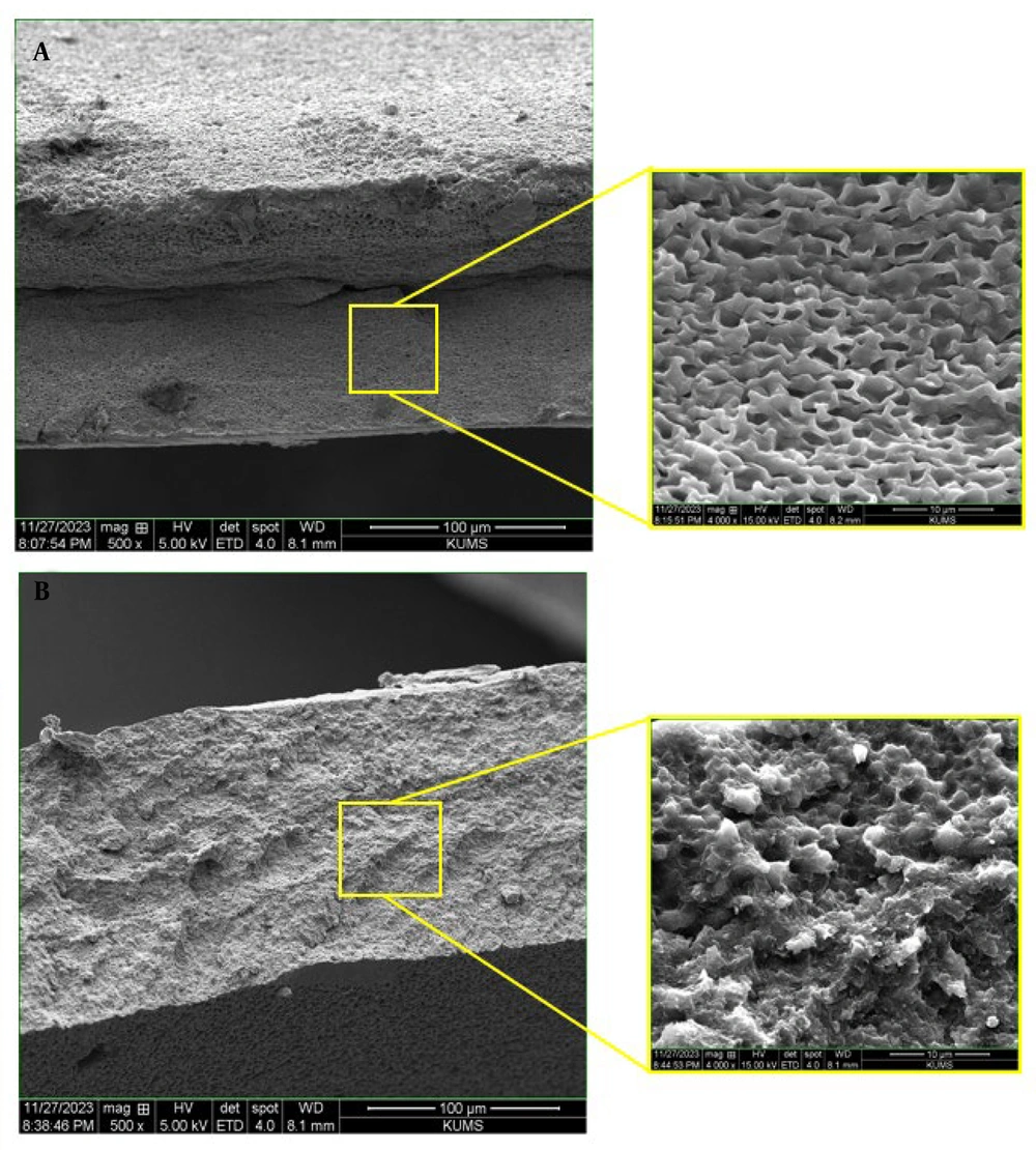
Figure 1.
Scanning electron microscopy images of the cross-section of (a) GEL/ALG and (b) GEL/ALG/SSD hydrogels at 3 magnifications of 50, 30, and 10
4.2. Swelling Assessment
As a hydrogel-based drug-delivery system absorbs fluid and swells, the polymer network expands, increasing mesh size and reducing the local drug concentration within the matrix. This decrease in the concentration gradient slows diffusion and promotes a more controlled and sustained release profile (19). The enhanced swelling capacity of both hydrogels may be attributed to the formation of larger, thinner pores, suggesting that silver is deposited on the pore walls. This facilitates rapid water infiltration and increases hydration capacity. The GEL/ALG hydrogel showed high swelling values, indicating a substantial capacity for retaining wound exudate and making it suitable for managing wounds with moderate to heavy exudation (20, 21). Over time, the swelling rate increased in both prepared hydrogels. The inclusion of SSD in the GEL/ALG hydrogel altered network interactions and may have affected the swelling rate (Figure 2). The swelling behavior of hydrogels is a critical factor affecting biodegradability. The swelling ratio indicates the amount of water the hydrogel can absorb, which directly affects its degradation rate. Higher swelling ratios are typically associated with greater mass loss during biodegradation because greater water absorption facilitates polymer-chain disintegration (22).
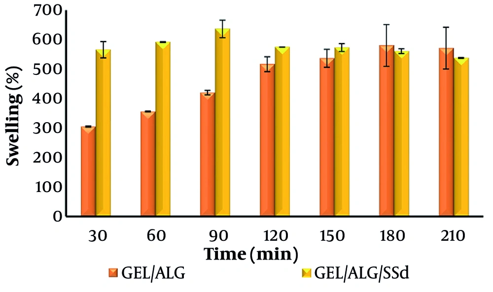
Figure 2.
Swelling rate of GEL/ALG and GEL/ALG/SSD hydrogels
4.3. Degradation Evaluation
Incorporation of SSD into GEL/ALG hydrogels substantially affected their biodegradability, which is crucial for wound-healing applications. Alginate and gelatin are biopolymers known for their biocompatibility and biodegradability. When SSD is added, it interacts with these polymers and may alter the physical and chemical properties of the hydrogel. Studies indicate that the presence of SSD can enhance the mechanical strength and stability of hydrogels, which is beneficial for maintaining structural integrity during healing (23). The results showed that the degradation rate gradually increased over time in both hydrogels (Figure 3). This rapid degradation may be attributed to the inherent properties of sodium ALG, which is soluble in aqueous environments. Gelatin also contributes to the overall degradation profile because it interacts with ALG to form a network that influences water absorption and mass loss over time. After 14 days, the degradation rates of the GEL/ALG and GEL/ALG/SSD hydrogels were 80.26% and 70.01%, respectively.
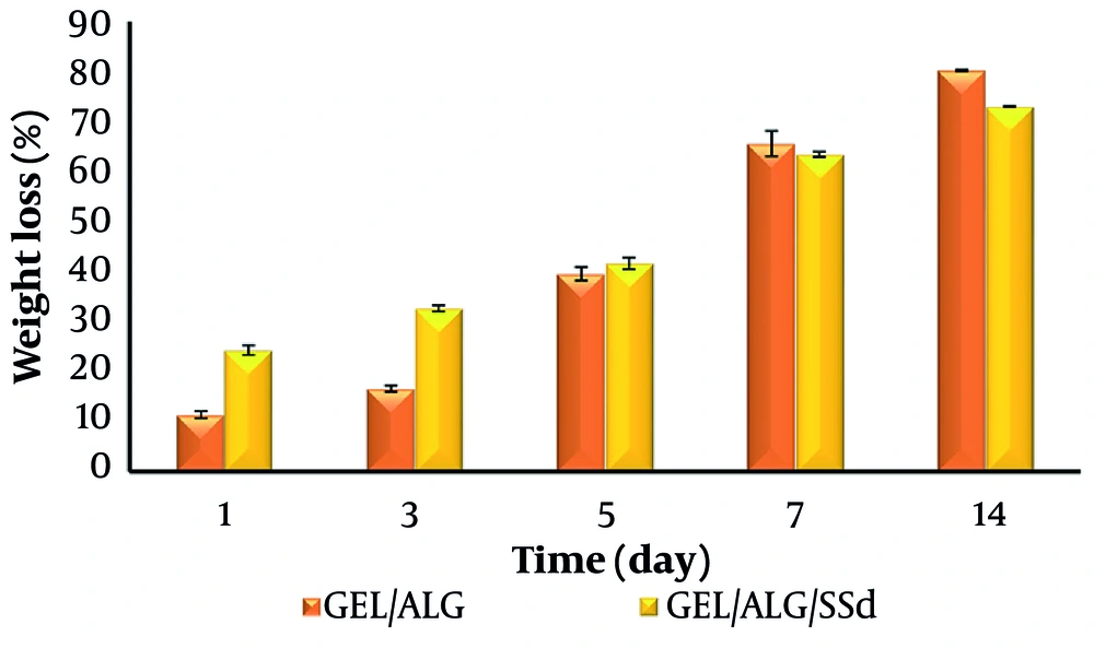
Figure 3.
Degradation of GEL/ALG and GEL/ALG/SSD hydrogels
4.4. Hemocompatibility Assay
Hemolysis refers to disruption of RBC membrane integrity, leading to the release of intracellular hemoglobin into plasma. This phenomenon highlights the importance of hemocompatibility in evaluating implantable scaffolds. According to ASTM standards, hemolysis is categorized as nonhemolytic at 0% - 2%, slightly hemolytic at 2% - 5%, and hemolytic at values exceeding 5% (24). The results presented in Figure 4 indicate that both samples showed hemolysis levels below 5% and therefore exhibited favorable blood compatibility.
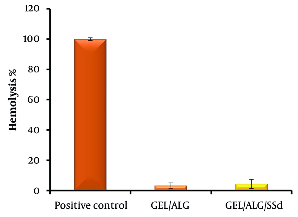
Figure 4.
Hemocompatibility of GEL/ALG and GEL/ALG/SSD hydrogels
4.5. Porosity
An ideal hydrogel for wound healing is characterized by a porous structure that significantly enhances nutrient and oxygen transfer within the hydrogel matrix. This porosity is crucial for creating an environment conducive to angiogenesis and promoting cell proliferation and migration, thereby facilitating effective nutrient and oxygen delivery within the hydrogel matrix (25). The porosity of the GEL/ALG and GEL/ALG/SSD hydrogels was measured using the liquid displacement technique. The GEL/ALG hydrogel showed substantial porosity of approximately 86.08%. Incorporation of SSD into the hydrogel decreased the porosity to 66.68% (Figure 5).
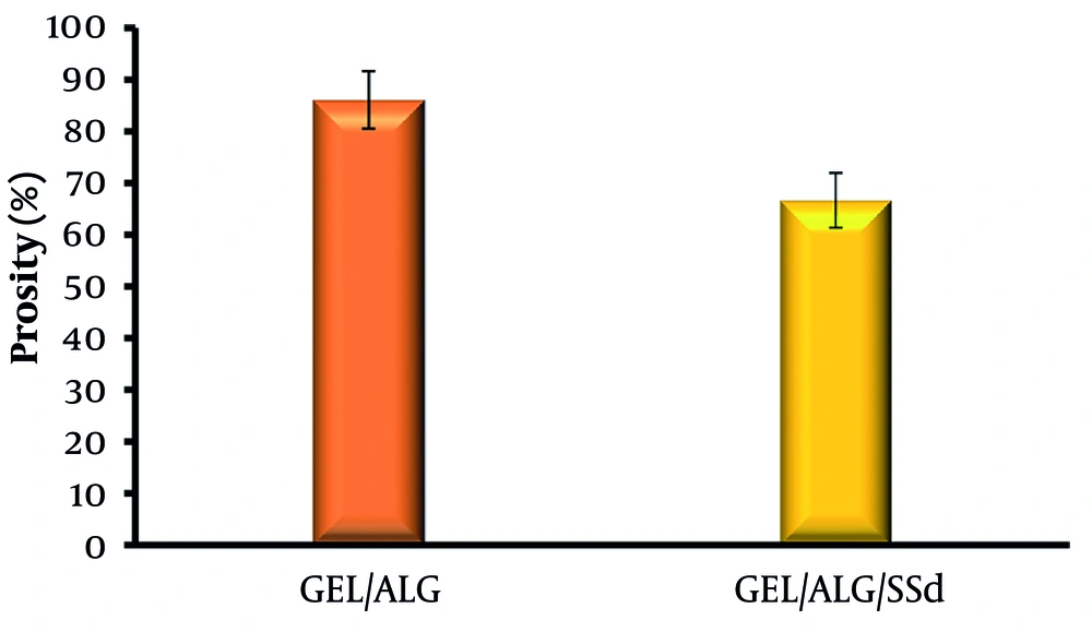
Figure 5.
Porosity of GEL/ALG and GEL/ALG/SSD hydrogels
4.6. Mechanical Properties
The mechanical properties of GEL/ALG hydrogels are substantially influenced by the GEL-to-sodium ALG ratio, the type and concentration of crosslinking agents, and the presence of additives such as SSD. Because hydrogen bonding and interactions between the GEL and ALG polymer chains strengthen the hydrogel network structure, adding GEL to ALG can increase the elastic modulus, tensile strength, and elasticity while reducing hydrogel fragility. The addition of SSD may affect hydrogel stability and mechanical properties through ionic interactions with ALG chains, which may alter hydrogel network integrity (26). Incorporation of SSD can influence mechanical properties by affecting crosslinking dynamics within the hydrogel matrix (Table 1). Silver ions may interact with calcium ions during gelation, potentially altering the network structure and mechanical performance (26).
Table 1.
Mechanical Study of the Hydrogels a
| Hydrogel | Tensile Strength (MPa) | Young's Modulus (MPa) | Elongation at Break (%) |
|---|---|---|---|
| GEL/ALG | 0.676 ± 0.0045 | 1.4977 ± 0.0525 | 46.1335 ± 1.241375 |
| GEL/ALG/SSD | 1.402 ± 0.0315 | 2.0312 ± 0.0533 | 69.023 ± 1.961875 |
a Values are expressed as mean ± SD.
4.7. Water Vapor Transmission Rate
The water vapor transmission rate (WVTR) of GEL/ALG hydrogels containing SSD is influenced by multiple factors, including composition ratios, crosslinking methods, and the presence of antimicrobial agents. Understanding these dynamics is essential for optimizing hydrogel formulations for wound care applications and ensuring adequate moisture while preventing infection. A substantial increase in WVTR was observed due to the more porous structure of the GEL/ALG hydrogel. Loading SSD into the hydrogel produced a slight reduction in WVTR (Figure 6).
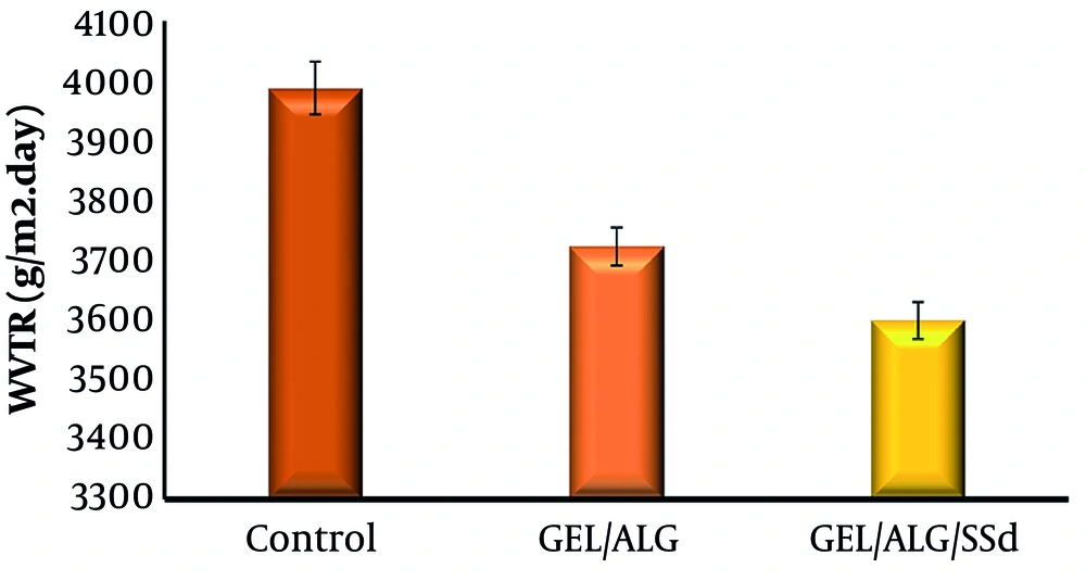
Figure 6.
Water vapor transmission rate of GEL/ALG and GEL/ALG/SSD hydrogels
4.8. Fourier-Transform Infrared Spectroscopy Analysis
Fourier-transform infrared spectroscopy can identify and characterize the functional groups and molecular interactions present in ALG, GEL, and SSD. This information is essential for understanding the chemical properties and potential interactions of these materials in the development of antimicrobial hydrogels. This analysis contributes to evaluating the compatibility and effectiveness of the components used in wound care formulations. The FT-IR spectra of the samples were recorded in the range of 500 - 4000 cm-1 (Figure 7). The analyzed samples included ALG, GEL, SSD, CaCl2, GEL/ALG, and GEL/ALG/SSD.
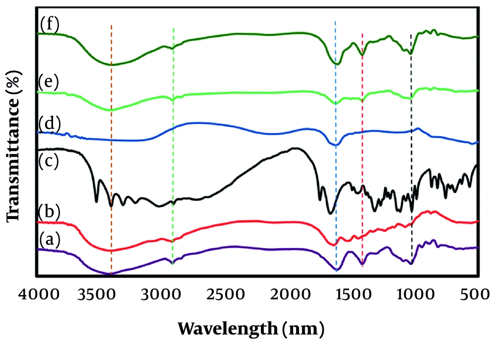
Figure 7.
Fourier-transform infrared spectra: (a) ALG, (b) GEL, (c) SSD, (d) CaCl2, (e) GEL/ALG, and (f) GEL/ALG/SSD
The FT-IR spectrum of ALG showed characteristic absorption bands at 3410 cm-1, corresponding to hydroxyl (OH) stretching vibrations and indicating the presence of alcohols and phenols in the polysaccharide structure. The band at 1604 cm-1 was associated with C=O stretching of the carboxylic group, demonstrating ALG functionality. Peaks at 1291 and 1051 - 1089 cm-1 were related to C-C and C-O stretching modes in the polysaccharide backbone. Additional peaks at 1028 cm-1 (C-C), 941 cm-1 (C-O), 880 cm-1 (C-H), and 810 cm-1 (Na-O) further characterized the molecular structure of ALG and confirmed its polysaccharide nature.
The spectrum of pure GEL powder showed a peak at 3435 cm-1, corresponding to NH and OH stretching and confirming the presence of amino and hydroxyl groups. Peaks at 1638 and 1514 cm-1 represented amide I and II bands. The amide I band was attributed to C=O stretching, and the amide II band was attributed to NH bending vibrations, both of which are key indicators of peptide linkages in proteins. The peak at 1155 cm-1 was assigned to C-O-C stretching, further highlighting the functional groups in GEL.
The FT-IR spectrum of pure SSD showed 4 characteristic absorption bands. The peak at 1660 cm-1 was attributed to vibrational stretching of the phenyl structure conjugated to the NH2 group, indicating aromatic characteristics in the SSD structure. The band at 1120 cm-1 corresponded to asymmetric stretching of the S=O bond, highlighting the sulfonamide functional group in the compound. The peaks at 3315 and 3215 cm-1 were assigned to NH2 stretching bands, confirming the presence of the amine group in the molecule (14). The absorption patterns observed in the SSD-containing hydrogel were notably similar to those in the hydrogel without SSD. This similarity indicates no substantial interaction between the hydrogel matrix and SSD.
4.9. Antibacterial Activity
Antibacterial activity against bacteria that cause wound infections, including P. aeruginosa and A. baumannii (gram-negative bacteria) and S. aureus (gram-positive bacterium), was examined and measured as inhibition zones in millimeters (Figure 8). The GEL/ALG/SSD hydrogel showed strong antibacterial activity, particularly against S. aureus and A. baumannii, with inhibition zones comparable to those of gentamicin. Gentamicin, an aminoglycoside antibiotic with broad-spectrum activity against gram-positive and gram-negative pathogens, was selected as the antibacterial positive control because it is a well-established reference standard in disk diffusion susceptibility testing according to Clinical and Laboratory Standards Institute guidelines. Activity against P. aeruginosa was weaker but still present, which is notable given the intrinsic resistance of this organism (Table 2).
Table 2.
Antibacterial Activity
| Organism | GEL/ALG/SSD Hydrogel (mm) | GEL/ALG Control (mm) | GM (20 μg/disk) (mm) |
|---|---|---|---|
| Staphylococcus aureus | 21 | 0 | 20 |
| Pseudomonas aeruginosa | 10 | 0 | 18 |
| Acinetobacter baumannii | 20 | 0 | 20 |
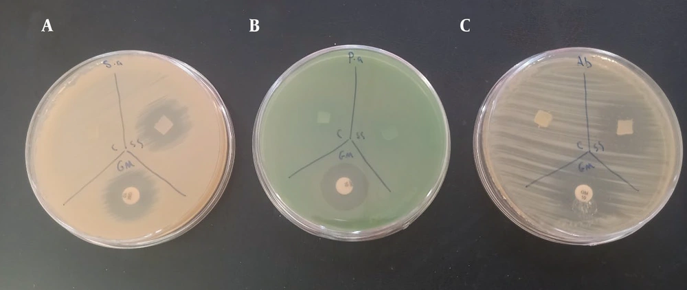
Figure 8.
Antibacterial activity of the hydrogel against (A) S. aureus, (B) P. aeruginosa, and (C) A. baumannii. Gentamicin disks (20 μg/disk) were used as positive controls.
Pseudomonas aeruginosa is resistant to many antibiotics and plays an important role in wound infections, particularly in patients with compromised immune systems or chronic wounds. Effective inhibition of this bacterium suggests that the hydrogels have suitable properties for preventing wound-care-associated infections. Staphylococcus aureus is known to cause health care-associated infections; therefore, hydrogel activity against this gram-positive organism is essential for comprehensive wound infection management. The addition of SSD as an antimicrobial agent substantially enhanced the antibacterial properties of the hydrogel, as shown by the increased inhibition zones compared with the GEL/ALG control. The GEL/ALG control hydrogel without SSD showed no antibacterial activity, suggesting that the antibacterial effects were primarily attributable to SSD.
The GEL/ALG/SSD hydrogel demonstrated strong antibacterial activity, particularly against S. aureus and A. baumannii, with activity comparable to gentamicin. Its activity against P. aeruginosa was weaker but still significant, as expected given this organism's resistance profile. The SSD component was critical for the observed antibacterial effects because the GEL/ALG control showed minimal or no activity. These findings support further development of these hydrogels for clinical applications, suggesting that they may enhance wound healing by reducing infection risk and contributing to a moist healing environment. Understanding the mechanisms of action and interactions between hydrogel components and bacterial strains will be critical for optimizing the formulation for maximum efficacy.
4.10. Antifungal Activity
The SSD component in the hydrogel is likely responsible for its antifungal activity. Silver sulfadiazine may disrupt fungal cell membranes, such as by interacting with ergosterol, a key component of fungal membranes; inhibit fungal cell wall synthesis, such as by targeting chitin or glucan synthesis; induce oxidative stress; or interfere with fungal metabolic pathways. The GEL/ALG matrix may also contribute by enhancing sustained SSD release, prolonging antifungal effects, and providing a physical barrier that prevents fungal adhesion or colonization.
The hydrogel showed stronger antibacterial activity, including 21 mm for S. aureus and 20 mm for A. baumannii, than antifungal activity, which was 12 mm. This finding is expected because C. albicans is a eukaryotic organism with a more complex cellular structure and greater resistance to antimicrobial agents than bacteria. The GEL/ALG/SSD hydrogel demonstrated moderate antifungal activity with a 12-mm inhibition zone. Although this activity was weaker than its antibacterial activity, it remains significant and suggests potential applications in preventing or treating fungal infections (Figure 9). Future work should include a free-SSD control, such as 1% SSD cream applied at an equivalent SSD dose, to determine whether hydrogel encapsulation alters the magnitude or kinetics of antimicrobial activity. An appropriate antifungal comparator should also be included to validate the observed activity.
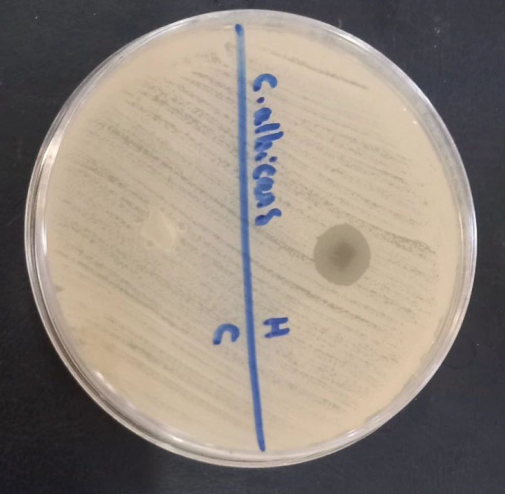
Figure 9.
Antifungal activity of the GEL/ALG/SSD hydrogel (12 mm)
5. Discussion
In recent decades, the use of hydrogels for wound healing and drug delivery has been optimized because of their effective fluid absorption, excellent water retention, and ability to support moist wound healing (27). The porous structure of hydrogels facilitates the absorption of wound exudate, reduces infection risk, and creates an environment conducive to accelerated healing (28). Incorporating a drug into a hydrogel increases viscosity and decreases diameter, which consequently reduces sample tensile strength. In this study, we aimed to develop an antibacterial hydrogel composed of GEL/ALG/SSD. The findings indicate that the combined properties of GEL and ALG within the hydrogel matrix enhance moisture retention, mechanical stability, and cellular response. This combination also creates an environment conducive to cell adhesion and proliferation.
Hydrogels composed of GEL, ALG, and SSD are gaining attention for potential wound-healing applications. These materials combine the beneficial properties of natural polymers with the antimicrobial effects of SSD, making them suitable for treating infected wounds. The interaction between GEL and ALG occurs through hydrogen bonding and electrostatic attraction, which can improve the consistency and performance of the hydrogel in biomedical applications (29). The integration of GEL/ALG and SSD into a single hydrogel system represents a promising approach for advanced wound care. By leveraging the unique properties of each component, these hydrogels not only facilitate healing but also provide a protective barrier against microbial infections, making them valuable in clinical and pharmaceutical settings.
5.1. Conclusions
This study focused on the development and optimization of an SSD-loaded hydrogel for topical antimicrobial applications. The formulated hydrogel showed favorable release behavior of the active agent, supported by swelling analysis, release-kinetic modeling, and antimicrobial performance testing. Because SSD release is governed by erosion of the polymeric hydrogel network, the system is well suited for extended use in occlusive wound dressings, in which gradual drug release is desirable. The material functions as a nonadherent dressing, maintaining a stable, moist gel layer over the wound surface without adhering to tissue or secondary bandages, provided that it remains hydrated. Future work will evaluate its clinical performance and explore its applicability across different wound types.