
1. Background
Biofilms are communities of bacterial cells embedded within a self-produced extracellular matrix that adheres to various surfaces (1). The reduced susceptibility of biofilm-associated bacteria to antibiotics is driven by both intrinsic biofilm characteristics and acquired resistance mechanisms. Bacteria in biofilms exhibit features that differ from those of planktonic cells, including high levels of antibiotic resistance (1, 2). These 2 growth modes are also associated with distinct gene expression profiles. Biofilm-associated traits, including altered gene expression patterns, properties of the extracellular polymeric matrix, and metabolic diversity among bacterial subpopulations, play a major role in promoting antibiotic resistance (3, 4).
Pseudomonas aeruginosa is an opportunistic Gram-negative pathogen frequently associated with healthcare-related infections (5). The global health threat posed by P. aeruginosa has intensified because of the increasing occurrence and spread of multidrug-resistant (MDR) strains (5, 6). This opportunistic pathogen is most frequently isolated from severe infections of the urinary tract, respiratory system, and wound sites. The therapeutic management of infections caused by MDR P. aeruginosa has become progressively more challenging (7). Numerous studies have demonstrated that biofilm-associated antibiotic resistance in P. aeruginosa is not primarily driven by genetic mutations. Instead, increased expression of various genes in biofilm cells, compared with their expression in planktonic counterparts, plays a significant role in conferring antibiotic resistance (8-10).
Among these genes are ndvB, tssC1, PA2070, and PA5033. The ndvB gene encodes a glycosyltransferase that synthesizes glucans, which trap antibiotics within biofilms and reduce drug efficacy. Disruption of ndvB increases biofilm sensitivity to multiple classes of antibiotics in vitro (8, 9). tssC1 is part of the type VI secretion system cluster (tssABC1 locus) in P. aeruginosa; deletion of tssC1 increases biofilm-specific antibiotic sensitivity, implicating a protective role independent of biofilm formation per se (10, 11). PA5033 is significantly overexpressed in biofilm cells and contributes to biofilm-specific antibiotic resistance (12). PA2070 is predicted to be involved in cell-surface signaling or metal uptake. PA2070 mutants display increased antibiotic susceptibility in biofilm cultures, indicating a distinct resistance mechanism (12, 13). Several studies have investigated the prevalence of biofilm-specific resistance genes in P. aeruginosa (14-16).
2. Objectives
The present study provides regional data from Eastern Anatolia, Türkiye, on the coexistence of multidrug resistance, biofilm formation, and biofilm-associated resistance genes in clinical P. aeruginosa isolates. In addition, the inclusion of isolates from diverse anatomical sources in a tertiary-care hospital setting enhances the clinical and regional relevance of the findings and addresses a notable gap in national molecular surveillance data. These findings may inform localized infection-control strategies and enrich the global dataset with molecular evidence from underrepresented geographic regions.
3. Methods
3.1. Clinical Isolates and Automated Antibiotic Susceptibility Testing
This study included 38 P. aeruginosa isolates cultured from blood, urine, sputum, and tracheal aspirate samples submitted to the microbiology laboratory from hospitalized patients at Erzurum Regional Training and Research Hospital between March 2021 and June 2021. All non-repeating P. aeruginosa isolates consecutively obtained from clinical samples during the study period and meeting the eligibility criteria were included. To ensure the independence of observations, only 1 isolate per patient was evaluated; if more than 1 isolate was obtained from the same patient, only the first isolate was processed. The inclusion criteria comprised clinically significant P. aeruginosa isolates obtained from blood, urine, sputum, and tracheal aspirate samples collected from hospitalized patients. Environmental isolates, surveillance cultures, colonization samples, and duplicate isolates from the same patient were excluded. Each sample was inoculated onto blood agar and eosin methylene blue agar under sterile conditions. Plates were incubated for 24 - 48 hours at 37°C under aerobic conditions.
The BD Phoenix M50 Automated Microbiology System (Becton Dickinson, Franklin Lakes, NJ, USA) was used for isolate identification and antimicrobial susceptibility testing. The tested antimicrobial agents were classified into the following antimicrobial classes for MDR assessment: aminoglycosides (amikacin and gentamicin), fluoroquinolones (ciprofloxacin and levofloxacin), carbapenems (imipenem and meropenem), cephalosporins (cefepime and ceftazidime), β-lactam/β-lactamase inhibitor combinations (piperacillin/tazobactam and ceftolozane/tazobactam), and polymyxins (colistin). Isolates categorized as increased exposure (I) according to EUCAST definitions were considered non-susceptible and were included in the resistant category for MDR classification and statistical analyses.
Multidrug resistance was defined as resistance to at least 1 antimicrobial agent in 3 or more different antibiotic classes according to established criteria. Pseudomonas aeruginosa ATCC 27853 was used as the reference strain. The Ethical Review Committee of Erzurum District Training and Research Hospital approved this study (01 February 2021/E-37732058 - 514.10-BEAH-KAEK 2021/03 - 50). The requirement for informed consent was waived by the Ethical Review Committee because of the retrospective laboratory-based design and the use of anonymized bacterial isolates and non-identifiable patient data.
3.2. Biofilm Formation Assay
As previously described, the biofilm-forming ability of the isolates was assessed using a crystal violet (CV) staining assay (17, 18). For biofilm quantification, 150 μL of a bacterial suspension adjusted to the 0.5 McFarland standard was dispensed into each well of sterile 96-well polystyrene microplates. After 48 hours of incubation at 37°C, the wells were washed with phosphate-buffered saline and stained with crystal violet. The remaining attached biofilms were stained with 0.1% crystal violet for 20 minutes. Crystal violet was removed, and the wells were rinsed thoroughly with tap water. Subsequently, bound crystal violet was solubilized using 30% acetic acid, and absorbance was measured at 595 nm. Pseudomonas aeruginosa PAO1, a well-characterized and robust biofilm-forming strain commonly used in biofilm research, was used as a positive control, and uninoculated Mueller-Hinton broth was used as a negative control. Based on their optical density (OD) values, isolates were categorized as follows: OD ≤ ODc = non-biofilm-forming, ODc < OD ≤ 2 × ODc = weak biofilm-forming, 2 × ODc < OD ≤ 4 × ODc = moderate biofilm-forming, and OD > 4 × ODc = strong biofilm-forming. In this classification, ODc refers to the cut-off optical density, calculated as the average OD of the negative control plus 3 times its standard deviation (ODc = OD negative control + 3 × SD) (19, 20). Based on the negative-control measurements, the calculated ODc value was 0.14, and the SD of the negative control was 0.069. For each isolate, optical density values from triplicate measurements were used for analysis.
3.3. Total DNA Isolation
The PCR assay was performed according to the kit instructions. Overnight-grown bacterial colonies were placed in a sterile Eppendorf tube containing 100 µL of distilled water and boiled for 10 minutes in a water bath. The tubes were subsequently centrifuged for 5 minutes at 1000 rpm. The supernatant was stored as DNA at -20°C. DNA concentration and purity were measured using a spectrometer (Thermo Scientific Multiskan GO) (21).
3.4. Detection of Biofilm-Specific Antibiotic Resistance Genes
PCR analysis was performed to detect the biofilm-associated antibiotic resistance genes ndvB, PA5033, PA2070, and tssC1. The primer sequences used in this study are provided in Table 1. PCR amplification was performed with an initial denaturation step at 95°C for 2 minutes, followed by 30 cycles of denaturation at 94°C for 30 seconds, annealing at 63°C for 30 seconds, and extension at 72°C for 50 seconds, with a final extension step at 72°C for 10 minutes. The amplified PCR products were resolved by electrophoresis on 1% agarose gels, stained with ethidium bromide, and visualized under ultraviolet illumination (22).
Table 1.
Primers Used in This Study
| Genes and Sequences | Reference | Amplicon Size (bp) |
|---|---|---|
| ndvB | Hall et al. (2018) (9) | 138 |
| F: GGCCTGAACATCTTCTTCACC | ||
| R: GATCTTGCCGACCTTGAAGAC | ||
| tssC1 | Beaudoin et al. (2011) (23) | 150 |
| F: CTCCAACGACGCGATCAAGT | ||
| R: TCGGTGTTGTTGACCAGGTA | ||
| PA5033 | Zhang et al. (2013) (11) | 127 |
| F: GGCGTTCTGGTAGGAACCTG | ||
| R: AGACCACGTTGCCGAAGCTG | ||
| PA2070 | Zhang et al. (2013) (11) | 151 |
| F: CTCCGCGGTGGATCTCAACA | ||
| R: GTCGAAGCGGCCTTCGTTCA |
3.5. Statistical Analysis
Each experiment was conducted 3 times, and results are reported as the mean with corresponding SDs. Statistical analyses were performed using IBM SPSS Statistics version 30.0 (IBM Corp., Armonk, NY, USA). Biofilm formation graphs were generated using GraphPad Prism 8. The association between the clinical sources of P. aeruginosa isolates and their biofilm-forming capacities was analyzed using the chi-square test. Cross-tabulation analyses were performed to evaluate the distribution of gene carriage among different biofilm phenotypes. The Pearson chi-square test was used to assess associations between categorical variables, and the Fisher exact test was applied when expected cell counts were lower than 5. Statistical significance was accepted at P < 0.05.
4. Results
4.1. Isolation and Identification of P. Aeruginosa
A total of 38 clinical P. aeruginosa isolates were obtained from various clinical sources, including 13 isolates from respiratory tract samples, 4 from sputum, 3 from blood, 13 from urine, and 5 from wound specimens.
4.2. Determination of the Antibiotic Resistance Profile
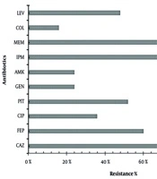
Antibiotic susceptibility analyses indicated that a substantial proportion of the P. aeruginosa isolates exhibited MDR phenotypes. Among the 38 clinical P. aeruginosa isolates, 35 (92.1%) were identified as MDR (Table S1 in Supplementary File). Notably, meropenem resistance was the most prevalent among the tested agents, as summarized in Table 2. Meropenem exhibited the highest resistance rate (81.6%), followed by ceftazidime, ciprofloxacin, and levofloxacin (all 73.7%). In contrast, gentamicin (65.8%) and amikacin (63.2%) showed the highest susceptibility rates, indicating their potential utility in treating infections caused by these strains. The relatively high colistin and meropenem resistance observed in our isolates may reflect local antimicrobial use patterns and the selective pressure associated with intensive-care settings.
Table 2.
Percentage of Antibiotic Susceptibility of 38 Strains of P. aeruginosaa
| Antibiotics | Susceptible | Intermediate | Resistant |
|---|---|---|---|
| Amikacin | 24 (63.2) | 2 (5.3) | 12 (31.6) |
| Cefepime | 6 (15.8) | 6 (15.8) | 26 (68.4) |
| Ceftazidime | 4 (10.5) | 6 (15.8) | 28 (73.7) |
| Colistin | 16 (42.1) | 1 (2.6) | 21 (55.3) |
| Gentamicin | 25 (65.8) | 1 (2.6) | 12 (31.6) |
| Imipenem | 15 (39.5) | 4 (10.5) | 19 (50.0) |
| Meropenem | 4 (10.5) | 3 (7.9) | 31 (81.6) |
| Ciprofloxacin | 7 (18.4) | 3 (7.9) | 28 (73.7) |
| Levofloxacin | 5 (13.2) | 5 (13.2) | 28 (73.7) |
| Piperacillin/Tazobactam | 5 (13.2) | 3 (7.9) | 30 (78.9) |
| Ceftolozane/Tazobactam | 6 (15.8) | 4 (10.5) | 28 (73.7) |
a Values are expressed as No. (%).
4.3. Biofilm Formation Capacity
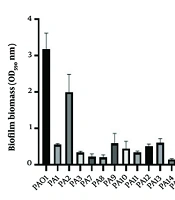
Each isolate was categorized as a non-biofilm producer or as a weak, moderate, or strong biofilm producer according to its OD values measured using the CV assay, as shown in Figure 1. Moderate biofilm producers were the most prevalent group, comprising 16 isolates (42.1%). Weak and strong biofilm producers accounted for 12 (31.6%) and 9 (23.7%) isolates, respectively, whereas only 1 isolate (2.6%), recovered from a respiratory sample, did not form any detectable biofilm, as summarized in Table 3. The relationship between the clinical source of P. aeruginosa isolates and their biofilm-forming capacity was assessed using the chi-square test. Although differences were observed in biofilm categories across sources, statistical analysis did not reveal a significant association (χ2 = 18.07, df = 12, P = 0.114). This finding was confirmed using the Fisher-Freeman-Halton exact test with Monte Carlo simulation, which also showed no significant association (P ≈ 0.13).
Table 3.
Biofilm-Forming Capacity of P. aeruginosa Clinical Isolates a
| Sources | Total Isolates | Non | Weak | Moderate | Strong |
|---|---|---|---|---|---|
| Respiratory | 13 | 1 | 2 | 6 | 4 |
| Sputum | 4 | 0 | 2 | 1 | 1 |
| Blood | 3 | 0 | 3 | 0 | 0 |
| Urine | 13 | 0 | 5 | 4 | 4 |
| Wound | 5 | 0 | 0 | 5 | 0 |
a Chi-square test: χ2 = 18.07, df = 12, P = 0.114.
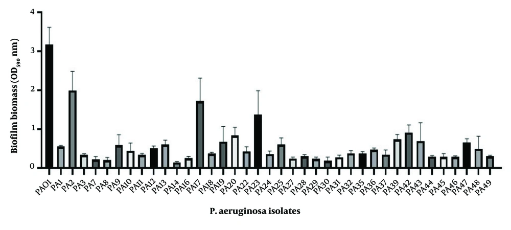
Figure 1.
Biofilm-forming capacity of clinical P. aeruginosa isolates determined by crystal violet assay. PAO1 was used as the reference strain. Values represent OD570 measurements obtained from triplicate experiments.
4.4. Analysis of Biofilm-Specific Antibiotic Resistance Genes via PCR
PCR analysis for the detection of biofilm-specific antibiotic resistance genes (ndvB, tssC1, PA5033, and PA2070) revealed that all P. aeruginosa isolates harbored at least 1 of these genes. Among the 38 isolates, ndvB and PA5033 were the most frequently detected, each present in 94.7% of isolates. The tssC1 gene was identified in 92.1% of the isolates, whereas PA2070 was found in 89.5%. Statistical analysis revealed no significant association between biofilm-forming capacity and the presence of ndvB (P = 0.127), tssC1 (P = 0.949), PA5033 (P = 0.699), or PA2070 (P = 0.478) among the clinical P. aeruginosa isolates. Representative agarose gel electrophoresis results for selected isolates are shown in Figure 2. The relationship between biofilm-associated resistance genes and biofilm-forming phenotypes in clinical P. aeruginosa isolates is shown in Table 4.
Table 4.
Distribution of Biofilm-Associated Genes According to Biofilm Phenotype a
| Genes | Weak (n = 12) | Moderate (n = 16) | Strong (n = 9) | Non-producer (n = 1) | P-Value |
|---|---|---|---|---|---|
| ndvB | 11 (91.7) | 16 (100) | 9 (100) | 1 (100) | 0.62 |
| tssC1 | 11 (91.7) | 15 (93.8) | 9 (100) | 1 (100) | 0.88 |
| PA5033 | 11 (91.7) | 16 (100) | 9 (100) | 1 (100) | 0.74 |
| PA2070 | 10 (83.3) | 15 (93.8) | 9 (100) | 1 (100) | 0.69 |
a Values are expressed as No. (%). P-values were calculated using the Fisher-Freeman-Halton exact test with Monte Carlo simulation (20,000 iterations). Biofilm status was analyzed as categorical phenotype groups derived from OD-based biofilm measurements.
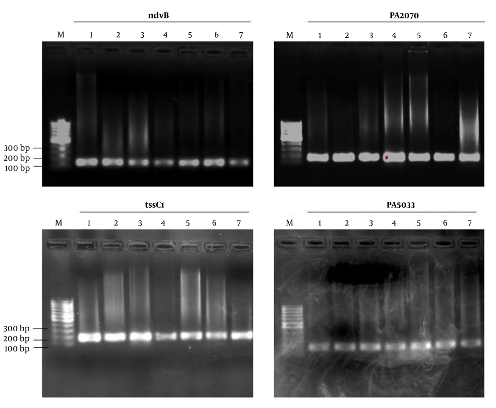
Figure 2.
Agarose gel electrophoresis of PCR amplification products of ndvB, PA2070, tssC1, and PA5033 genes in representative clinical Pseudomonas aeruginosa isolates. M: molecular weight marker; lanes 1 - 7: representative clinical isolates showing positive amplification bands
Biofilm formation was initially quantified as continuous OD values obtained from the CV microtiter plate assay. Subsequently, isolates were categorized as non-, weak-, moderate-, and strong-biofilm producers according to established OD cut-off criteria, and the comparative analyses in Table 4 were performed using these categorical biofilm phenotypes. Owing to the limited sample size, the presence of only 1 non-biofilm-producing isolate, and the high prevalence of the studied genes across all groups, the statistical power to detect phenotype-associated differences was limited. No statistically significant association was observed between the investigated genes and biofilm phenotype. However, interpretation of these findings is limited by the small sample size, the presence of only 1 non-biofilm-producing isolate, and the high prevalence of the investigated genes across all phenotype groups.
5. Discussion
Among nosocomial pathogens, P. aeruginosa is one of the most clinically significant species (5, 7). Its antibiotic resistance develops through interactions among multiple pathways, including biofilm formation (5-7). Multiple studies have shown that the resistance mechanisms of biofilm-associated P. aeruginosa differ markedly from those of planktonic cells (10, 11, 23). Polysaccharides, extracellular DNA, and proteins in the biofilm matrix play a central role in this resistance, collectively acting as a barrier that limits antibiotic penetration. In addition to matrix components, gene expression profiles unique to the biofilm state also contribute significantly to antibiotic resistance (24). In our previous studies, we investigated how antibiotic exposure affects the expression profiles of biofilm-associated antibiotic resistance genes in the P. aeruginosa PAO1 strain.
We observed that sub-inhibitory concentrations of antibiotics were sufficient to induce the expression of these genes, highlighting the potential risk of low-dose antibiotic exposure in promoting biofilm-associated resistance (25). These findings support the notion that biofilm formation not only serves as a physical barrier but also induces transcriptional reprogramming, which together complicate the therapeutic management of P. aeruginosa infections. In this study, we investigated the biofilm-forming capacity, antibiotic resistance profiles, and presence of biofilm-specific resistance genes in 38 clinical P. aeruginosa isolates. Our findings confirm the clinical significance of P. aeruginosa as an MDR pathogen with a high tendency for biofilm formation, consistent with previous reports (26-28). A high proportion of isolates exhibited resistance to key antipseudomonal agents, including meropenem, ciprofloxacin, and colistin. Meropenem resistance was the most prevalent (Table 2), in agreement with recent reports highlighting high levels of carbapenem resistance in P. aeruginosa clinical isolates that complicate treatment strategies (29, 30).
The ability of P. aeruginosa to form biofilms contributes significantly to chronicity and reduced treatment efficacy in clinical infections (6, 7). In our study, 97.4% of the isolates were able to form biofilms, with 66% categorized as moderate or strong biofilm producers. These findings are consistent with previous reports emphasizing the widespread biofilm-forming capacity of clinical P. aeruginosa strains (7). For instance, Saffari et al. (22) reported that all 92 isolates obtained from ocular infections were biofilm producers. These results align with earlier reports indicating that moderate biofilm formation is the predominant phenotype among MDR P. aeruginosa isolates (12). In addition, these patterns suggest a frequent co-occurrence of biofilm formation and multidrug resistance, as also documented by Kamali et al. (31), El-sayed et al. (32), and Brandão et al. (33). Overall, the present findings support a possible relationship between multidrug resistance and biofilm production while highlighting the need for further research to clarify the precise biological mechanisms underpinning this relationship.
PCR analysis revealed that nearly all isolates harbored at least 1 of the biofilm-specific resistance genes (ndvB, tssC1, PA5033, and PA2070), with ndvB and PA5033 most frequently detected, followed by tssC1 and PA2070. Although the investigated biofilm-associated resistance genes were highly prevalent among the isolates, no statistically significant relationship was observed between gene carriage and biofilm-forming phenotype. This finding may be partially explained by the limited number of gene-negative isolates, which reduced the statistical power of the comparative analysis. These findings are consistent with previous reports indicating a high prevalence of these genes in biofilm-forming P. aeruginosa isolates (12). Functionally, these genes are associated with distinct biological processes, including cyclic periplasmic glucan synthesis (ndvB), type VI secretion system activity (tssC1), and putative membrane transport or secretion pathways (PA2070 and PA5033) (24). In addition, the PCR-based approach used in this study only confirmed the presence of the investigated genes and did not provide information on their transcriptional activity or functional contribution under planktonic or biofilm conditions. Therefore, the detection of these genes alone is insufficient to establish direct mechanistic relationships with biofilm formation or antimicrobial resistance. Future studies involving transcriptional analyses, such as RT-qPCR, together with functional approaches, including gene knockout, overexpression, and mutant comparison studies, are required to better clarify the biological roles of these genes in biofilm-associated resistance mechanisms.
Previous studies have also demonstrated significant upregulation of these genes under biofilm conditions, supporting the notion that their contribution to resistance is likely context-dependent and regulated at the transcriptional level (23, 31). Overall, these findings suggest that the high prevalence of these genes, combined with strong biofilm-forming capacity and multidrug resistance, reflects the multifactorial nature of P. aeruginosa persistence in clinical settings. Although no statistically significant association was identified between gene carriage and biofilm phenotype, these results should not be interpreted as evidence of an absence of association. The limited sample size, uneven phenotype distribution, and near-universal prevalence of several investigated genes substantially reduced the statistical power of the analysis. Therefore, larger studies incorporating quantitative expression analyses are required to clarify the contribution of these genes to biofilm formation.
5.1. Conclusions
In this study, clinical isolates of P. aeruginosa were evaluated with respect to antibiotic susceptibility patterns, biofilm formation potential, and the occurrence of biofilm-associated resistance genes. Although biofilm-specific resistance genes were found in more than 90% of the isolates, biofilm formation capacity was moderate or strong. The investigated biofilm-associated resistance genes were commonly detected among P. aeruginosa isolates, which also exhibited varying biofilm-forming capacities. In conclusion, this study presents descriptive data on biofilm formation, antimicrobial resistance, and the presence of selected resistance genes in clinical P. aeruginosa isolates, which may serve as a basis for further investigation into their biological significance.