
1. Introduction
Myocarditis in neonates is an uncommon but life-threatening condition that may progress rapidly to dilated cardiomyopathy and heart failure (1). Among infectious etiologies, enteroviruses are a significant but underrecognized cause. Clinical diagnosis is often delayed because early symptoms overlap with those of bacterial sepsis or other nonspecific neonatal illnesses (2). Enteroviruses (EVs) are RNA viruses within the Picornavirus family. These pathogens primarily proliferate in the oropharynx and gastrointestinal tract, resulting in diverse clinical manifestations ranging from asymptomatic infection to severe systemic disease (3, 4). Severe complications associated with enterovirus infections, particularly in neonates and pediatric populations, include encephalitis, myocarditis, neonatal sepsis, acute flaccid paralysis, and meningitis (5). Here, we present a life-threatening case of enterovirus-associated myocarditis in a newborn. By highlighting the diagnostic and therapeutic challenges, we aim to raise awareness among clinicians caring for febrile infants.
2. Case Presentation
A 5-day-old female neonate was admitted to the Neonatal Intensive Care Unit (NICU) with fever and grunting. Initially, the neonate had been hospitalized in the Neonatal Special Care Unit (NSCU); however, owing to the onset of fever and poor feeding, she was transferred to the NICU for further evaluation and management. She was born at 37 weeks and 5 days of gestation by cesarean section, with a birth weight of 2580 g. Her 30-year-old mother, who was otherwise healthy and had no chronic conditions or regular medication use, developed symptoms of an upper respiratory tract infection, including fever and sore throat, beginning 2 days postpartum. The family history was unremarkable. The postpartum period was uneventful, and the infant was discharged in good health 24 hours after birth.
On physical examination at admission, the neonate appeared clinically stable. She was afebrile, had no signs of respiratory distress, and was alert, responding appropriately to auditory and visual stimuli. Her respiratory rate was 50 breaths/min, body temperature was 36.5 °C, oxygen saturation was 98%, and pulse rate was 160 beats/min. On auscultation, mild bilateral rales were present in the lung fields. Peripheral pulses were palpable, strong, symmetrical, and well perfused. Cardiac auscultation revealed a gallop rhythm. Electrocardiography demonstrated sinus tachycardia with a heart rate of 160 beats/min and normal P-wave morphology. The ST segment showed low voltage (total amplitude < 10 mm in limb leads), accompanied by T-wave suppression. The PR interval was 0.12 seconds, which is marginally prolonged when adjusted for the patient's postnatal age and heart rate (normal upper limit, 0.11 seconds for 160 - 180 beats/min in neonates younger than 1 month). No cyanosis was observed in the extremities. Neurological examination showed intact age-appropriate reflexes, including Moro and rooting reflexes. On abdominal examination, the liver was palpated approximately 4 cm below the right costal margin.
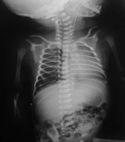
At admission, initial investigations were requested. The arterial blood gas results were as follows: pH, 7.55; PCO2, 48; HCO3, 30; base excess, +5; and PO2, 50. The initial complete blood count showed the following: white blood cell count, 7400/mm3; neutrophils, 70%; hemoglobin, 16.1 g/dL; and platelets, 237 × 103/µL. Blood, urine, and stool cultures were obtained on admission, and all showed no growth. A chest X-ray was requested, as shown in Figure 1. One day after admission, the patient underwent a lumbar puncture, and the results were as follows: cerebrospinal fluid culture, no growth after 24 hours; cerebrospinal fluid analysis, white blood cell count, 45/mm3; lymphocytes, 1.7%; polymorphonuclear cells, 98.3%; glucose, 81 mg/dL; and protein, 128.8 mg/dL. A cerebrospinal fluid sample was also sent for a multiplex meningitis/encephalitis polymerase chain reaction panel. The patient was initially admitted with a presumptive diagnosis of neonatal sepsis and was started on empirical antibiotic therapy with ampicillin, cefotaxime, and vancomycin.
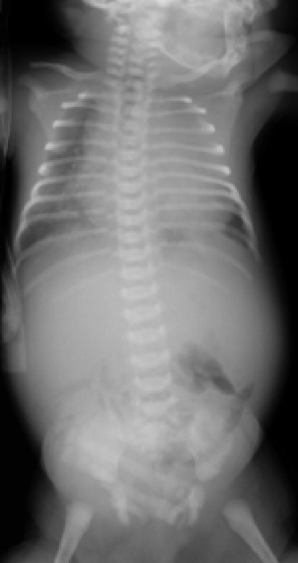
Figure 1.
Pediatric anteroposterior chest X-ray showing cardiomegaly in the newborn.
During the hospital course, the neonate developed recurrent fever and grunting 2 days after admission. At that time, respiratory support with continuous positive airway pressure was initiated because of signs of evolving respiratory distress. Laboratory evaluation revealed thrombocytopenia with a platelet count of 40000/µL. The patient received supportive therapy in addition to empirical management for presumed neonatal sepsis. Supportive measures included a single dose of intravenous immunoglobulin (1 g/kg) and a platelet transfusion. Later laboratory tests demonstrated elevated liver enzymes (aspartate aminotransferase, 177 U/L; alanine aminotransferase, 56 U/L; and alkaline phosphatase, 423 U/L) and hyperbilirubinemia (total bilirubin, 8.6 mg/dL; direct bilirubin, 1.1 mg/dL). The neonate was subsequently treated with phototherapy for hyperbilirubinemia. Despite these interventions, her overall clinical condition continued to deteriorate.
During hospitalization, the neonate developed generalized edema, raising suspicion of cardiac involvement and prompting a cardiology consultation. Abdominal examination revealed hepatomegaly, which was initially considered multifactorial and possibly related to viral infection and congestive hepatopathy, prompting a gastroenterology consultation. Based on specialist recommendations, supportive hepatoprotective therapy, including ursodeoxycholic acid, was initiated. Echocardiography revealed severe tricuspid regurgitation, moderate mitral regurgitation, reduced left ventricular ejection fraction (40%), and biatrial enlargement, consistent with dilated cardiomyopathy secondary to myocarditis. Concurrently, polymerase chain reaction testing of cerebrospinal fluid confirmed enterovirus infection, supporting the clinical suspicion of viral myocarditis.
Despite supportive interventions, the neonate's condition deteriorated. She developed acute respiratory distress requiring endotracheal intubation and mechanical ventilation. Shortly thereafter, she experienced cardiorespiratory arrest. Resuscitation was attempted according to standard neonatal advanced life support protocols, but these efforts were unsuccessful, and the patient did not survive. Given the mother's history of respiratory symptoms and the neonate's presentation with fever, neonatal sepsis was initially suspected. Cerebrospinal fluid analysis revealed pleocytosis (white blood cell count, 45/mm3), with a predominance of polymorphonuclear cells, elevated protein, and normal glucose levels, while the culture remained negative. Importantly, polymerase chain reaction testing of the cerebrospinal fluid was positive for enterovirus, strongly supporting a viral etiology. Congenital heart disease was also considered; however, the absence of a family history of cardiac or genetic disorders, together with normal structural findings on chest radiography and echocardiography, made this diagnosis less likely. To exclude inborn errors of metabolism, a comprehensive metabolic screening panel was performed and returned normal. Altogether, the combination of cerebrospinal fluid polymerase chain reaction positivity for enterovirus and echocardiographic findings of dilated cardiomyopathy confirmed the final diagnosis of neonatal viral myocarditis secondary to enterovirus infection.
3. Discussion
Myocarditis in neonates is an uncommon but life-threatening condition, most often triggered by viral infections such as enteroviruses (5, 6). Early recognition is challenging because initial manifestations are nonspecific and may mimic neonatal sepsis. In our patient, fever and poor feeding initially suggested sepsis; however, the subsequent development of hepatomegaly, thrombocytopenia, cardiomegaly, and reduced ejection fraction shifted attention toward cardiac involvement. Confirmation of enterovirus infection by cerebrospinal fluid polymerase chain reaction testing ultimately established the diagnosis of viral myocarditis. It is well recognized that cerebrospinal fluid findings in enteroviral meningitis may show neutrophil predominance during the first 48 hours of illness, followed by a transition to lymphocytic predominance. Therefore, the initial polymorphonuclear-predominant cerebrospinal fluid profile observed in our patient is fully compatible with the early phase of enteroviral meningitis and should not be interpreted as evidence against a viral etiology. This characteristic temporal evolution of cerebrospinal fluid cellular composition has been consistently described in previous studies (7).
Several reports in the literature describe enterovirus myocarditis in neonates, often with rapid progression to dilated cardiomyopathy and high mortality despite intensive care (8). Similar to our case, supportive treatment, including intravenous immunoglobulin and mechanical ventilation, was insufficient to alter the fatal outcome. This finding underscores the poor prognosis associated with neonatal enterovirus myocarditis and the urgent need for more effective therapeutic strategies (9, 10). The differential diagnosis in this case included bacterial sepsis, congenital heart disease, and inborn errors of metabolism. Sepsis was initially suspected; however, negative cultures and polymerase chain reaction positivity for enterovirus supported a viral etiology. Structural heart disease was excluded by echocardiography, and metabolic screening results were normal. These evaluations emphasize the importance of systematic assessment in critically ill neonates, as overlapping features can obscure the underlying cause.
Limitations of this case include the absence of maternal virological testing; therefore, maternal enterovirus infection could not be confirmed. However, given that the neonate was less than 1 week old at the time of symptom onset (5 days), perinatal/peripartum transmission remains suspected rather than confirmed (11, 12). In addition, enterovirus genotyping or serotyping was not performed because the multiplex meningitis/encephalitis polymerase chain reaction panel used at our institution identifies only the presence of enterovirus and does not determine the viral subtype. From a preventive standpoint, early recognition of maternal infection, appropriate infection control measures during the peripartum period, and careful monitoring of neonates born to symptomatic mothers may help reduce the risk of neonatal enterovirus infection. Even mild maternal respiratory illnesses can have severe consequences for newborns because of their immature immune systems. Strengthening awareness of viral etiologies in febrile neonates and incorporating early viral testing into diagnostic protocols may facilitate timely diagnosis and potentially improve outcomes.