1. Background
Based on the results of molecular phylogenetic analysis, the Mucorales, the core group of the traditional Zygomycota, have been reclassified into the subphylum Mucoromycotina of the new division, Glomeromycota (1).
These fungi are ubiquitous saprophytes that are scattered widely in nature, food, soil, and air (2). The Mucorales are regarded as important animal and human pathogens, and they are responsible for mucormycosis (formerly known as zygomycosis) (3, 4), the third most common invasive fungal infection (5-7). The Mucorales are increasingly recognized as opportunistic pathogens in immunocompromised or immunosuppressed patients (8, 9).
Mucormycosis is a very aggressive invasive fungal disease. It is a serious condition that affects a variety of patient groups. Mucormycosis can be caused by any of the six families of Mucorales, although the members of the mucoraceae including Rhizopus, Mucor, Absidia (Lichtheimia), and Rhizomucor are more frequently isolated in human infections (5, 7, 10, 11).
Members of the genus Rhizopus are the most common isolates recovered in a clinical setting, with Rhizopus arrhizus occurring the most frequently. Members of the genus Mucor come second to Rhizopus with respect to frequency, while Cunninghamella, Apophysomyces, Lichtheimia, Saksenaea, Rhizomucor, Cokeromyces, and Syncephalastrum each constitute a significantly lower percentage of clinical isolates (7, 12, 13).
Due to the infection’s rapidly progressive course and unexpectedness, many mucormycosis cases are only diagnosed postmortem. The infection therefore constitutes a huge diagnostic and therapeutic challenge (9). The number of cases of mucormycosis has increased over the last few decades, making the fast and accurate diagnosis of these infections imperative. Unfortunately for patients who are affected by mucormycosis, no routine laboratory tests are available to diagnose this disease. Clinical diagnosis instead relies on histopathology and the isolation of etiologic fungi from infected tissues (2, 14). The identification of Mucorales is primarily based on standard mycological methods. The Mucorales are characterized by anamorph structures. The mycelium is typically non-septate (coenocytic) or irregularly septate. Sporangiospores are produced in multi-sporangia. The sporangia are characterized by the inclusion of a variously shaped columella (15).
The growth of the Mucorales in media is rapid, with mycelia elements expanding to cover the entire plate in only a few (one to seven) days. However, culture-based identification is often difficult and time-consuming, and it relies entirely on the experience of the technician or expertise mycologist. Conventional phenotypic methods usually identify isolates to only the genus level, and sometimes only as Mucorales. If the diagnosis is not made early enough, dissemination often occurs (8, 16, 17).
Based on the above, the development and validation of a new detection system, perhaps involving polymerase chain reaction (PCR) methods, for the fast and accurate diagnosis of mucormycosis is necessary. PCR-restriction fragment length polymorphism (RFLP) is a rapid and highly reliable method for the identification and differentiation of important common Mucorales species (16).
2. Objectives
This study aimed to identify the Mucor and Lichtheimia (formerly Absidia) species among other genera of Mucorales isolated in culture media using the PCR-RFLP method.
3. Materials and Methods
3.1. Isolation of Fungi
Of the course of a year, 500 samples were taken from different city districts, parks, hospitals, greengrocers, food stores, laboratories, public restrooms, mosques, and cattle houses. The samples were cultured on sabouraud dextrose agar (SDA; Merck KGaA, Darmstadt, Germany) and potato dextrose agar (PDA; Merck KGaA, Darmstadt, Germany) supplemented with chloramphenicol. A diversity of different types of filamentous fungi and yeasts were identified in the media. Finally, 120 pure cultures belonging to the Mucorales were gained after the purification of the colonies. Primary identification of the Mucorales was performed on the basis of the macroscopic and microscopic features of the colonies.
In the next step, a mixture of two genus-specific sense primers and one degenerate anti-sense primer from the 18S rRNA gene region were selected to determine the two genera of Lichtheimia and Mucor within the Mucorales.
3.2. DNA Extraction From Pure Fungal Cultures
All of the pure colonies obtained were sub-cultured on 2% SDA and PDA and then incubated for one to seven days at 27°C in stable conditions. The genomic DNA was extracted and purified from each colony using the phenol-chloroform method as follows. Briefly, hyphae (without sporangia) from fresh 48 hours cultures on SDA or PDA were suspended in a 200 µL lysis buffer (100 mM Tris-Hcl, 10 mM EDTA (pH = 8), 2% Triton X-100, 1% SDS, 100 mM Nacl) (Merck KGaA, Darmstadt, Germany) in 2 ml Eppendorf tubes and grinded well. Then, the suspension (without the hyphal elements) of each tube was transferred to a new Eppendorf tube. Next, 200 µL Phenol-Chloroform (1:1) was added and vortexed (or shaken by hand for 5 minutes) rigorously with 200 µL of glass beads (0.5 mm in diameter) to release the DNA. After centrifugation for 5 minutes at 5,000 rpm, the supernatant was mixed with an equal volume of 2-propanol and 0.1 volume of 3 M sodium acetate (pH = 5.2), vortexed, and incubated at -20°C for 10 min. It was then centrifuged for 12 min at 12,000 rpm. All of the solution was gently removed and then 100 µL of 70% ethanol was gently added to the tube and centrifuged for 5 minutes at 5000 rpm. The ethanol was then removed from the tube, dried, and resuspended in 50 µL of distilled deionized water, and it was kept at -20°C as the purified DNA until use.
3.3. PCR
Selected primers targeted an 830 bp sequence in the 18S fungal ribosomal gene, which excludes the amplification of human DNA and other filamentous fungi. The specific sense primers were MucL1: 5’ TGATCTACGTGACATATTTCT 3’ and AbsL1: 5’ TGA TCTACACGGCATCAAAT 3’ (Bioneer, Daejeon, South Korea), which corresponded to the sequences of Mucor sp. and Absidia (Lichtheimia), respectively. A degenerate anti-sense primer (MR1: 5’ AGTAGTTTGTCTTCGGTCAA 3’) was applied for the Mucorales. The sense primers annealed to the region of the template starting at position 75, while the antisense primer annealed to the region at position 901 (18).
Each amplification reaction included 12.5 µL of premix (containing 2.5 U Taq DNA polymerase, PCR buffer, 1.5 mM Mgcl2, and 200 µM dNTP) (Ampliqon, Odense, Denmark), 2 µL (about 10 ng) of template DNA, 0.5 µL of each primer (3 × 0.5 = 1.5 µL), and 9 µL of distilled deionized water in a final volume of 25 µL. Amplification was performed on a 2700 thermocycler (Applied Biosystems, Singapore) as follows. One cycle of 1 minute at 94°C (primary denaturation), 30 cycles of 1 minute at 94°C (denaturation), 1 minute at 60°C (annealing), 1 minute at 72°C (extension), and finally one cycle of 5 minute at 72°C. Negative controls (no DNA template) were included in each run to detect the presence of any DNA contamination in the reagents and reaction mixtures.
3.4. Restriction Enzymes
The PCR products of 120 Mucorales were individually digested with selected restriction enzymes including AfIII, XmnI (Thermo Fisher Scientific, Vilnius, Lithuania), and AcII (New England BioLabs, USA), which were specified to the Mucor sp.: Mucor circinelloides, M. racemosus, M. ramosissimus, M. plumbeus, L. corymbifera, and L. blakesleeana.
3.5. RFLP
Eighteen s amplified fragments of the 120 Mucorales were separately digested by the three abovementioned enzymes. The amplicons were digested for 1 hour at 37°C in a total volume of 25 µL (containing 2 µL of the enzyme, 2.5 µL of related buffer, 10 µL of PCR product, and 10.5 µL of distilled water). The digested amplification products were subjected to electrophoresis, and the sizes of the restriction fragments were determined by comparison with a 100 bp ladder standard DNA molecular weight marker (Fermentas, Vilnius, Lithuania). The restriction site, specificity, and fragment size of each enzyme are detailed in Table 1.
Table 1.
The Restriction Site, Specificity, and Fragment Size of Each Enzyme
| Enzyme (Restriction Site) | Specificity | Fragment Size, bp |
|---|---|---|
| AfIII (5’ CTTAAG 3’) | Mucor sp. | 750 + 87 |
| XmnI (5’GAATAGCTTC 3’ or 5’ AGCTTCGGT 3’) | Mucor circinelloides, M. racemosus, M. ramosissimus, M. plumbeus | 613 + 224 |
| AcII (5’ AACGTT 3’) | Lichtheimia corymbifera, L. blakesleeana | 518 + 306 |
3.6. Electrophoresis
Agarose gel in a TBE buffer (90 mM Tris, 90 mM boric acid, and 2 mM EDTA) at 100 V for 45 - 120 minutes in a 1%, 1.5%, and 2% gel was used for the electrophoresis of the extracted DNA, PCR products, and RFLP products, respectively.
3.7. Characteristic Features of the Mucor and Lichtheimia Species
The Mucor sp. typically exhibits rapid growth, producing globose sporangia on sporangiophores that are either solitary or branched. The sporangia contain the entire columella and spores that are mucus bound. The sporangial wall collapses irregularly, if at all. The sporangia may also be deliquescent (dissolving). Rhizoids and stolons are absent. These features distinguish the Mucor spp. from the other producers of globose sporangia (8). The genus Lichtheimia is defined by its pyriform, apophysate sporangia (15). The characteristic features of some Mucor and Lichtheimia species are summarized in Table 2 (8, 15, 19-23).
Table 2.
Characteristic Features of the Some Mucor and Lichtheimia Species
| Organism name | Colony Morphology | Sporangium Morphology, µm | Columella Morphology, µm | Sporangiospore Morphology, µm | Rhizoids and Apophysis | Sporangiophore Morphology | Other |
|---|---|---|---|---|---|---|---|
| Mucor ramosissimus | Rapidly growing low colonies; gray to buff | Globose, 25 - 80 | Round to flattened, 20 - 37 by 17 - 30; collars may be seen; smaller sporangia lack columella | Oval to round, smoothwalled, brownish; 3.3 - 5.5 by 3.5 - 8 | Absent | Sympodially branched; may have racquet shaped swellings | Optimal growth at 24°C, poor growth at 37°C |
| Mucor circinelloides | Floccose with rapid growth; pale gray to yellowish, brown at 37°C | Globose, up to 60 | Spherical, up to 50 in diameter; collars may be present | Smooth walled and oval; 4.4 - 7 | Absent | Sympodially branched and circinate | Growth from 5°C to 37°C |
| Mucor plumbeus | Grey to light olive-green | Globose up to 80, Hyaline, dark brown to brownish grey with age, with spinulose walls | Pyriform, ovoid with a truncate base, up to 25 - 50 | Globose, sometimes more or less ellipsoidal or irregularly shaped, 7 - 8 | Absent | With slightly encrusted walls, branching | Optimal growth at 5 - 20°C |
| Mucor racemosus | Low to medium-high colonies; light to medium brown | Globose, light brown, encrusted walls, up to 80 | Ellipsoidal to pyriform, up to 40 long | Oval to subspherical, smooth walled; 5 - 8 | Absent | Branched | Optimal growth at 25°C, poor or no growth over 32°C |
| Lichtheimia corymbifera | Floccose; first white, turning brown togreenish brown with age | Pyriform 20 - 80 | Usually with an apical projection, Columella: dome shaped | Single-celled, hyaline, subglobose to broadly ellipsoidal | Present apophysis: flask shaped | Erect, simple or slightly branched, typically rising along the stolon but not opposite the rhizoids, apically with a well-developed, funnel shaped or swollen apophysis | Capable of growth at 48°C to 52°C |
| Lichtheimia blakesleeana | Wooly, white, and grey-brown to olive green with age | Pyriform 20 - 80 | Usually with a apical projection, Columella: dome shaped | Single-celled, hyaline, subglobose or more rarely, broadly ellipsoidal | Present apophysis: flask shaped | Apically with a well-developed, funnel shaped or swollen apophysis | No growth at 48°C |
4. Results
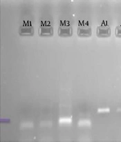
The genomic DNA belonging to the Mucorales was successfully amplified with the selected primers and a product of approximately 830 base pair (bp) was amplified for all Mucorales. The PCR products were electrophoresed in 1.5% agarose gels in the presence of ethidium bromide and then visualized under UV light (Figures 1 and 2). The PCR amplicons were separately digested by AfIII, XmnI, and AcII enzymes. The obtained sizes of the RFLP products were exactly comparable with the sizes detailed in Table 1.
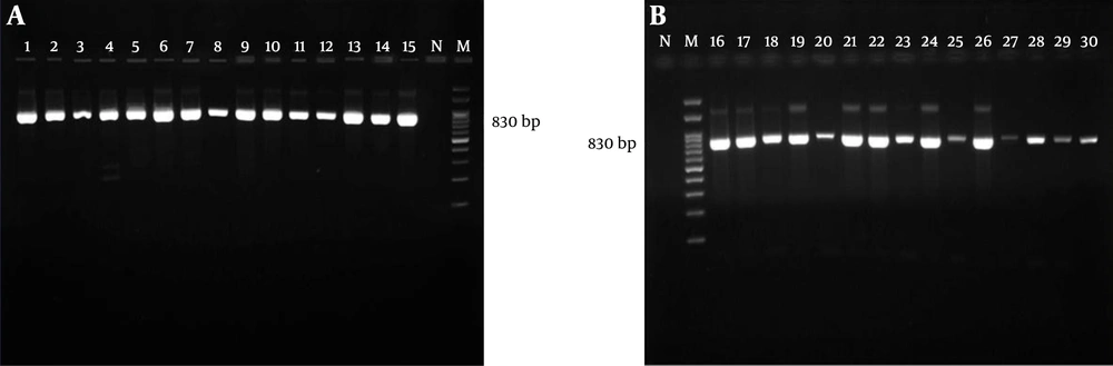
Figure 1.
All samples yielded a single band of approximately 830 bp in lanes 1 to 30. Lanes N, negative control; M, 100 bp molecular size marker.
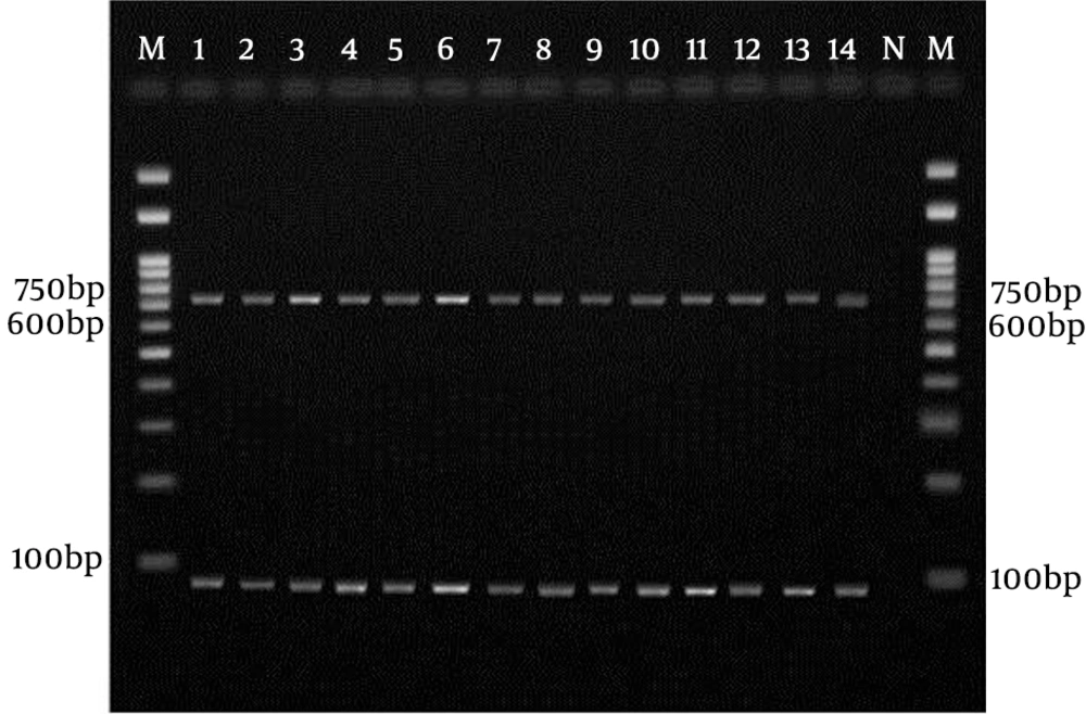
Figure 2.
Lanes 1 to 13, Mucor sp.; N, negative control; M, 100 bp molecular size marker.
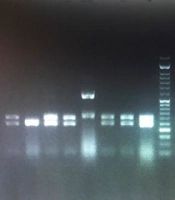
Two fragments (750 + 78) were visualized following the AfIII restriction. The specific restriction pattern of M. circinelloides, M. racemosus, M. ramosissimus, and M. plumbeus was established using XmnI (613 + 224). Additionally, two fragments (518 + 306) were visualized following digestion with AcII, which represented the specific pattern for Lichtheimia corymbifera and L. blakesleeana (Figures 3 and 4).
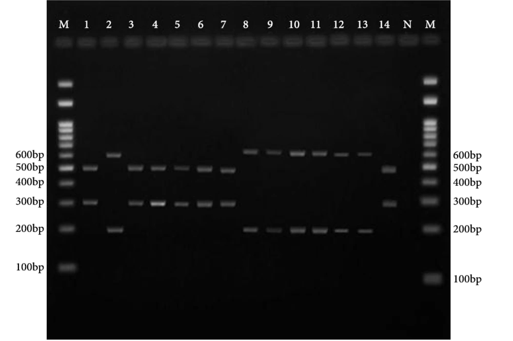
Figure 3.
Lanes 12, 11, 10, 9, 8, 2 and 13, M. circinelloides, M. racemosus, M. ramosissimus or M. plumbeus; Lanes 3, 1, 7, 6, 5, 4, and 14, L. corymbifera or L. blakesleeana; N, negative control; M, 100 bp molecular size marker.
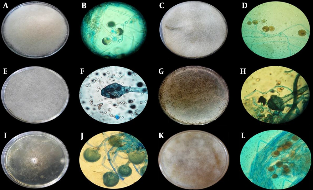
Figure 4.
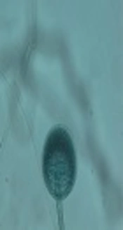
A, Colony of M. plumbeus; B, Microscopic morphology of M. plumbeus; globos sporangia with spinolus walls and pyriform columella; C, colony of M. circinelloides; D, microscopic morphology of M. circinelloides with globos sporangia and spherical columella; E, colony of L. corymbifera; F, microscopic morphology of L. corymbifera with pyriform sporangia and flask like apophysis; G, colony of L. Blackesleenana; H, microscopic morphology of L. Blackesleenana with pyriform sporangia and flask like apophysis; I, colony of M. ramosissimus; J, microscopic morphology of M. ramosissimus with globos sporangia and round columella; K, colony of M. racemosus; L, microscopic morphology of M. racemosus with globos, light brown and encrusted walls sporangia, and ellipsoidal columella.
The discrimination of these species was conducted based on the macroscopic and microscopic features as well as the growth ability in different temperatures (Figure 4). Finally, Mucor sp. and Lichtheimia sp represented approximately 39.17% of all the pure colonies. Thirty-five colonies (29.17%) were identified as Mucor sp., namely M. circinelloides (9.17%), M. racemosus (5.83%), M. plumbeus (10.83%), and M. ramosissimus (3.33%). Twelve colonies (10%) were identified as Lichtheimia, which belonged to L. corymbifera (9.17%) and L. blakesleeana (0.83%).
5. Discussion
The identification of Zygomycetes is mainly based on macroscopic and microscopic characteristics, which is a difficult and time-consuming process that sometimes needs the expertise of a reference laboratory (14). Additionally, the precise identification of the Mucorales down to a species level may hold great importance for further research on antifungal effectiveness (18, 24, 25). Molecular techniques have showed enormous potential for rapidly and accurately identifying the ecological agents of mucormycosis, which helps in conducting epidemiologic investigations. Molecular detection assays for the Mucorales are, however, not yet widely available (18, 26).
Different regions of the rRNA operon have most frequently been the targets for the detection of Zygomycetes, with previously reported PCRs for zygomycosis. Several prior reports have described the utilization of universal fungal primers from the 18S, 28S, or ITS rRNA gene regions for PCR amplification followed by the sequencing or hybridization of the product to specific probes (2, 13, 27, 28).
PCR-RFLP is a reliable and easy to perform technique that can be used in epidemiological and research studies. PCR-RFLP-based methods target the 18S ribosomal gene of Zygomycetes on DNA extracted from human specimens and may therefore provide clinicians with a rapid and definitive diagnosis of mucormycosis (29). In the present study, a PCR-RFLP method based on the 18S ribosomal gene of Zygomycetes, which had been previously developed by Machouart et al. was used to identify the Mucor and Absidia species from pure cultures. The identification of this region by PCR amplification with selective primers has proved to be reliable for the identification of Zygomycetes (18).
Bialek et al. (30) developed a PCR-based method targeting the 18S rRNA gene for the identification of mucormycosis and aspergillosis agents in paraffin wax embedded tissue. Piancastelli et al. (31) identified L. corymbifera among other fungi with PCR-RFLP using the ITS region as a target sequence and the AcII restriction enzyme. In our study, AfIII was used for Mucor identification to the genus level and, after that, the XmnI restriction enzyme was applied to distinguish M. circinelloides, M. racemosus, M. ramosissimus, and M. plumbeus. The AcII restriction enzyme was used to identify L. corymbifera and L. blakesleeana. Digestion with AfIII, XmnI, and AcII generated two fragments of 750 + 87, 613 + 224, and 518+306 bp, respectively. The restriction site and fragment sizes can be seen in Table 1. XmnI does not cut the amplicons obtained from M. hiemalis or M. indicus. Therefore, following RFLP, the differentiation of the four abovementioned species was performed based on comparing the macroscopic, microscopic, and other features. These features are summarized in Table 2.
Iwen et al. (19) identified M. circinelloides as a cause of primary zygomycosis using a sequence analysis of the ITS region as well as phenotyping methods. The Mucor species are considered to be a distant third behind the Rhizopus species and Absidia corymbifera in terms of causing zygomycosis. Only five species are suspected of causing human disease. These include the thermotolerant species M. racemosus, which either does not grow or else grows poorly at 37°C. The presence of fungal species has been considered to be an environmental microbiological indicator, and some of the fungi have been found to cause fungal infection (26).
A considerable number of mucormycosis cases have been associated with M. circinelloides, which appears to be the most common cause of the disease. There have been a few mucormycosis cases associated with M. ramosissimus. However, no mycosis cases associated with M. plumbeus have yet been reported (32). In this study, the genera of Mucor and Lichtheimia were represented in 29.17% and 10% of the 120 pure Mucorales cultures, respectively. Alvarez et al. (33) studied 190 isolates morphologically identified as Zygomycetes using sequencing of the ITS region of the rDNA, which revealed that M. circinelloides, L. corymbifera, and M. indicus represented approximately 9.5%, 5.3%, and 2.6% of these isolates, respectively.
Among the Absidia species, the most important species associated with mucormycosis is A. corymbifera. Based on physiological, phylogenetic, and morphological data, it was proposed that three Absidia species, namely A. corymbifera, A. blakesleeana, and A. hyalospora, should be reclassified as a separate family, the Lichteimiaceae fam. nov., and the three species renamed as L. corymbifera, L. blakesleeana, and L. hylospora. L. blakesleeana was subsequently reduced to a synonym of L. hyalospora (34, 35). In Dannaoui et al. (24) study, almost all of the PCR results for M. circinelloides were negative. However, in the present study, PCR amplification of all the pure cultures using specific primers was carried out successfully, and the obtained bands were fully sharp.
Finally, our findings revealed that molecular methods can be used for the rapid detection and differentiation of species that are responsible for infection, and they can hence help in conducting epidemiologic investigations. In contrast to other methods, a PCR-based approach has the potential to be time efficient, highly specific, and endowed with a good sensitivity (36).
In summary, the present study maintains that the diagnosis of Zygomycetes to a species level based on macroscopic and microscopic features is very difficult. At the same time, there are several limitations to the method used in our study. Therefore, it seems that more research is needed to modify the present molecular approaches. In a future study, we intend to design a PCR-RFLP method that needs to lower specific primers and enzymes in order to identify the genera and species belonging to Zygomycetes.