
1. Background
Wound healing is a complex biological process involving coordinated inflammatory, proliferative, and tissue-remodeling responses that collectively restore skin integrity following injury (1, 2). Disruption of these processes may result in chronic or non-healing wounds, which represent a major clinical challenge associated with prolonged hospitalization, increased healthcare costs, reduced quality of life, and an elevated risk of morbidity (3). Chronic wounds are particularly common in patients with diabetes mellitus, vascular insufficiency, trauma, prolonged inflammation, and impaired immune function. Among chronic wound types, diabetic foot ulcers (DFUs) remain among the most difficult conditions to manage clinically because of delayed surface coverage, impaired angiogenesis, persistent inflammation, neuropathy, and increased susceptibility to microbial colonization and biofilm formation (4). Similar challenges are encountered in post-surgical wounds, traumatic injuries, and chronic non-healing ulcers, in which delayed tissue regeneration and recurrent infection frequently complicate wound management and prolong recovery (5). Although current wound-care strategies include debridement, antimicrobial therapy, dressings, and moisture-retention approaches, many available treatments remain insufficient to simultaneously address infection control, inflammatory modulation, and tissue regeneration (6, 7). Furthermore, increasing antimicrobial resistance (AMR) has reduced the effectiveness of conventional antibiotic-based therapies, emphasizing the need for alternative or adjunctive wound-healing formulations with both antimicrobial and regenerative properties (8, 9). In recent years, multifunctional biomaterial-based wound-care systems have attracted increasing attention because of their potential to provide antimicrobial protection while simultaneously supporting tissue repair and maintaining a favorable wound-healing environment (10). Chitosan-based biomaterials, in particular, have demonstrated promising antimicrobial, biocompatible, and wound-supportive properties in experimental and translational wound-healing studies (11, 12).
Based on these considerations, Chitagel, a multifunctional bioactive wound-healing hydrogel, was developed as a complementary wound-care formulation for managing chronic and difficult-to-heal wounds. The formulation combines quaternary ammonium-modified chitosan with PHMB, E. purpurea extract, glycerin, dexpanthenol, and sorbitol to provide antimicrobial protection, moisture retention, and support for tissue-regeneration processes. Previous preclinical investigations demonstrated promising antimicrobial and wound-healing activity of Chitagel in experimental models, including inhibition of methicillin-resistant Staphylococcus aureus (MRSA) growth and accelerated wound closure in infected wound models (13). However, despite these encouraging preclinical findings, clinical evidence regarding the wound-healing performance of Chitagel in patients with chronic and non-healing wounds remains limited.
2. Objectives
This prospective, single-arm clinical study was conducted to evaluate wound-healing progression associated with topical application of Chitagel in patients with diabetic, traumatic, post-surgical, and chronic non-healing wounds treated at the Sepahan Wound Care Specialty Clinic (Isfahan, Iran) between 2022 and 2023 (IRCT20210510051243N2). The primary objective was to assess clinical wound-healing outcomes, including wound contraction, epithelialization, fibrovascular tissue development, and overall wound-bed improvement, following treatment with Chitagel in conjunction with standard wound care.
3. Methods
3.1. Study Design and Clinical Setting
This study was a prospective, single-arm interventional clinical study designed to evaluate the wound-healing performance of Chitagel in patients with chronic and non-healing wounds. The study was conducted at the Sepahan Wound Care Specialty Clinic, Isfahan, Iran, between 2022 and 2023. The trial was registered in the Iranian Registry of Clinical Trials (IRCT20210510051243N2).
The primary objective was to evaluate wound-healing progression following topical application of Chitagel in addition to standard wound care. Healing outcomes were monitored through clinical assessments, serial wound imaging, and wound measurements during follow-up evaluations. The study was conducted in accordance with ethical principles for clinical research involving human participants, and all patients provided written informed consent before enrollment. The study was designed as a descriptive, single-arm feasibility assessment. No control group was included, and no blinding was performed. Therefore, the results are reported as clinical observations and representative cases only.
3.2. Patient Selection
A total of 80 patients with chronic or non-healing wounds were enrolled after providing written informed consent. Eligible patients included individuals with chronic, difficult-to-heal wounds persisting for more than six weeks despite routine wound management. Wound etiologies included diabetic wounds, post-surgical wounds, traumatic wounds, and chronic non-healing wounds. Vascular and pressure ulcers were categorized as chronic non-healing wounds. Patients with active malignancy, severe systemic infection, known hypersensitivity to any component of Chitagel, autoimmune disorders, or ongoing immunosuppressive therapy were excluded. Patients were recruited consecutively from among all eligible individuals presenting to the clinic during the study period. No formal sample size calculation was performed. The risk of selection bias is acknowledged because of the single-arm design.
Baseline demographic and clinical characteristics, including wound type, wound duration, wound dimensions, infection status, and relevant comorbidities, were recorded before treatment initiation. Because diabetic and chronic wounds are often associated with prolonged inflammation, impaired vascularization, and delayed tissue regeneration, careful clinical monitoring was performed throughout the study period (Table 1).
Table 1.
Baseline Clinical Characteristics of Patients Included in the Study a
| Variables | Patients | Clinical Characteristics |
|---|---|---|
| Total enrolled patients | 80 (100) | Patients with chronic and difficult-to-heal wounds treated with Chitagel in conjunction with standard wound care |
| Diabetic wounds | 43 (53.8) | Included diabetic foot ulcers and chronic diabetic wounds associated with delayed healing, inflammation, and infection risk |
| Traumatic wounds | 12 (15.0) | Included traumatic soft-tissue injuries and head trauma wounds with tissue disruption and inflammatory burden |
| Chronic non-healing wounds | 11 (13.8) | Included wounds persisting despite routine wound management and associated with impaired tissue repair |
| Post-surgical wounds | 14 (17.5) | Included surgical wounds with delayed closure, tissue disruption, and exudative wound beds |
a Values are expressed as No. (%).
3.3. Treatment Protocol
All patients received standard wound care procedures, including wound cleansing, debridement when necessary, infection control measures, and appropriate dressing selection. Chitagel was applied topically to the wound surface at each dressing change. After gel application, wounds were covered with sterile occlusive or semi-occlusive dressings depending on wound condition and exudate level. Dressing changes were performed every 48 - 72 hours according to clinical assessment. Chitagel contains quaternary ammonium-modified chitosan, PHMB, glycerin, sorbitol, dexpanthenol, and E. purpurea extract. Quaternary ammonium-modified chitosan and PHMB may contribute broad-spectrum antimicrobial activity, whereas glycerin and sorbitol support the maintenance of a moist wound-healing environment (14). In addition, E. purpurea extract and dexpanthenol may contribute to inflammation modulation and tissue regeneration (15). Patients were monitored throughout the study for wound progression, signs of infection, exudate level, local irritation, allergic reactions, and other potential adverse events. Standard wound care was delivered by the same trained clinical team using a predefined checklist to ensure consistency. However, specific components, such as off-loading devices or glycemic control targets, were not uniformly standardized across all patients because of the pragmatic clinical setting.
3.4. Outcome Measures
The primary outcome was wound-healing progression, assessed through changes in wound area and wound closure over the treatment period. Secondary outcomes included reductions in wound depth, wound-bed appearance, infection control, and treatment tolerability. Wound measurements were obtained using standardized digital photography at baseline and during follow-up visits (weeks 1, 2, 3, and 4). Measurements were performed using manual linear measurements from standardized photographs, without software planimetry or reliability testing. Wound area reduction was calculated using the following formula (6):
Clinical wound assessments were performed by trained wound-care clinicians throughout the study period. Particular attention was paid to tissue granulation, epithelial coverage, wound contraction, inflammatory signs, and infection control, which are critical parameters in chronic wound management. Wound dimensions were measured manually using a sterile ruler and caliper from standardized photographs. Digital planimetry software was not used. Image calibration was not performed. Intra- and interobserver reliability were not assessed. All measurements were performed by a single unblinded clinician, which may introduce observer bias.
3.5. Statistical Analysis
No inferential statistical tests were performed because quantitative cohort-level data (eg, wound area at each time point for every patient) were not collected. Analyses were limited to descriptive summaries of clinical observations and representative cases. Confidence intervals and within-subject repeated-measures tests were not applicable.
3.6. Ethical Considerations
Individual patient-level quantitative data are not available because of the descriptive nature of the study. Only aggregated clinical observations and de-identified photographs are reported. The study protocol was conducted in accordance with the ethical principles outlined in the Declaration of Helsinki and was approved by the relevant institutional ethics review board before patient enrollment. The clinical trial was also registered in the Iranian Registry of Clinical Trials (IRCT20210510051243N2). Written informed consent was obtained from all participants before inclusion in the study, after a comprehensive explanation of the study objectives, treatment procedures, potential risks, anticipated benefits, and the voluntary nature of participation. Patient confidentiality and data privacy were strictly maintained throughout the study. All clinical information and wound images were anonymized before analysis and publication to protect participant identity. Participants were informed of their right to withdraw from the study at any stage without affecting their routine clinical care. Patients were monitored throughout the treatment period for potential adverse events, including local irritation, allergic reactions, infection progression, delayed healing, or other treatment-related complications. Any safety concerns identified during sequential wound assessments were documented and managed according to standard clinical practice. Because of the exploratory single-arm design, the findings were interpreted as preliminary clinical observations intended to evaluate the feasibility, tolerability, and potential wound-healing performance of Chitagel in chronic wound management.
The study methodology was designed to provide a standardized clinical framework for assessing wound-healing progression through serial clinical evaluation, digital imaging, and wound measurement. Integration of clinical observations with future mechanistic and biomaterial-based investigations may contribute to an improved understanding of multifunctional wound-healing formulations and support the development of advanced therapeutic strategies for chronic wound care.
4. Results
Among 80 patients with diabetic (n = 43), traumatic (n = 12), chronic non-healing (n = 11), and post-surgical (n = 14) wounds (Table 1), topical Chitagel plus standard wound care was associated with consistent clinical improvement across all categories. Serial assessments revealed progressive wound contraction, improved wound-bed appearance, and enhanced tissue repair. Notably, diabetic foot ulcers demonstrated substantial surface area reduction, with several wounds achieving near-complete or complete closure. These clinically meaningful observations, derived from a representative cohort, provide a strong foundation for further investigation (16). In contrast, chronic non-healing wounds exhibited slower healing progression, likely reflecting the underlying pathological conditions commonly associated with chronic wounds, including impaired circulation, prolonged inflammation, metabolic dysregulation, and increased susceptibility to microbial colonization (17). Nevertheless, observable tissue repair and wound-size reduction were identified across these wound categories.
Because the present investigation was conducted as a prospective, single-arm clinical study without a concurrent control group, the findings should be interpreted cautiously as preliminary descriptive clinical observations rather than definitive evidence of comparative therapeutic efficacy.
Table 2.
Descriptive Clinical Outcomes According to Wound Category
| Wound Categories | Healing Trend Observed During Follow-up | Estimated Change in Wound Depth | Overall Healing Progression |
|---|---|---|---|
| Diabetic wounds (n = 43) | Most diabetic wounds demonstrated progressive reduction in wound area throughout wound monitoring, with several cases approaching near-complete closure by Week 4 | Moderate reduction in wound depth with progressive tissue filling | Gradual wound contraction and epithelial coverage observed in several cases, with some wounds approaching near-complete closure |
| Chronic non-healing wounds (n = 11) | Chronic wounds generally demonstrated slower but observable tissue repair over extended treatment periods | Gradual reduction in tissue depth over prolonged treatment periods | Slower but consistent tissue repair and wound-bed improvement observed during serial assessments |
| Post-surgical wounds (n = 14) | Post-surgical wounds frequently demonstrated early tissue approximation and marked wound closure during follow-up | Rapid reduction in wound depth and improved tissue approximation | Early wound contraction and progressive closure frequently observed during treatment |
| Traumatic wounds (n = 12) | Traumatic wounds demonstrated progressive tissue stabilization and wound closure during treatment | Marked reduction in exposed tissue surfaces and wound depth | Progressive tissue stabilization and wound closure observed with reduced inflammatory signs |
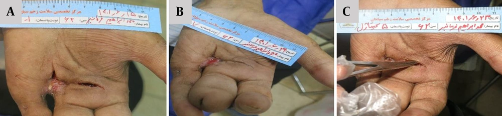
Figure 1.
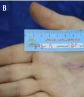
Clinical progression of diabetic wound healing in a 52-year-old female patient treated with Chitagel. A, Baseline presentation of a diabetic ulcer measuring approximately 4 × 1 cm with inflammatory changes and delayed surface coverage. B, Day 10 demonstrating wound closure, improved epithelial coverage, and reduced inflammatory signs.
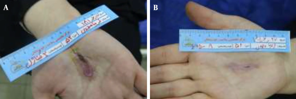
Figure 2.
Sequential clinical evaluation of diabetic foot ulcer healing in a 53-year-old female patient treated with Chitagel over a 10-day follow-up period. A, Baseline wound presentation showing an ulcer measuring approximately 10 × 6 cm with extensive tissue disruption and inflammatory changes. B, Day 4 demonstrating partial wound contraction and increased healthy granulation tissue development. C, Day 7 showing reduction in wound size and progressive surface coverage. D, Day 10 demonstrating near-complete wound closure with improved wound continuity.
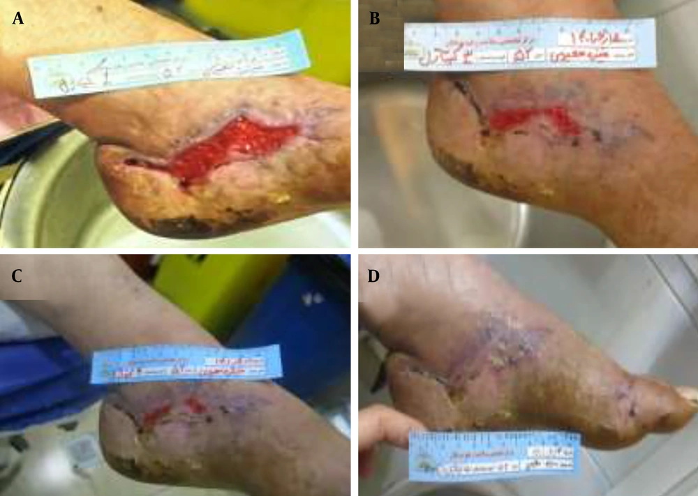
Figure 3.
Clinical progression of wound healing in a diabetic male patient treated with Chitagel. A, Baseline wound presentation demonstrating an approximately 4 cm2 ulcerative lesion with granulation tissue and inflammatory changes. B, Intermediate healing stage showing decreased wound size and improvement in tissue organization. C, Advanced healing stage demonstrating marked wound contraction and improved surface coverage.
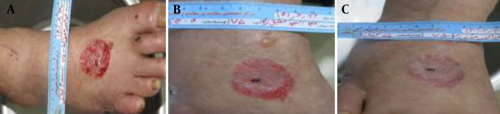
Figure 4.
Sequential wound-healing progression in a 60-year-old diabetic patient treated with Chitagel. A, Baseline wound presentation showing an approximately 4 × 3 cm ulcer with a depth of 0.5 cm associated with granulation tissue and mild inflammation. B-C, Intermediate healing stages demonstrating reduced wound depth, improved tissue continuity, and decreased inflammatory signs. D, Late healing stage showing progressive surface coverage and reduced wound dimensions. E, Final healing stage demonstrating near-complete wound closure.

Figure 5.
Clinical progression of diabetic wound healing in a 60-year-old patient treated with Chitagel. A, Baseline wound presentation demonstrating a 4 × 3 cm ulcerative lesion with irregular wound margins, inflammatory changes, and granulation tissue. B - C, Intermediate healing stages showing reduction in wound area and decreased inflammatory signs. D, Follow-up evaluation after three months demonstrating complete surface closure with minimal residual tissue disruption.
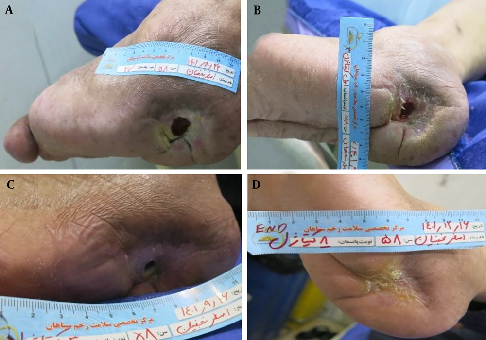
Figure 6.
Sequential wound-healing progression in a 69-year-old diabetic female patient treated with Chitagel. A, Baseline wound presentation demonstrating a 2 × 2 cm ulcer with a depth of approximately 0.5 cm associated with exposed tissue and mild inflammation. B, Day 7 demonstrating reduced wound area and progressive surface coverage. C-D, Day 11 showing complete wound closure with re-established tissue continuity and minimal residual scarring.
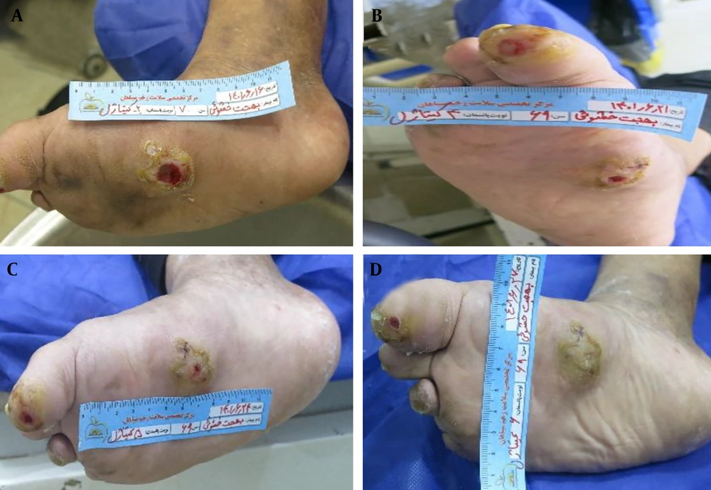
Figure 7.
Clinical progression of wound healing in a 62-year-old diabetic female patient treated with Chitagel. A, Baseline wound presentation demonstrating an ulcerative lesion with an approximate depth of 1 cm associated with inflammation and exposed tissue. B, Day 7 demonstrating granulation tissue formation, reduction in inflammatory signs, and partial wound contraction. C, Day 14 showing complete wound closure and improved wound continuity with minimal residual scarring.
4.1. Efficiency of Chitagel on Diabetic Wounds
Healing progression in diabetic wounds varied according to wound severity, vascular condition, metabolic status, and other patient-specific clinical factors (18). Despite these inherent challenges, consistent and clinically meaningful healing progression was observed in the majority of diabetic wounds during monitoring. Many wounds demonstrated notable reductions in inflammatory signs, robust granulation tissue formation, and progressive surface coverage. The following representative cases illustrate the favorable responses observed with Chitagel as an adjunct to standard wound care in this traditionally difficult-to-treat patient population (Figures 1-7). Serial evaluations demonstrated decreased inflammatory signs, improved wound-bed appearance, and progressive surface coverage in several cases. Healing progression varied between patients, with some wounds approaching complete closure within shorter treatment periods, whereas others required prolonged management, likely reflecting differences in wound chronicity and underlying metabolic status.
4.2. Efficiency of Chitagel on Non-Healing Wounds
Chronic non-healing wounds remain clinically challenging because of persistent inflammation, impaired vascularization, delayed tissue repair, and increased susceptibility to microbial colonization (19, 20). In the present study, elderly patients with chronic non-healing wounds treated with Chitagel in conjunction with standard wound care demonstrated gradual wound improvement during serial evaluations (Figures 8 and 9). Clinical observations included decreased inflammatory signs, progressive surface coverage, and improved wound-bed appearance over extended follow-up periods. Healing progression was generally slower than that observed in diabetic and post-surgical wounds, likely reflecting the chronic nature and underlying pathology of these lesions.

Figure 8.
Clinical progression of wound healing in an 80-year-old male patient with a chronic non-healing wound treated with Chitagel. A, Baseline wound presentation demonstrating an approximately 4 × 3 cm ulcerative lesion with granulation tissue, inflammatory changes, and irregular wound margins. B, Day 10 demonstrating reduction in wound dimensions, improved tissue continuity, and decreased inflammatory signs. C - D, Day 16 showing complete wound closure with minimal residual scarring.
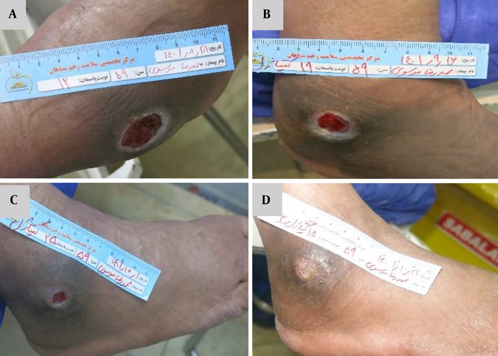
Figure 9.
Sequential clinical evaluation of wound healing in a 59-year-old male patient with a chronic non-healing wound treated with Chitagel. A, Baseline wound presentation demonstrating an approximately 4 × 4 cm ulcerative lesion with a depth of 0.5 cm associated with chronic inflammatory changes and irregular wound margins. B, Day 10 showing wound contraction and decreased inflammatory signs. C, Day 40 demonstrating further wound contraction with near-complete surface coverage. D, Day 54 showing complete wound closure with minimal residual scarring.
4.3. Efficiency of Chitagel on Post-Surgical Wounds
Post-surgical wounds are frequently associated with tissue disruption, inflammatory responses, exudation, and an increased risk of microbial contamination, all of which may delay tissue repair (21). In the present study, post-surgical wounds treated with Chitagel demonstrated encouraging, progressive tissue approximation and clear improvement in wound appearance. The following representative cases from 14 post-surgical wounds highlight early and robust tissue approximation, suggesting that Chitagel may be particularly beneficial in the post-surgical setting (Figures 10 and 11). Serial assessments demonstrated gradual wound contraction, decreased inflammatory signs, and improved wound-bed appearance over the treatment period. Earlier healing progression was generally observed in post-surgical wounds than in chronic non-healing wounds.
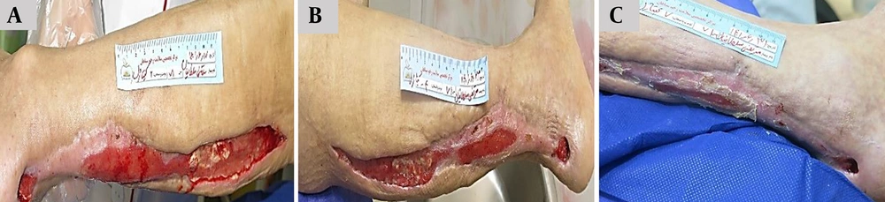
Figure 10.
Sequential clinical evaluation of post-surgical wound healing in a 71-year-old male patient treated with Chitagel. A, Baseline wound presentation demonstrating an approximately 22 × 5 cm post-surgical wound with a depth of 2 cm associated with exposed tissue surfaces and mild exudate. B, Day 10 demonstrating decreased wound size to approximately 18 × 3 cm with a depth of 1 cm, accompanied by decreased exudation and tissue approximation. C, Day 14 showing further wound reduction to approximately 10 × 1 cm with a depth of 0.7 cm and improved wound continuity.
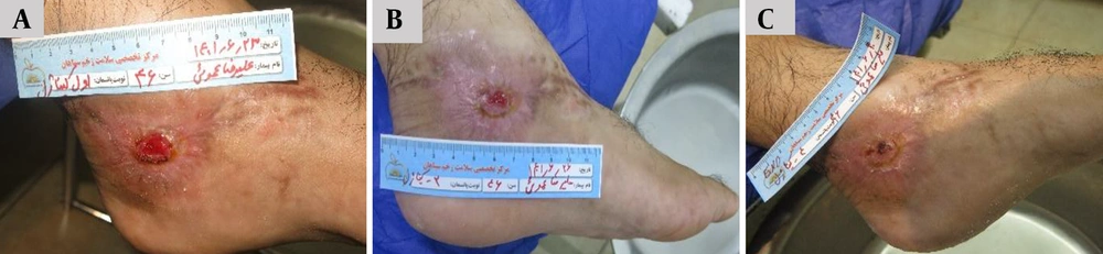
Figure 11.
Sequential clinical evaluation of post-surgical wound healing in a 46-year-old male patient treated with Chitagel. A, Baseline wound presentation demonstrating an approximately 2 × 2 cm surgical wound with visible granulation tissue, moderate inflammatory changes, and irregular wound margins. B, Day 4 demonstrating reduced wound area and decreased inflammatory signs. C, Day 7 showing complete wound closure with improved surface continuity and minimal residual scarring.
4.4. Efficiency of Chitagel on Trauma Wounds
Traumatic wounds, particularly in elderly patients, are frequently associated with extensive tissue disruption, an elevated inflammatory burden, increased infection risk, and delayed tissue repair (22). Effective management therefore requires infection control together with support for tissue recovery and wound-remodeling processes. A single illustrative case of traumatic wound healing is presented in this study. The clinical case presented in Figure 12 demonstrated progressive tissue repair in a 76-year-old male patient with a severe traumatic head injury treated with Chitagel in conjunction with standard wound care. Baseline evaluation demonstrated extensive soft-tissue injury with exposed tissue surfaces and disrupted skin integrity. Subsequent evaluations demonstrated reduced wound size and improved tissue continuity, with wound closure observed within 8 days of treatment.
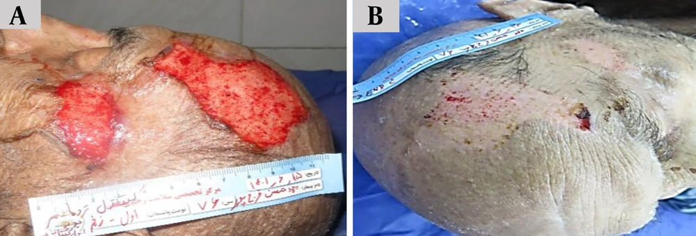
Figure 12.
Sequential clinical evaluation of traumatic wound healing in a 76-year-old male patient with severe head trauma treated with Chitagel. A, Baseline wound presentation demonstrating extensive soft-tissue injury with exposed tissue surfaces, active bleeding, and severe disruption of skin continuity. B, Follow-up evaluation demonstrating progressive wound closure and improved tissue continuity, with complete surface healing observed within 8 days of treatment.
5. Discussion
Chronic, difficult-to-heal wounds remain a major clinical challenge because of persistent inflammation, impaired vascularization, microbial colonization, delayed epithelial coverage, and disruption of normal tissue repair processes (23). These complications are particularly pronounced in diabetic, traumatic, post-surgical, and chronic non-healing wounds, in which delayed recovery frequently increases the risk of infection, prolongs hospitalization, and reduces quality of life. Therefore, the development of multifunctional wound-care formulations capable of simultaneously supporting antimicrobial protection, moisture balance, and tissue repair represents an important and timely area of clinical investigation.
In this prospective, single-arm clinical study, clinically meaningful wound improvement was observed across all wound categories following topical application of Chitagel in conjunction with standard wound care. Serial clinical assessments consistently showed progressive reductions in wound dimensions, notable improvements in wound-bed appearance, clear reductions in inflammatory signs, and visible progressive tissue repair during follow-up examinations. Although healing progression varied among patients and wound types, consistent and encouraging wound improvement was observed throughout the treatment period across the entire cohort of 80 patients.
The observed clinical responses are likely related to the unique multifunctional composition of Chitagel, which combines antimicrobial, moisturizing, anti-inflammatory, antioxidant, and tissue-supportive components within a single hydrogel formulation. Maintaining a moist wound-healing environment together with local antimicrobial protection may support tissue repair by facilitating cellular migration, reducing excessive exudation, and limiting microbial burden within the wound microenvironment. Furthermore, previous studies have reported that chitosan-based biomaterials and E. purpurea extracts contribute favorably to inflammatory modulation and tissue remodeling processes involved in wound healing (13), aligning with our observations.
These findings are broadly consistent with previous experimental and biomaterial-based wound-healing studies reporting beneficial effects of multifunctional hydrogel systems in supporting tissue repair, granulation tissue development, and wound closure in chronic wounds. Although direct comparisons among studies remain challenging because of differences in wound etiology, patient characteristics, wound severity, treatment duration, and outcome assessment methodologies, the convergence of evidence strengthens the rationale for further investigation of Chitagel.
Healing progression varied among wound categories, with diabetic and post-surgical wounds generally showing earlier tissue repair than chronic non-healing wounds, reflecting the underlying pathophysiology rather than a limitation of the intervention. Importantly, reductions in inflammatory signs, exudation, and wound-bed irregularity were observed in most patients during serial follow-up evaluations (Table 2), underscoring the broad applicability of Chitagel across different chronic wound types.
Consecutive recruitment was used to minimize selection bias; however, the absence of a control group and blinding means that residual selection and observer bias cannot be excluded. In summary, consistent and clinically observable wound improvement was noted across multiple representative cases treated with Chitagel plus standard care. Although the uncontrolled, unblinded, descriptive design does not permit causal attribution, these encouraging and reproducible observations provide a strong, evidence-based rationale for further investigation. These findings represent valuable, hypothesis-generating evidence that can meaningfully inform the design of future randomized controlled trials. We are confident that this preliminary work will serve as a solid foundation for the rigorous evaluation of Chitagel as a promising adjunctive therapy for chronic and difficult-to-heal wounds.
5.1. Conclusions
This prospective, single-arm, descriptive clinical study demonstrated clinically meaningful, progressive wound healing in representative cases of diabetic, chronic non-healing, post-surgical, and traumatic wounds following topical application of Chitagel together with standard wound care. Although the study was uncontrolled and hypothesis-generating by design, the consistent and observable improvements across multiple difficult-to-heal wound types are highly encouraging. These findings strongly suggest that Chitagel has promising therapeutic potential as an adjunctive wound-care formulation. The results provide a solid rationale for advancing to larger, randomized, controlled, blinded trials with standardized quantitative outcome measures to confirm efficacy and establish the clinical value of Chitagel in chronic wound management.
5.2. Clinical Implications and Future Directions
These observations, despite their descriptive and uncontrolled nature, provide encouraging preliminary evidence that Chitagel may be a valuable candidate for further investigation in rigorous controlled trials. Progressive wound contraction, improved wound-bed appearance, and gradual surface coverage were consistently observed across multiple chronic and difficult-to-heal wound categories, including diabetic foot ulcers, post-surgical wounds, traumatic injuries, and chronic non-healing wounds of various etiologies. These findings are particularly noteworthy given the well-recognized challenges of these wound types, including persistent inflammation, impaired vascularization, delayed epithelialization, and a high risk of infection.
It is essential to interpret these results in the context of the study design limitations. This investigation was conducted as a prospective, single-arm, descriptive clinical study without a concurrent control group, without blinding of outcome assessors, and without systematic collection of quantitative, cohort-level wound measurement data.
Nevertheless, these limitations do not diminish the clinical value of the observations. Rather, they position this work as a legitimate hypothesis-generating study that serves an essential role in the translational research pathway. The consistent pattern of improvement across a substantial number of patients (n = 80) with diverse, real-world chronic wounds provides a strong empirical rationale for advancing to more definitive study designs. Future investigations should focus on large, multicenter, randomized controlled trials with blinded outcome assessment, standardized quantitative measures such as calibrated digital planimetry, longer follow-up periods, and, where possible, comparative effectiveness arms. In summary, while this preliminary study does not provide definitive evidence of efficacy, it provides clinically meaningful signals that strongly support the continued development of Chitagel as a potentially valuable adjunctive wound-care formulation for chronic and difficult-to-heal wounds. We are optimistic that the wound-care research community will build upon these observations in well-designed, adequately powered trials.