
1. Introduction
Chronic exposure to excess glucocorticoids results in a set of clinical pictures first described by Harvey Cushing as Cushing's syndrome (CS) (1). This rare disease occurs in 2 to 3 cases per 1000000 people every year (2). Glucocorticoids, either exogenous or endogenous, can suppress luteinizing hormone, affect follicular development, and impair ovulation, making pregnancy in CS unlikely (3, 4). CS mainly affects women (5), especially those of childbearing age (6). Although this case is rare, pregnancy can happen in women with CS (6), and to date, only 220 cases of pregnancies with CS have been reported (7). CS imposes a significant risk of morbidity and mortality on both the mother and the fetus (8). As medical or surgical interventions can improve outcomes, CS must be detected early (6). Here we report a case of CS with adrenocortical adenoma in pregnancy with the very rapid presentation of signs and symptoms of a typical CS with a review of current literature.
2. Case Presentation
A 22-year-old patient was diagnosed with hypertension at GA 18, hypothyroidism at GA 18, and GDM at 18 weeks of gestation and was referred to our nephrology department to control hypertension. Preeclampsia was previously ruled out; liver function tests and rheumatologic workups were regular; she was on insulin, levothyroxine, ASA, and enoxaparin. Nothing was remarkable in her previous medical, obstetrical, social, and family history. She was obese (BMI = 30) and had generalized muscle weakness on physical examination with no other abnormal findings. Her blood pressure was 150/90, and on her laboratory workup, she had potassium of 2.8, magnesium of 1.7, FBS of 110, pH of 7.,5, and HCO3 of 29. Due to hypokalemia and metabolic alkalosis besides hypertension, renin, and aldosterone levels were checked, and both were suppressed.
Her hypertension was corrected with Methyldopa and Diltiazem (Amiloride was unavailable). In addition, hypokalemia and hypomagnesemia were treated with K and Mg supplementations. The patient was discharged in good condition while receiving anti-hypertensive medications, insulin, and OPD follow-up with 24-hour urine free cortisol (UFC) test result.
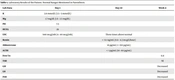
Ten days later, she had a UFC report three times above the upper limit of the normal range. During this 10-day interval, she had gained a significant amount of weight and developed a moon face, acne, coracoclavicular fat pad, and purple striae on her abdomen, all typical of Cushing's syndrome. She had developed a fungal infection in her armpit and inframammary fold and complained of right-sided hip pain and ipsilateral limb weakness. Preeclampsia was again ruled out, and renin, aldosterone, ACTH, and UFC were checked (Table 1).
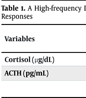
Table 1.
Laboratory Results of the Patient a
| Lab Data | Day 1 | Day 10 | Week 4 |
|---|---|---|---|
| K | 2.8 mmol/L (3.5 - 5 mmol/L) | ||
| Mg | 1.7 mg/dL (1.6 - 2.5 mg/dL) | ||
| pH | 7.5 | ||
| HCO3 | 29 | ||
| UFC | 940 mcg/24h (4 - 40 mcg/24h) | Three times above normal | |
| Renin | < 0.1 ng/mL (0.6 - 4.3 mcg/L/hour) | ||
| Aldosterone | 16 pg/mL (< 150 pg/mL) | ||
| ACTH | < 1 pg/mL (10 - 60 pg/mL) | ||
| Free T4 | 0.8 | ||
| TSH | Normal | ||
| GH | Decreased | ||
| LH | Decreased | ||
| FSH | Decreased |
a Normal ranges mentioned in parenthesis.
A hypoechoic solid mass measuring 41.5 mm by 30 mm without vascular flow was observed on abdominal sonography at the anatomic site of the right adrenal gland. The left adrenal gland was normal on sonography. Adrenolytic agent Mitotane was started, but she developed hypertension, thrombocytopenia, elevated liver enzymes, elevated LDH levels, headache, hemoptysis, alveolar hemorrhage, and blurred vision. The fetus's FHR also decreased and became oligohydramnios. HELLP was diagnosed at 22 weeks gestation, resulting in an emergency cesarean section.
The newborn developed respiratory distress, got intubated, and died a few hours later. The mother was admitted to ICU, but she was alert and conscious. She received antibiotics, platelet transfusion, and IVIg. After the treatment, thrombocytopenia, pulmonary symptoms, and liver function tests got corrected. PCR for COVID-19 was negative two times. Her hip MRI showed no evidence of avascular necrosis or fracture. Brain MRI, neurologic examinations, and color Doppler sonography of both lower extremities were regular, and the ophthalmologic test was compatible with hypertension. Her echocardiography showed left ventricular hypertrophy but a good ejection fraction. TSH and T4 levels were normal, but GH, LH, and FSH were decreased. Abdomen and pelvic CT scan showed a well-defined hypo dense mass with diameters of 41 × 31 mm in the right adrenal gland with HF = 30 and washout = 33%, suggesting adrenocortical carcinoma or pheochromocytoma. 24-hour urinary catecholamines, metanephrine, and normetanephrine were within normal range. Therefore, pheochromocytoma was ruled out, and she underwent right-side adrenalectomy. After the operation, she was treated with hydrocortisone, spironolactone, and insulin. Her hypertension, hypokalemia, and hyperglycemia were all corrected, and she was discharged in good condition. The 24h UFC was average seven days later in the outpatient follow-up. She was admitted again to the hospital due to a wound infection but was discharged on oral antibiotics in good condition.
3. Discussion
According to the American College of Obstetricians and Gynecologists (ACOG), hypertension detected in pregnancy before 20 weeks of gestation is considered chronic hypertension (9). Chronic hypertension complicates about 1 - 2% of pregnancies (10). More than 90% of chronic hypertension in pregnancy is due to primary hypertension, which is genetic or lifestyle-related, and only 10% of cases are due to secondary causes, including renovascular, endocrine, and respiratory (11). Although prudent, screening secondary causes in all pregnancies complicated with hypertension is not cost-effective. Still, such etiologies must be considered, especially when accompanied by related signs and symptoms (11).
A triad of hypertension, hypokalemia, and metabolic alkalosis is mineralocorticoid hypertension. This condition has a broad endocrine differential diagnosis: (1) hyperaldosteronism; due to primary aldosteronism, adrenal adenoma, bilateral adrenal hyperplasia, and adrenal carcinoma. (2) hypercortisolemia, as cortisol has a high affinity for mineralocorticoid receptors because of: Cushing’s syndrome, syndrome of apparent mineralocorticoid excess (SAME), congenital adrenal hyperplasia (CAH) (12), and (3) receptor dysfunctions, Liddle syndrome, Chrousos syndrome, and Geller syndrome (13).
3.1. Primary Hyperaldosteronism
Primary aldosteronism (PA) is a genetic condition with continuous, autonomous excessive production of aldosterone and is the most common cause of secondary hypertension. The excess aldosterone causes mineralocorticoid hypertension (14). There have been courses of normotensive pregnancies followed by postpartum hypertension, further diagnosed as PA (15). PA type 1 is a familial autosomal dominant disease that results from the fusion of genes encoding cortisol in response to ACTH and aldosterone in response to angiotensin. Therefore, aldosterone is produced in response to ACTH, and this type of PA is responsive to glucocorticoids, named glucocorticoid remediable aldosteronism (GRA) (16).
3.2. Cushing's Syndrome
Cushing's syndrome results from primary pituitary disease, adrenal hyperplasia, or adenoma. Despite the first two etiologies, androgen excess does not occur in adrenal adenoma. Therefore, pregnancy is likely to happen (8), and recent reviews have demonstrated that most cases of Cushing syndrome in pregnancy are due to adrenal adenoma (40 - 60%) (17). Another explanation is that subclinical LH receptor-bearing adrenal adenomas get stimulated by placental HCG and become clinically apparent (17).
Although abdominal obesity, wide purple striae, especially on the abdomen, proximal muscle weakness, and easy bruising suggest cortisol excess, they are not always present in CS (2, 18). Non-specific signs and symptoms of CS include hypertension, weight gain, glucose intolerance, back pain, and lethargy, which could be separated or as a part of other clinical entities, including pregnancy (18, 19).
Hypothalamic-pituitary-adrenal axis over activity normally happens in pregnancy (8). Together with placental CRH (17), hypercortisolemia becomes evident from the first trimester and remains so until the end of pregnancy. Therefore, a misinterpretation of overnight high-dose dexamethasone suppression test and UFC results is created. But still, salivary cortisol level remains the best diagnostic tool as salivary cortisol level does not change during pregnancy (8).
Complications that hypercortisolism imposes on pregnancy include the following: hypertension (68%), diabetes (25%), preeclampsia (14%), osteoporosis and fractures (5%), heart failure (3%), and maternal death (2%). The most common fetal complications are prematurity (43%), intrauterine growth restriction (21%), stillbirth (6%), spontaneous abortion or intrauterine death (5%), and hypoadrenalism (2%) (17). However, higher fetal survival rates were observed in women treated during pregnancy(3).
All these mean that Cushing’s syndrome should be considered in any pregnant patient with hypertension, hypokalemia, easy bruising, and other related symptoms.
3.3. Syndrome of Apparent Mineralocorticoid Excess
Syndrome of apparent mineralocorticoid excess is caused by either genetic inactivation mutation of 11ß-hydroxysteroid dehydrogenase (11β-HSD2) (16), converting cortisol to its inactive form, or by acquired inhibition of 11β-HSD2 in chronic or high dose licorice ingestion. Elevated cortisol binds to mineralocorticoid receptors for a long time, producing hypertension, hypernatremia, and hypokalemia (16, 20).
3.4. Congenital Adrenal Hyperplasia
The more common enzyme deficiencies that can cause congenital adrenal hyperplasia are 21-hydroxylase deficiency, 11β-hydroxylase deficiency, and 17α-hydroxylase deficiency, which the latter can cause hypertension, hypernatremia, and hypokalemia. This effect is brought by excess amounts of deoxycorticosterone (DOC) with mineralocorticoid properties. In opposition to 17α-hydroxylase deficiency, 11β-hydroxylase deficiency is associated with androgen excess, in amounts to cause ambiguous genitalia in females. However, non-classic forms of 11β-hydroxylase deficiency have less androgen excess (16).
3.5. Liddle's Syndrome
Liddle's syndrome is a familial autosomal dominant disorder causing hypertension, hypernatremia, and hypokalemia. The disease is caused by activating the mutation of ENaC epithelial channels. The disease is usually diagnosed in early childhood, but patients may not be recognized until early adulthood (16). Cases of Liddle's syndrome first identified during pregnancy have also been reported, and both were heterozygotes for the disease (21). As mineralocorticoids do not activate ENaC, spironolactone is ineffective for treatment, and the disease is managed by amiloride. Management of Liddle syndrome in pregnancy with amiloride has been reported (22, 23).
3.6. Geller Syndrome
Geller syndrome is an excessively rare gain of function mutation in mineralocorticoid receptors. These receptors are normally activated by aldosterone and inactivated by progesterone. In Geller syndrome, these receptors are activated by progesterone, causing hypertension in a situation like pregnancy. These patients have characteristic features of mineralocorticoid hypertension (16).
3.7. Chrousos Syndrome
Chrousos syndrome is an inherited condition with generalized resistance to glucocorticoids. As a result of this resistance, these patients have high levels of ACTH and, therefore, aldosterone and androgens, presenting as hypertension, hypokalemia, and sign and symptoms of androgen excess (24), similar to patients with 11β-hydroxylase deficiency. These patients do not respond to the dexamethasone suppression test (24), contrary to GRA.
3.8. Conclusions
Based on the observations, maternal and fetal complications of a pregnancy complicated by Cushing syndrome remain a significant concern. Our patient presented only with hypertension, obesity, and GDM, and then only in 10 days showed other more characteristic signs and symptoms of Cushing syndrome. Although this case is rare, it is essential to consider Cushing syndrome as a cause of uncontrolled hypertension, GDM, and obesity.