1. Introduction
Central giant cell granuloma (CGCG) is an uncommon intraosseous jaw lesion composed of cellular fibrous tissue with multiple areas of hemorrhage, accumulations of multinucleated giant cells, and occasional immature bone trabeculae (1). It is predominantly found in the jawbones, particularly the mandible, and accounts for approximately 7% of benign mandibular tumors. It typically affects individuals younger than 30 years and is more common in females. Clinically, CGCG may be asymptomatic or may present with edema, pain, and, in some cases, tooth displacement or loosening (2).
The pathogenesis of jaw CGCG is unknown and controversial; however, infection, local reactive responses to trauma, growth factors, and a neoplastic origin have been proposed. According to the updated World Health Organization definition, CGCG is a benign but sometimes invasive osteolytic proliferation (3). Clinically and radiographically, this lesion can present with a wide range of features, mimic other lesions, and complicate diagnosis. In terms of behavior, CGCG is classified as invasive or noninvasive. The invasive type may grow rapidly, invade surrounding tissues, and have a higher risk of recurrence. Typically, this lesion arises in the anterior mandible and commonly crosses the midline, whereas maxillary involvement is much less common (4).
Extensive maxillary involvement by CGCG is very rare and can create diagnostic and therapeutic challenges. This report describes an unusual case of CGCG with extensive maxillary extension, reviews its clinical, radiographic, and pathological findings, and discusses diagnostic and therapeutic considerations.
2. Case Presentation
The patient was a 40-year-old woman with no history of underlying disease and no specific family history who presented with progressive swelling on the left side of the maxilla.
Written informed consent was obtained from the patient for treatment and publication of this case report. All clinical images were anonymized before submission, and no patient name or identifying personal information was included. The patient reported that symptoms began approximately five years before the current presentation, initially as pain in the left maxillary region. The symptoms were initially considered odontogenic in origin, and dental treatment, including endodontic therapy and antibiotic administration, was performed. Approximately three years before the current presentation, an incisional biopsy established the diagnosis of CGCG. However, definitive surgical excision was not performed, and only limited tissue sampling was obtained. Regular follow-up was not maintained after diagnosis. The patient subsequently developed progressive swelling and facial asymmetry, prompting the current presentation. Previous radiographic records and histopathological materials were unavailable for review.
2.1. Intraoral Examination
Intraoral examination revealed a nontender swelling measuring 4 x 6 cm, with no discharge and no change in the overlying skin. The patient did not report neurological symptoms, such as numbness, or a history of trauma (Figure 1).
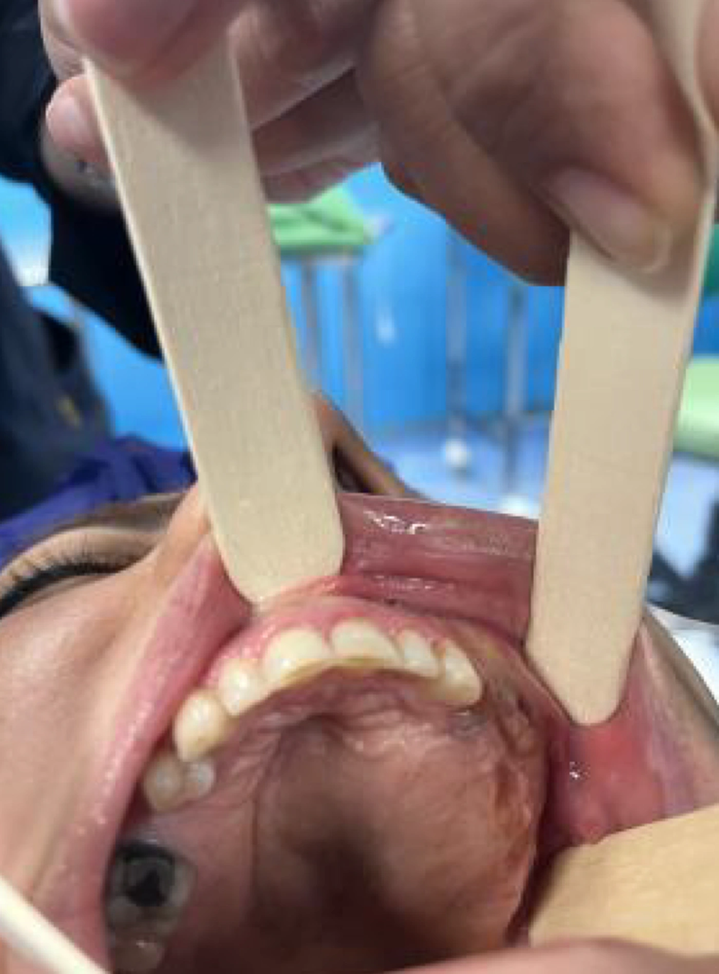
Figure 1.
Intraoral clinical photograph showing buccal swelling and expansion in the left maxillary region. The image was anonymized and contains no identifying patient information.
Extraoral examination revealed facial asymmetry manifested as left-sided prominence. Clinical examination showed partial obstruction of the left nostril. No ulceration, discharge, or skin discoloration was observed. Lymphadenopathy was absent. Laboratory tests, including calcium, phosphorus, parathyroid hormone, and alkaline phosphatase levels, were all normal, ruling out a metabolic lesion or hyperparathyroidism.
2.2. Radiographic Features
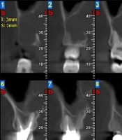
Cone-beam computed tomography revealed a large, radiolucent, ill-defined, space-occupying lesion in the maxilla that extended bilaterally (Figure 2). The lesion originated from the left maxillary canine and extended posteriorly to the left pterygoid plates. It also horizontally encompassed the entire palate and nasal floor and involved the posterior right maxilla. The lesion was associated with complete involvement of the right and left maxillary sinuses, destruction of the nasal septum, destruction of the alveolar ridge crest and the left buccal and palatal cortical plates, and extension into the adjacent soft tissue. Resorption of the left canine and right second molar roots was also observed (Figure 3). A notable finding was the presence of scattered calcifications in the lesion bed and thin, wispy septa within the lesion (Figure 4).
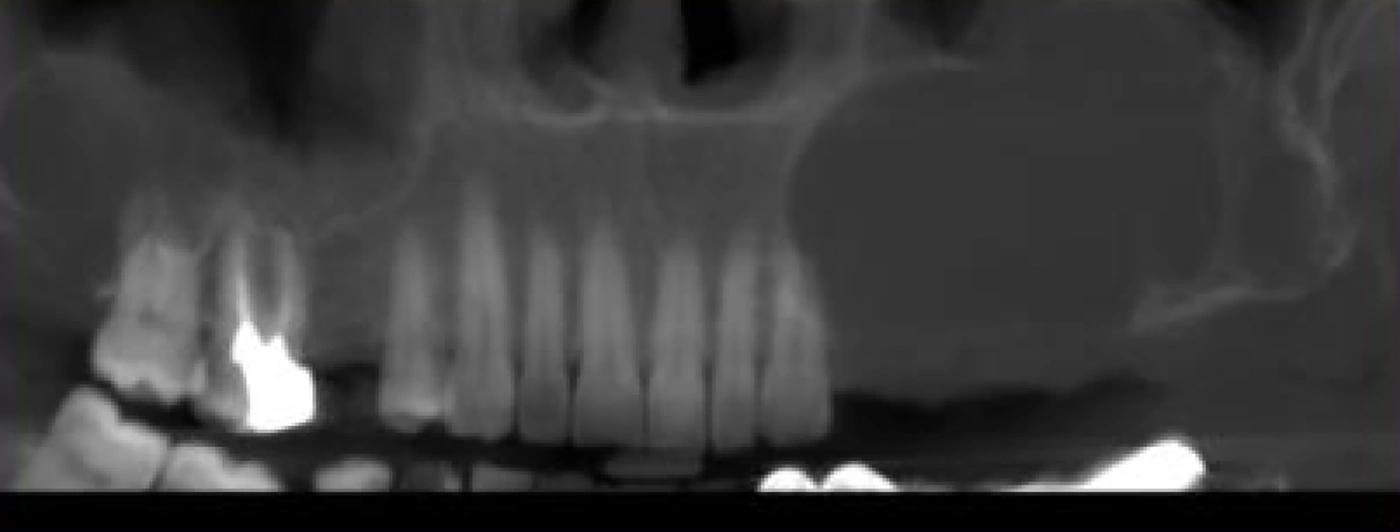
Figure 2.
Panoramic radiograph demonstrating bilateral maxillary expansion caused by the lesion, with involvement extending across the maxillary region.
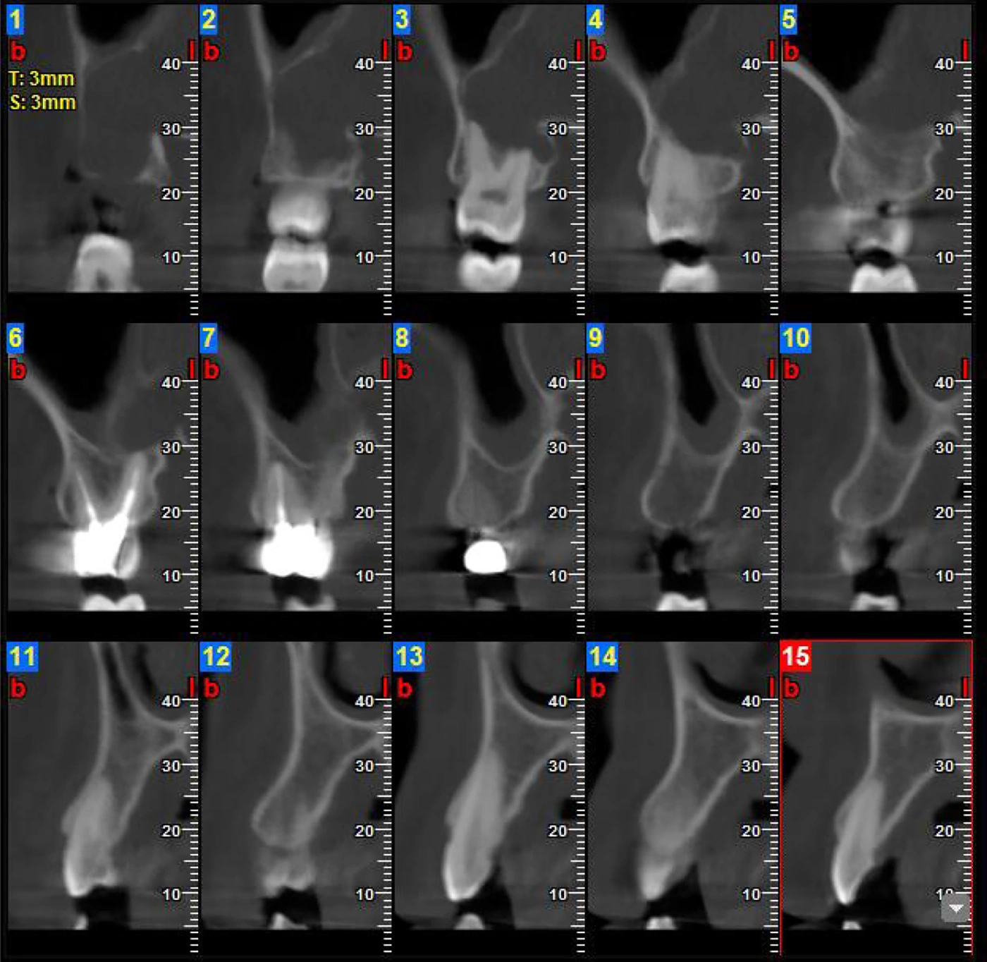
Figure 3.
Selected CBCT cross-sectional images showing root resorption of the maxillary second molar adjacent to the lesion.
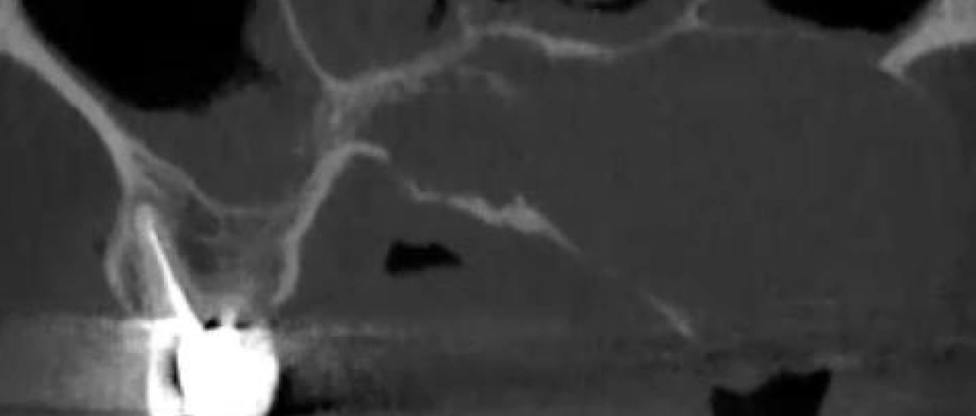
Figure 4.
Radiographic image demonstrating a thin wispy internal septum within the maxillary lesion, a characteristic internal radiographic feature of the lesion.
The suggested differential diagnoses included infected ameloblastoma, CGCG, and malignancy.
Although CBCT enabled detailed evaluation of the lesion’s osseous extent, including cortical destruction and involvement of adjacent anatomical structures, assessment of soft-tissue extension was limited. Additional imaging modalities, such as MRI or CT, could have provided a more comprehensive evaluation of soft-tissue and vital-structure involvement; however, these studies were not performed in the present case and represent a limitation of this report. Nevertheless, the extensive osseous involvement observed on CBCT played a major role in treatment planning and contributed to the selection of a conservative surgical approach to minimize morbidity.
2.3. Pathological Findings
Previous histopathological slides from the biopsy performed three years earlier were not available for review.
The samples sent for microscopic examination included multiple gray-brown tissue fragments with a total size of 1.8 x 1.5 x 0.5 cm and a beige-brown color.
Microscopic examination of hematoxylin and eosin-stained sections revealed numerous multinucleated giant cells diffusely distributed throughout the lesion. These cells varied in size and shape and were present within a background of ovoid to spindle-shaped mononuclear stromal cells. Areas of erythrocyte extravasation and hemosiderin deposition were also observed (Figure 5 and 6). Based on the histopathological findings, together with clinical, radiographic, and laboratory correlation, a final diagnosis of CGCG was established.
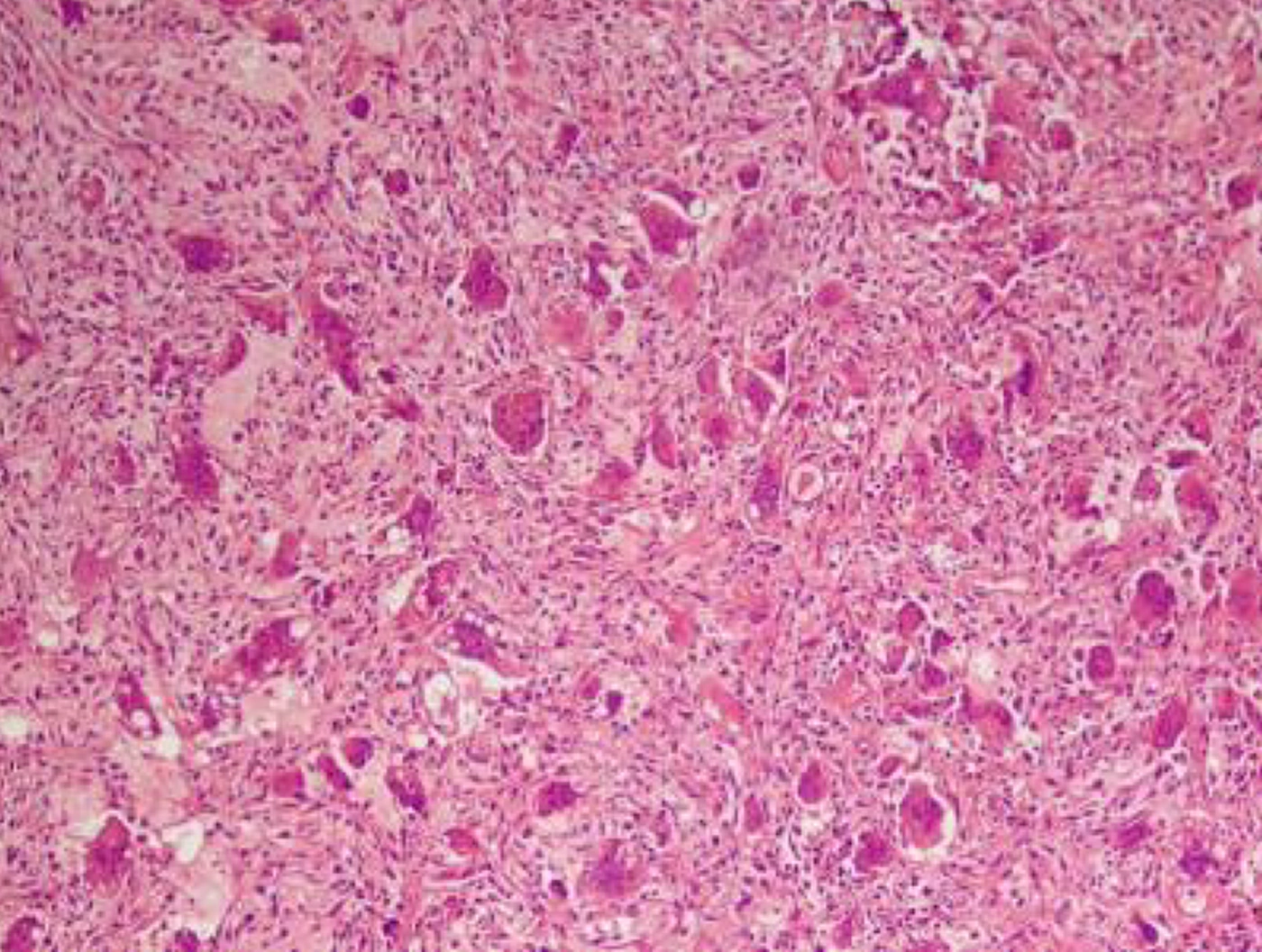
Figure 5.
Histopathological section showing multinucleated giant cells distributed within a fibrocellular stroma with erythrocyte extravasation and hemosiderin deposition (hematoxylin and eosin staining, original magnification x100).
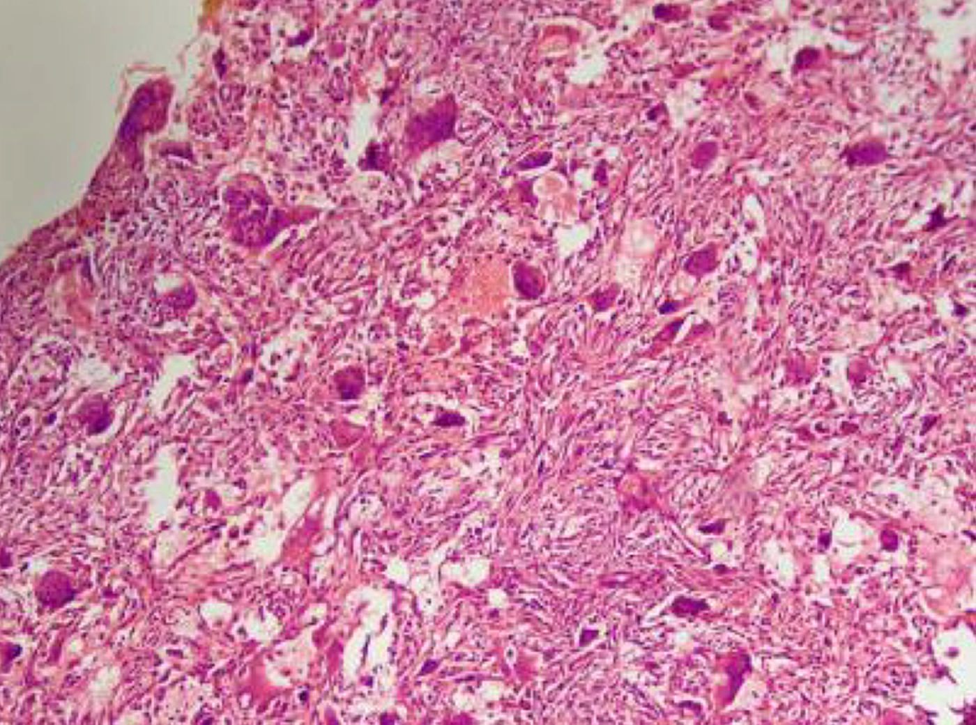
Figure 6.
Histopathological section at higher magnification showing multinucleated giant cells within a fibrocellular stromal background with hemorrhagic areas (hematoxylin and eosin staining, original magnification x200).
Hyperparathyroidism-associated brown tumor was excluded because serum calcium, phosphorus, alkaline phosphatase, and parathyroid hormone levels were within normal limits. Immunohistochemical analysis was not considered necessary because the diagnosis could be established by clinicopathological correlation. Other giant-cell-rich lesions, including aneurysmal bone cyst and malignant lesions, were considered less likely based on overall correlation of the histopathological findings with the clinical and radiographic features.
Malignancy was considered in the initial differential diagnosis because the lesion showed aggressive radiographic features, including ill-defined borders, extensive bone destruction, cortical perforation, root resorption, soft-tissue extension, and involvement of adjacent anatomical regions. These findings can overlap with aggressive odontogenic tumors and malignant maxillofacial lesions. Nevertheless, malignancy was considered less likely after clinicopathological correlation. Clinically, there was no cervical lymphadenopathy, ulceration, skin discoloration, or systemic sign suggestive of malignancy. Laboratory findings, including calcium, phosphorus, alkaline phosphatase, and parathyroid hormone levels, were within normal limits. Histopathological findings showed multinucleated giant cells in a fibrocellular stromal background with erythrocyte extravasation and hemosiderin deposition. Based on the combination of clinical, radiographic, laboratory, and histopathological findings, the final diagnosis of central giant cell granuloma was established.
2.4. Treatment and Follow-Up
Treatment planning was performed through discussion between the surgical and radiology teams. Due to the aggressive behavior and large size of the lesion, complete surgical excision with adequate margins was not feasible because of its proximity to critical anatomical structures, including the eyeball. Therefore, surgical debulking was performed to reduce lesion volume while preserving surrounding structures. Based on the operating surgeon’s clinical judgment, a single postoperative intralesional injection of triamcinolone acetonide (40 mg/mL; total dose, 40 mg; 1 mL) was administered as adjunctive therapy to decrease residual lesion activity and minimize the risk of further progression. Intralesional triamcinolone was preferred over alternative medical approaches, such as calcitonin, interferon-alpha, and denosumab, because of its less invasive nature, availability, cost-effectiveness, and lower risk of systemic adverse effects. The lesion was accessed through the left maxilla, the initial site of tumor initiation, and the mass was evacuated as much as possible. Postoperative follow-up evaluations at two weeks and one month demonstrated satisfactory healing, reduced facial swelling, improved nasal obstruction, and partial restoration of facial symmetry. No postoperative pain, infection, wound complications, or impairment of oral function was observed. Radiographic follow-up imaging was not obtained during this early postoperative period. Further follow-up, including a planned six-month evaluation, was scheduled but had not been completed at the time of manuscript preparation.
3. Discussion
Central giant cell granuloma is a rare, non-neoplastic lesion that occurs predominantly in the mandible and at a younger age. Approximately 70% of cases occur in the mandible, most often in the anterior region, whereas maxillary lesions are less common and are usually confined to the anterior region. The present case is noteworthy because it showed extensive, bilateral, and aggressive maxillary involvement extending to the maxillary sinuses, nasal septum, and pterygoid plates and involving large areas of the palate, features that have rarely been described in previous reports (2).
The imaging findings of this lesion, including extensive radiolucency, scattered calcifications, wispy septa, and multiple root resorptions, suggested a classic pattern of invasive CGCG; however, there was considerable overlap with lesions such as ameloblastoma, fibro-osseous lesions, and malignant neoplasms. Therefore, an accurate differential diagnosis is possible only through a combination of clinical, radiographic, and histopathological findings (2, 5).
Pathophysiologically, the exact mechanism of CGCG development remains unclear; however, several hypotheses have been proposed, including inflammatory responses, trauma, and vascular disorders (3). Given the high recurrence rate of this lesion, long-term follow-up and selection of an appropriate treatment plan are essential to prevent lesion re-expansion (6).
According to the seminal classification proposed by Chuong et al. (7), CGCG can be divided into invasive and noninvasive forms. This classification continues to be widely referenced and is supported by more recent literature on the biological behavior and management of CGCG. The aggressive type is usually associated with rapid growth, bone destruction, pain, and root displacement or resorption and carries a higher risk of recurrence (7). Treatment of these lesions remains controversial. Although simple curettage is sufficient for small and nonaggressive CGCGs, in cases such as the present case, in which the lesion is invasive and extensive, wide en bloc resection with a safe margin of healthy tissue is recommended. Some studies have also suggested that microperforation of the bone margin with a diamond bur can reduce the risk of recurrence (8, 9).
Although the present report focuses specifically on central giant cell granuloma of the maxilla, recent reports from related medical and dental fields highlight the importance of considering systemic, inflammatory, and multidisciplinary aspects when evaluating extensive maxillofacial lesions. Large facial lesions may present considerable diagnostic and cosmetic challenges, as shown in recent case-based literature (10). In addition, systemic and immune-related factors have been discussed in relation to oral and medical conditions, emphasizing the need for careful medical history assessment and comprehensive clinical evaluation (11, 12). These considerations support the importance of a multidisciplinary approach to the diagnosis and management of extensive maxillofacial lesions such as the present case.
In recent years, pharmacological treatments have received increasing attention. Intralesional corticosteroids, calcitonin, interferon-alpha, and, more recently, denosumab have been proposed as antiosteoclast drugs for larger lesions. However, medium- and long-term studies on the efficacy of these drugs are limited, and most of these treatments require adjunctive surgical intervention (13).
The present case demonstrated an unusually extensive and aggressive CGCG of the maxilla that posed considerable diagnostic and therapeutic challenges. Given the associated destruction of vital structures and the history of prior swelling, this case is rare and valuable for improving understanding of the clinical behavior of CGCG and its treatment challenges. It also emphasizes the importance of thorough multidisciplinary clinical, radiographic, and histopathological evaluation for selecting an appropriate treatment plan.
3.1. Conclusions
Central giant cell granuloma is a lesion with variable biological behavior that, in rare cases, can occur aggressively with extensive involvement of the maxilla. The present case, with bilateral extension, destruction of vital maxillary structures, and a history of recurrence, represents a rare example of this disease, for which diagnosis and treatment required a multidisciplinary approach, including careful clinical, radiographic, and histopathological examination.
Early postoperative clinical follow-up showed improvement in swelling and nasal obstruction without treatment-related adverse effects; however, long-term recurrence, progression, stabilization, or regression could not yet be determined because the planned six-month follow-up had not been completed at the time of manuscript preparation.
Given the aggressive nature of the lesion, appropriate treatment planning and regular follow-up are essential to prevent recurrence.