The etiology of osteoma is not clearly understood, and embryologic, traumatic, and infectious theories are proposed (
8,
9). Osteoma may arise as a result of infection stimulating osteoblasts, which in turn may become secondarily calcified (
10). Our patient had chronic infection symptoms related to maxillary sinusitis. Long-standing chronic maxillary sinusitis in pediatric patients results in a decrease in the volume of the maxillary sinuses and an increase in bony thickness of paranasal sinuses (
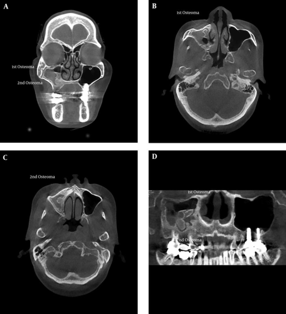
11). In this case, bony changes due to chronic maxillary sinusitis were seen. CBCT revealed sclerotic bony thickening and an irregular border of the inner wall of the right maxillary sinus, which indicated periosteal new bone formation due to sinusitis (
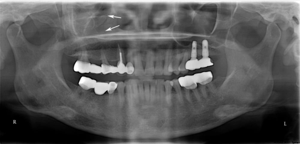
Figure 2). Two radiopacities with a partially corticated border in the right maxillary sinus were also detected.
The differential diagnosis of antral radiopaque lesions in this case included antrolith, osteoma, complex odontoma, and calcification in chronic maxillary sinusitis.
Retrospective studies of antroliths showed that most cases were related to sinus infection. Antrolith may occur around a nidus that continues to grow by deposition of mineral salts (
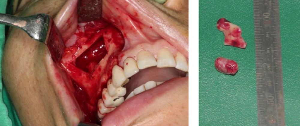
12). It could have a pitted or laminated surface. In this study, the removed specimens had a smooth surface (
Figure 3), which indicated a possible osteoma, complex odontoma, or intrasinus calcification with fungal sinusitis.
In a previoius study (
13), the prevalence of intrasinus calcifications on CT scans in patients with sinonasal fungus ball has been reported as 67.2% (116 cases). The calcifications were located mostly in the center of the maxillary sinus (85.9%), followed by near the sinus wall (7.7%). Nodular (53.8%), linear (20.5%), mixed (11.5%), round (10.3%), and fine punctate (3.8%) calcifications are seen with fungal sinusitis. A round-shaped, intrasinus calcification with maxillary sinusitis may be similar in appearance to an osteoma, but dystrophic calcification in sinusitis is separated from the sinus wall.
In this case, heterogeneous radiopacities with partially corticated shell were attached to the medial wall of the right maxillary sinus on coronal CBCT images, which was helpful in the differential diagnosis from an intrasinus calcification due to maxillary sinusitis.
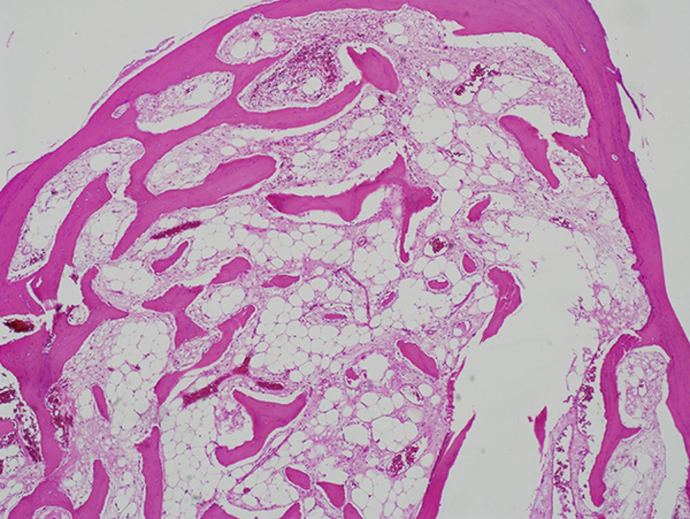
Histologically, osteoma has three variants: compact osteoma (dense bone with minimal marrow space), cancellous osteoma (bony trabeculae and fibrofatty marrow), and mixed type osteoma (
1,
2,
14). The present case was of a mixed type osteoma.
Symptoms of paranasal sinus osteoma are variable, and are associated with the size, location, and growth rate of the tumor. Symptomatic osteomas and large (extending to more than 50% of the sinus) or growing osteomas should be surgically removed (
15). However, management of asymptomatic osteoma is controversial (
10).
The management options for paranasal sinus osteomas include surgical endoscopic surgery, an external approach, or a combined approach using both procedures. The type of surgical procedure is determined by lesion size and location (
16). Recurrence of osteomas after surgical removal is rare, but incomplete resection of an osteoma could lead to regrowth (
17). There are no guidelines on postoperative follow-up. However, periodic radiological evaluation is recommended after incomplete resection (
18).
Other than in Gardner’s syndrome, which is characterized by intestinal polyposis, multiple osteomas, cutaneous fibromas, epidermal cysts, and impacted supernumerary teeth, osteomas of the craniofacial bones are solitary lesions (
7,
19).
Buyuklu et al (
2). reported that only two patients had multiple osteoma in 17,154 paranasal sinus CT scans of 14,137 patients. However, the lesions were not found in the maxillary sinus but in frontal and ethmoidal sinuses (
2).
To the best of our knowledge, double osteoma of a single maxillary sinus is very rare. Our patient did not have Gardner’s syndrome but double osteoma coexisting with sinusitis was detected. Osteoma should be considered in the differential diagnosis of a radiopaque paranasal lesion. CBCT images may be helpful in diagnosing osteoma and peripheral bone changes due to maxillary sinusitis not clearly seen on panoramic radiograph.