
1. Background
Acquired resistance to anticancer drugs limits their efficacy in treating different types of cancer, exacerbated by the complex, incompletely understood mechanisms of resistance and the diminishing availability of novel effective anticancer agents (1). This issue has become increasingly critical as the development pipeline for new effective anticancer agents struggles to keep pace with the rapid evolution of emerging resistance and the increasing number of cancer cases worldwide (2). Among the most concerning are carcinomas, the most common types of cancer, characterized by developing from epithelial tissues that line internal organs and glands. For instance, hepatocellular carcinoma HCC is the most common form of liver cancer and represents more than 90% of liver cancer cases (3). Sorafenib is considered the first-line option for the treatment of advanced HCC (4). However, the sensitivity of HCC to sorafenib is decreased in some cases due to an efflux resistance mechanism through autophagic flux (5). The urgent need for alternative treatment options has driven research initiatives aimed at discovering innovative compounds that can effectively combat resistant HCC (6). One promising area of exploration involves azo compounds, which are water-soluble substances characterized by vivid colors. Chemically, azo compounds contain nitrogen-nitrogen double bonds -N=N- that are usually linked to aromatic groups, giving them unique chemical properties (7, 8). These compounds have garnered significant interest due to their versatility and wide range of applications, particularly in diagnostic dyes and therapeutic agents. In pharmaceuticals, azo compounds have a wide range of pharmacological applications, including antioxidant, antimicrobial, antitubercular, anti-inflammatory, antiviral, antifungal, anthelmintic, and anticancer properties (7-10). The discovery of prontosil, an azo compound, as an effective antimicrobial agent significantly heightened researchers' interest in this class of compounds (11). The diverse biological activities of azo compounds, combined with their ability to be linked to various chemical groups, make them highly versatile and promising candidates for developing new drugs with different therapeutic effects (12). Azo compounds are primarily synthesized through two key methods: either by coupling diazo compounds with electron-rich aromatic compounds or by reacting nitroso-acid compounds with primary aromatic amines (13, 14).
2. Objectives
After the promising anticancer activity of 5-methyl-3-phenyl-pyrazole, KA5, against HepG2 cells (15), in this study, seven novel azo compounds were synthesized by coupling various phenyl diazonium salts with benzoyl acetone and pyrazole Figure 1, followed by in vitro cytotoxicity evaluation using normal human skin fibroblast HSF cells and HepG2 cancer cells.
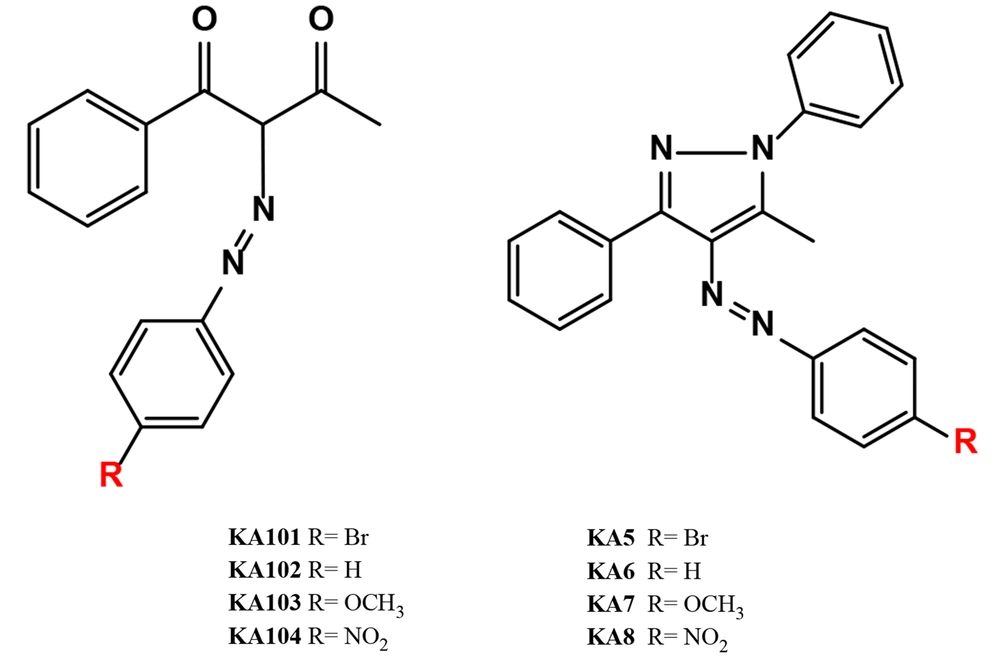
Figure 1.
Synthesized compounds, KA101-104, and KA5-8
3. Methods
All chemicals were purchased from Sigma Chemical Co. and used as received without further purification. The ^1H and ^13C NMR spectra were obtained using a Bruker AVANCE and a Varian Unity INOVA spectrometer, operating at 500 MHz for ^1H NMR and 126 MHz for ^13C NMR, respectively. Deuterated chloroform CDCl3 was used as the solvent, and the residual solvent peaks were used as the internal standard; δ = 7.27 ppm for ^1H NMR and δ = 77.23 ppm for ^13C NMR. The signal multiplicities were characterized using the standard notation: s singlet, d doublet, t triplet, q quartet, dd doublet of doublets, m multiplet, or br broad, with J-coupling constants measured in hertz Hz. To complement the NMR data, Liquid Chromatography-Mass Spectrometry LC-MS analyses were also performed to verify the purity and molecular weight of the compounds. The LC-MS analysis of pure compounds was conducted using a Shimadzu Nexera Lite HPLC system 2050c 3D coupled with an LCMS-2050 mass spectrometer Shimadzu, Japan. For the HPLC separation, an Acclaim™ 120 C8 column 4.6 mm I.D. × 150 mm, particle size 5 µm was employed. A gradient elution method was used, with methanol as the mobile phase, to ensure the effective separation of the compounds before mass spectrometric detection. For the LC-MS analysis, the nebulizing gas flow rate was set to 0.3 L/min, with the drying gas and heating gas flow rates at 6.0 L/min and 7.0 L/min, respectively. The desolvation temperature was maintained at 400°C to enhance solvent evaporation efficiency. Detection was carried out over a mass-to-charge ratio m/z range of 100 - 500, with a scan time of 0.5 seconds per scan, ensuring accurate and efficient analysis of the target compounds. The data were collected in the positive ion mode at a detector voltage of 2.0 kV. Thin-layer chromatography TLC was utilized for monitoring the reactions using pre-coated silica gel 60 F254 sheets Merck as the stationary phase. The developed TLC plates were visualized under ultraviolet light at a wavelength of 254 nm to assess the progress of the reactions. For the purification of synthesized compounds, recrystallization from methanol was carried out to achieve higher purity. Biological assays were performed according to standard methods (16, 17).
3.1. General Method for the Synthesis of Azo Benzoyl Acetone Derivatives KA 101-104 and Structure Characterization
In the first flask, diazonium salts were prepared by slow addition of 2.5 mL of conc. HCl to 10 mmol of substituted aromatic amines aniline, para-bromoaniline, para-methoxyaniline, and para-nitroaniline, followed by the addition of 5 mL of 15 % NaNO2 solution at 0°C, and the reaction was stirred for 15 min at 0°C. In the other flask, 10 mmol of benzoyl acetone and 30 mmol of sodium acetate in 20 mL ethanol were stirred for 10 min at room temperature to dissolve. Then, the diazonium salt obtained from the first flask was added to the ice-cold solution of benzoyl acetone in the second flask and stirred for another 10 min at 0°C. The obtained crude product was filtered, washed with alcohol/water solution 9:1, 3 mL, and crystallized in methanol to afford azo compounds. The structure of azo derivatives was characterized using ^1H NMR, ^13C NMR, and mass spectrometry Figures S1 - 4 (Figure 2).
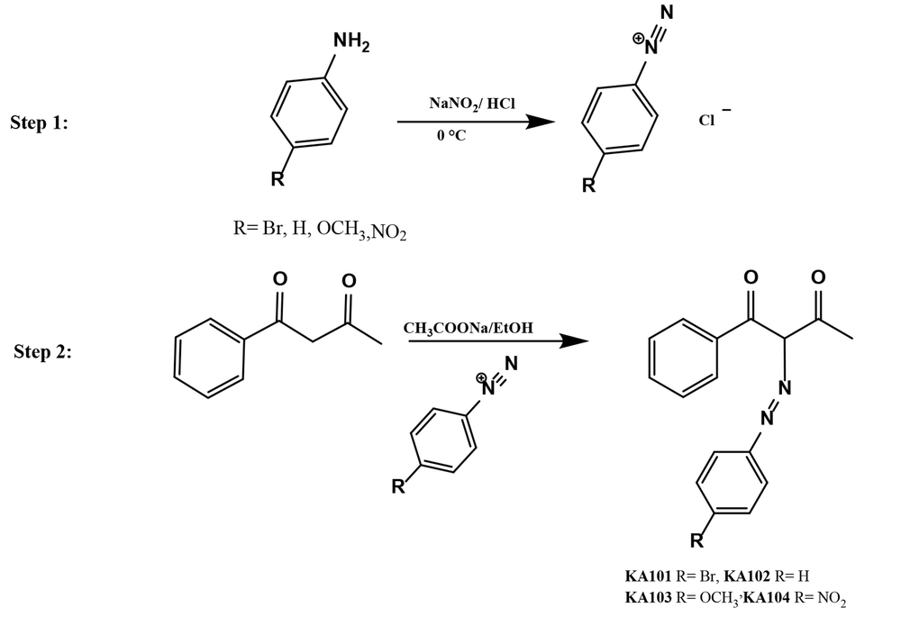
Figure 2.
Synthetic route of the synthesized compounds KA 101-104: KA101 R= Br Yield: 72%, KA102 R= H Yield: 68 %, KA103 R= OCH3 Yield: 61%, KA104 R= NO2 Yield: 44 %.
3.1.1. KA101 [E-2-[4-bromophenyldiazinyl]-1-phenylbutane-1,3-dione]
^1H NMR 500 MHz, CDCl3 δ 14.59 s, 1H, 7.86 d, J = 8.1 Hz, 1H, 7.63-7.49 m, 1H, 7.49-7.38 m, 3H, 7.26 s, OH, 7.07 d, J = 8.9 Hz, 1H, 2.62 s, 2H. ^13C NMR 126 MHz, CDCl3 δ 198.68, 192.08, 186.67, 140.90, 138.35, 132.77, 132.73, 132.27, 130.41, 128.51, 128.33, 127.97, 118.39, 117.67, 117.24, 30.56. MS calculated for C16H12O2N2Br [M+H]+ and [M+Na]+ yield 345, 347 and 367, 369. Reaction Percentage Yield: 72%.
3.1.2. KA102 [E-1-phenyl-2-phenyldiazenyl butane-1,3-dione]
^1H NMR 500 MHz, CDCl3 δ 14.67 s, 1H, 7.89-7.86 m, 1H, 7.63-7.51 m, 2H, 7.49-7.45 m, 2H, 7.44-7.40 m, 1H, 7.34-7.30 m, 2H, 7.26-7.19 m, 2H, 7.15-7.11 m, 1H, 2.63 s, 3H, 2.58 s, 1H. ^13C NMR 126 MHz, CDCl3 δ 198.53, 192.29, 141.76, 138.63, 132.85, 132.06, 130.45, 129.77, 129.70, 128.46, 128.27, 127.90, 125.67, 116.30, 115.87, 30.58. MS calculated for C16H14O2N2 [M+H]+ and [M+Na]+ yield 267, 289. Reaction Percentage Yield: 68 %.
3.1.3. KA103 [E-2-[4-methoxyphenyldiazinyl]-1-phenylbutane-1,3-dione]
^1H NMR 500 MHz, CDCl3 δ 15.26-14.63 m, 1H, 7.91-7.79 m, 1H, 7.66-7.33 m, 3H, 7.16 d, J = 9.0 Hz, 1H, 6.86 d, J = 9.1 Hz, 1H, 3.78 s, 2H, 2.63 s, 2H. ^13C NMR 126 MHz, CDCl3 δ 198.29, 192.34, 157.94, 139.03, 135.31, 132.24, 131.76, 130.35, 127.97, 117.69, 117.35, 100.00, 55.65, 30.52. MS calculated for C17H16O3N2 [M+H]+ and [M+Na]+ yield 297, and 319. Reaction Percentage Yield: 61%.
3.2. General Method for the Synthesis of KA5-KA8 and Structure Characterization
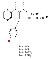
To obtain the tri-substituted pyrazole, KA5-KA8, the purified intermediates from the previous reactions KA101-KA104 were refluxed with phenyl hydrazine in ethanol at 80°C for 1 hr using acetic acid as a catalyst to result in precipitate that was filtered and crystallized in methanol, and the two structures were characterized using Liquid Chromatography High Resolution Mass Spectrometry LC/HRMS Figures S5 – 9 (Figure 3).
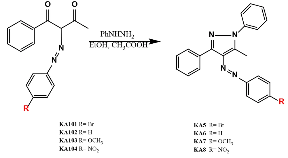
Figure 3.
Synthetic route of the synthesized compounds KA5-8: KA5 R= Br Yield: 70%, KA6 R= H Yield: 78 %, KA7 R= OCH3 Yield:53 %, KA8 R= NO2 Yield: 59 %.
3.2.1. KA5 [E-4-[4-bromophenyldiazinyl]-5-methyl-1,3-diphenyl-1H-pyrazole]
^1H NMR 400 MHz, DMSO-d6 δ 8.04 d, J = 7.5 Hz, 1H, 7.73 d, J = 8.4 Hz, 1H, 7.68 d, J = 5.7 Hz, 2H, 7.65 s, 2H, 7.60 d, J = 7.3 Hz, 1H, 7.55 d, J = 8.2 Hz, 2H, 7.48 d, J = 7.3 Hz, 1H, 7.41 d, J = 7.0 Hz, 2H, 7.37 d, J = 3.9 Hz, 2H, 7.30 d, J = 7.2 Hz, 1H, 2.52 s, 2H. ^13C NMR 101 MHz, DMSO δ 152.22, 152.19, 149.51, 144.44, 141.08, 139.40, 138.74, 135.99, 135.18, 132.90, 132.85, 132.73, 132.40, 130.96, 129.82, 129.63, 129.53, 129.22, 128.98, 128.92, 128.85, 128.71, 128.46, 128.36, 125.83, 125.58, 124.06, 123.98, 123.90, 123.74, 40.61, 40.40, 40.19, 39.98, 39.77, 39.56, 39.36, 15.41, 13.74. MS calculated for C22H17BrN4 is 417.0637 and the two isotopes [M+H]+ are reported. Reaction Percentage Yield: 70%.
3.2.2. KA6 [E-5-methyl-1,3-diphenyl-4-phenyldiazenyl-1H-pyrazole]
^1H NMR 400 MHz, DMSO-d6 δ 7.67 d, J = 1.6 Hz, 1H, 7.65 d, J = 1.1 Hz, 1H, 7.53–7.48 m, 3H, 7.46 t, J = 1.4 Hz, 1H, 7.43 d, J = 2.0 Hz, 1H, 7.43–7.42 m, 1H, 7.41 d, J = 2.2 Hz, 2H, 7.40 d, J = 1.9 Hz, 2H, 7.38 dd, J = 4.3, 2.0 Hz, 2H, 7.32 d, J = 1.9 Hz, 1H, 7.31 t, J = 1.6 Hz, 1H, 2.55 s, 2H. ^13C NMR 101 MHz, DMSO δ 153.33, 144.21, 140.92, 139.49, 135.98, 131.00, 130.63, 129.75, 129.59, 129.56, 128.72, 128.50, 128.45, 125.64, 122.15, 40.62, 40.41, 40.20, 39.99, 39.79, 39.58, 39.37, 15.39. MS calculated for C22H18N4 is 338.1531 and reported as [M+H]+. Reaction Percentage Yield: 78%.
3.2.3. KA7 [E-4-[4-methoxyphenyldiazenyl]-5-methyl-1,3-diphenyl-1H-pyrazole]
^1H NMR 400 MHz, DMSO-d6 δ 8.07–8.05 m, 1H, 7.80–7.77 m, 2H, 7.68 d, J = 5.7 Hz, 2H, 7.64 d, J = 3.4 Hz, 1H, 7.61 d, J = 7.5 Hz, 1H, 7.56–7.54 m, 1H, 7.50 t, J = 7.5 Hz, 2H, 7.42 d, J = 3.2 Hz, 1H, 7.40–7.39 m, 1H, 7.14–7.11 m, 2H, 7.08–7.05 m, 1H, 3.86 s, 2H, 2.61 s, 3H. ^13C NMR 101 MHz, DMSO δ 161.61, 161.46, 148.86, 147.56, 147.54, 143.29, 140.85, 139.59, 137.76, 135.77, 132.71, 132.05, 130.95, 129.83, 129.53, 129.43, 129.08, 128.85, 128.79, 128.68, 128.32, 125.84, 125.57, 123.98, 123.89, 115.05, 114.91, 113.31, 113.30, 109.44, 56.03, 55.98, 40.56, 40.36, 40.15, 39.94, 39.73, 39.52, 39.31, 15.34, 13.61. MS calculated for C22H18N4 is 368.1637 and reported as [M+H]+. Reaction Percentage Yield: 53 %.
3.2.4. KA8 [E-5-methyl-4-[4-nitrophenyldiazenyl]-1,3-diphenyl-1H-pyrazole]
^1H NMR 400 MHz, DMSO-d6 δ 8.41–8.38 m, 2H, 8.35–8.33 m, 1H, 8.03 d, J = 7.0 Hz, 1H, 7.95–7.93 m, 1H, 7.81 d, J = 9.0 Hz, 1H, 7.68 d, J = 7.6 Hz, 1H, 7.63 t, J = 7.5 Hz, 2H, 7.58–7.56 m, 1H, 7.50 d, J = 7.5 Hz, 2H, 7.42 d, J = 8.0 Hz, 2H, 7.32 dd, J = 6.1, 2.1 Hz, 1H, 2.64 s, 3H. ^13C NMR 101 MHz, DMSO δ 156.76, 147.92, 145.76, 141.36, 139.14, 136.57, 130.98, 129.99, 129.68, 129.03, 128.86, 127.91, 125.73, 125.48, 123.00, 40.26, 40.06, 39.85, 39.64, 39.43, 39.22, 39.01, 15.41. MS calculated for C22H18N4 is 383.1382 and reported as [M+H]+. Reaction Percentage Yield: 59%.
3.3. Biological Evaluation
3.3.1. HepG2 and HSF Cytotoxicity Assays
3.3.1.1. Cell Culture
Human skin fibroblast HSF and hepatocellular carcinoma HepG2 cell lines were obtained from Nawah Scientific Inc. Mokatam, Cairo, Egypt. The cells were maintained in Dulbecco's Modified Eagle Medium DMEM supplemented with 100 mg/mL of streptomycin, 100 units/mL of penicillin, and 10% heat-inactivated fetal bovine serum FBS. The cultures were incubated in a humidified atmosphere containing 5% CO2 v/v at a temperature of 37°C to ensure optimal growth conditions.
3.3.2. Cytotoxicity Assay
Cell viability was evaluated using the Sulforhodamine B SRB assay. A total of 100 μL aliquots of a cell suspension containing 5 × 10⁵ cells were seeded in 96-well plates and incubated in complete medium for 24 hours to allow for cell attachment and growth. After the incubation period, cells were treated with an additional 100 μL of medium containing the drugs at various concentrations. Following drug exposure, the medium was replaced with 150 μL of 10% trichloroacetic acid TCA to fix the cells, and the plates were incubated at 4 °C for 1 hour. After fixation, the TCA solution was carefully removed, and the cells were washed five times with distilled water to eliminate any residual TCA. Subsequently, 70 μL of a 0.4% w/v SRB solution was added to each well, and the plates were incubated in the dark at room temperature for 10 minutes to allow the SRB dye to bind to the protein in the cells. Following incubation, the plates were washed three times with 1% acetic acid to remove unbound dye and allowed to air-dry overnight. To quantify the protein-bound SRB stain, 150 μL of a 10 mM TRIS buffer was added to each well to dissolve the dye. The absorbance was then measured at a wavelength of 540 nm using an Infinite F50 microplate reader TECAN, Switzerland 16,17.
4. Results and Discussion
A series of novel azo-benzoyl acetone derivatives, KA101–KA104, were successfully synthesized via azo coupling reactions between the corresponding diazonium salts of aromatic amines and benzoyl acetone in ethanolic sodium acetate, as outlined in Figure 2. Reaction progress and completion were monitored by thin-layer chromatography TLC to ensure full consumption of the starting materials. Following purification by crystallization, the obtained compounds, KA101–KA104, were comprehensively characterized using mass spectrometry MS, proton nuclear magnetic resonance ^1H NMR, and carbon-13 nuclear magnetic resonance ^13C NMR. The corresponding spectral data are provided in the Supporting Information Figures S1 - 4.
In addition, pyrazole derivatives of selected azo precursors were synthesized to evaluate the influence of heterocyclic incorporation on anticancer activity. Specifically, KA5 and KA6 were obtained as pyrazole analogues of KA102 and KA103, respectively, and were designed for comparison with the previously reported anticancer lead compound KA5 15. Structural elucidation of the pyrazole derivatives was confirmed using high-resolution mass spectrometry HRMS, ^1H NMR, and ^13C NMR analyses Figures S5 - 9.
The cytotoxic activities of the synthesized azo compounds and their corresponding pyrazole derivatives were evaluated in vitro against human hepatocellular carcinoma cells HepG2 and human skin fibroblasts HSF as a representative normal cell line, with sorafenib employed as a reference anticancer agent. HepG2 cells were selected due to their relevance as a hepatic model system capable of reflecting drug metabolism and potential hepatotoxic effects, while evaluation against HSF cells provided insight into cytotoxicity toward normal cells. Treatment of HepG2 and HSF cells with the synthesized compounds revealed that the parent azo-benzoyl acetone derivatives exhibited markedly lower cytotoxicity compared to their corresponding pyrazole analogues and sorafenib Figures 4. and 5.
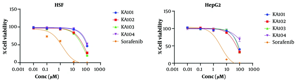
Figure 4.
Dose-response curve of cytotoxicity assay of KA 101-104 and Sorafenib on human skin fibroblast cells HSF and hepatocellular carcinoma cells HepG2.
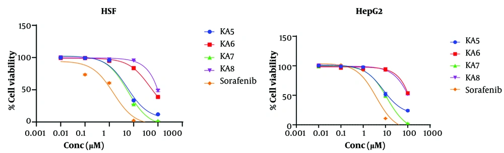
Figure 5.
Dose-response curve of cytotoxicity assay of KA 5-8 and Sorafenib on human skin fibroblast cells HSF and hepatocellular carcinoma cells HepG2.
These findings highlight the enhanced anticancer potential associated with pyrazole incorporation. Notably, compounds KA5 and KA7 induced pronounced cytotoxic effects in both HepG2 and HSF cell lines Table 1, with IC₅₀ values ranging from 9.75 to 10.37 µM, approaching that of sorafenib IC₅₀ = 4.8 µM. This observation indicates that azo-pyrazole hybrids possess substantial cytotoxic potency and may represent promising anticancer scaffolds.
Table 1.
IC50 Values of KA 101-103 and KA 5-8 Against HepG2 and HSF Cell Lines a
| Compound | HepG2 [IC50 µM] | HSF [IC50 µM] |
|---|---|---|
| KA101 | > 50 | 42.29 ± 1.00 |
| KA102 | > 50 | > 50 |
| KA103 | > 50 | 32.74 ± 0.96 |
| KA104 | > 50 | > 50 |
| KA5 | 10.37 ± 0.96 | 5.84 ± 0.82 |
| KA6 | > 50 | > 50 |
| KA7 | 9.75 ± 0.97 | 5.03 ± 0.27 |
| KA8 | > 50 | > 50 |
| Sorafenib | 4.8 ± 0.26 | 4.00 ± 0.44 |
a Data are expressed as the means ± SEM from three independent experiments
Although the phenyl-substituted azo-benzoyl acetone derivatives exhibited limited anticancer activity, the cytotoxicity profiles of KA101 and KA103 against HSF cells suggest that para-substituted phenyl moieties, particularly para-methoxy and para-bromo groups, may contribute to enhanced biological activity. These results underscore the importance of electronic effects and substituent variation in modulating cytotoxic behavior. Despite KA5 and KA7 demonstrating cytotoxic activities comparable to sorafenib, their selectivity indices SI, defined as the ratio of IC₅₀ values in normal cells to that in cancer cells, were calculated to be 0.56 and 0.52, respectively, indicating lower selectivity toward cancer cells relative to sorafenib SI = 0.83. Nevertheless, the observed SI values remain within a range reported for certain clinically used anticancer agents. For instance, 4-hydroxytamoxifen, a widely used breast cancer therapeutic, has been reported to exhibit SI values of approximately 1.29 in selected in vitro assays, demonstrating that modest selectivity indices do not necessarily preclude clinical relevance (18). Collectively, these findings provide valuable insights into the structure-activity relationships of azo-benzoyl acetone and azo-pyrazole derivatives and support their further optimization as potential anticancer agents. Based on the current findings and previously reported cytotoxicity data for azo-pyrazole derivatives (15), the presence of a pyrazole moiety appears to be a critical structural requirement for anticancer activity. However, the lack of activity observed for KA6, which bears an unsubstituted phenyl group, indicates that incorporation of the pyrazole ring alone is insufficient to confer cytotoxic potency. In contrast, KA5 and KA7, featuring para-bromo and para-methoxy substituents, respectively, displayed pronounced anticancer activity, underscoring the importance of aromatic substitution. In the case of KA7, the observed activity may be attributed to the electron-donating nature of the methoxy group, which enhances electron density within the aromatic system through both inductive and resonance effects. Although halogens are generally classified as electron-withdrawing substituents via inductive effects, they are known to donate electron density through resonance, which may explain the retained activity observed for the para-bromo-substituted compound KA5. To further validate the role of electron donation, the nitro-substituted analogue KA8, bearing a strong electron-withdrawing group, was synthesized and evaluated against the same cell lines. The absence of significant cytotoxic activity for KA8 Table 1 strongly supports the hypothesis that resonance-mediated electron donation within the aromatic system plays a pivotal role in modulating anticancer activity. Comparative analysis of the resonance structures of the pyrazole derivatives Figure 6, together with the biological assay results, allows the following conclusions regarding the structure-activity relationships of azo-substituted pyrazoles:
- The presence of a pyrazole moiety is essential for anticancer activity, as evidenced by the substantial approximately seven-fold loss of activity observed for the parent azo compounds KA101 and KA103.
- The ability to favor hydrazone tautomer formation is critical, as demonstrated by the diminished activity of azo-pyrazole derivatives with a dominant azo tautomeric form, such as KA6 and KA8.
- Planarity of the pyrazole ring system is a key determinant of activity, inferred from the lack of cytotoxicity exhibited by KA8, where disruption of aromaticity and loss of planarity, potentially due to carbocation formation within the pyrazole ring, negatively impact biological performance.
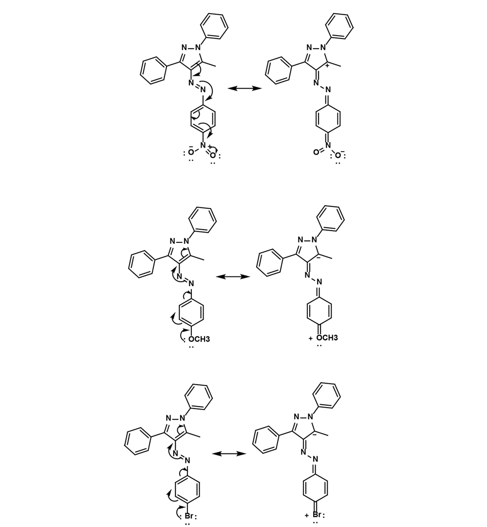
Figure 6.
The resonance structures of KA5, KA7, and KA8 indicating the effect of nitro groups on the geometry of the pyrazole ring, in contrast to bromo and methoxy groups
4.1. Conclusions
In conclusion, seven azo-based derivatives were synthesized via diazo coupling of aromatic amines with benzoyl acetone, followed by cyclization to afford selected pyrazole analogues. Structural characterization was accomplished using ^1H NMR, ^13C NMR, and mass spectrometric analyses. The synthesized compounds were evaluated for their in vitro anticancer activity against HepG2 hepatocellular carcinoma cells and HSF normal fibroblasts. Among the tested compounds, KA5 and KA7 demonstrated the most promising cytotoxic activity, with IC₅₀ values comparable to that of sorafenib, highlighting their potential as lead candidates for further investigation. The present study underscores the importance of hydrazone-linked aromatic pyrazole scaffolds and electron-donating para-substituents in modulating anticancer activity, thereby providing a rational basis for further optimization and in vivo evaluation of this class of compounds.
![Synthesis and Cytotoxic Evaluation of 6-Amino-4-Aryl-3-Methyl- 2,4-Dihydropyrano[2,3-C]Pyrazole-Carbonitrile Derivatives Using Borax with Potential Anticancer Effects](https://brieflands.com/journals/jrps/articles/147742/figures/thumbnail.webp)