


1. Background
2. Methods
3. Results
| Dermoscopic Features | Plantar wart (n = 56) | Corn (n = 22) | Callus (n = 14) | P Value |
|---|---|---|---|---|
| Red dots | 50 (89.28) | - | - | - |
| Linear hemorrhages or streaks | 10 (10.71) | - | - | - |
| Yellow halo | 46 (82.14) | - | - | - |
| White halo | 10 (17.85) | - | ||
| Translucent central core | - | 22(100) | - | - |
| Yellowish areas | - | 16 (72.72) | 14 (100) | 0.08 |
| Preserved dermatoglyphics | - | 22 (100) | 14 (100) | |
| Absence of dermatoglyphics | 56 (100) | - | - | - |
| Whitish annular ring | - | 18 (81.81) | - | - |
| Focal white areas | - | - | 6 (42.85) | - |
aValues are expressed as No. (%) unless otherwise indicated.
![A, Clinical image of plantar wart showing hyperkeratotic papule on the sole. B, Dermoscopy shows dotted (yellow arrow) and linear (red arrow) vessels. Dermatoglyphics (black arrows) are absent abruptly in the involved skin [Illuco 1100 IDS, Polarized, 10×]. C, Clinical image of plantar wart showing grouped hyperkeratotic papules on the sole. D, Dermoscopy shows dotted vessels (yellow arrow) with the absence of dermatoglyphics, which are appreciated in adjacent normal skin (black arrows). E, Dermoscopic image of plantar wart shows dotted (yellow arrow) and globular (red arrow) vessels with absent dermatoglyphics [Illuco 1100 IDS, Polarized, 10×]. F, After paring bleeding, points are well appreciated in the plantar wart [Illuco 1100 IDS, Polarized, 10×].](https://brieflands.com/journals/jssc/articles/116806/figures/jssc-116806-g001-F1-preview.webp "A, Clinical image of plantar wart showing hyperkeratotic papule on the sole. B, Dermoscopy shows dotted (yellow arrow) and linear (red arrow) vessels. Dermatoglyphics (black arrows) are absent abruptly in the involved skin [Illuco 1100 IDS, Polarized, 10×]. C, Clinical image of plantar wart showing grouped hyperkeratotic papules on the sole. D, Dermoscopy shows dotted vessels (yellow arrow) with the absence of dermatoglyphics, which are appreciated in adjacent normal skin (black arrows). E, Dermoscopic image of plantar wart shows dotted (yellow arrow) and globular (red arrow) vessels with absent dermatoglyphics [Illuco 1100 IDS, Polarized, 10×]. F, After paring bleeding, points are well appreciated in the plantar wart [Illuco 1100 IDS, Polarized, 10×].")
A, Clinical image of plantar wart showing hyperkeratotic papule on the sole. B, Dermoscopy shows dotted (yellow arrow) and linear (red arrow) vessels. Dermatoglyphics (black arrows) are absent abruptly in the involved skin [Illuco 1100 IDS, Polarized, 10×]. C, Clinical image of plantar wart showing grouped hyperkeratotic papules on the sole. D, Dermoscopy shows dotted vessels (yellow arrow) with the absence of dermatoglyphics, which are appreciated in adjacent normal skin (black arrows). E, Dermoscopic image of plantar wart shows dotted (yellow arrow) and globular (red arrow) vessels with absent dermatoglyphics [Illuco 1100 IDS, Polarized, 10×]. F, After paring bleeding, points are well appreciated in the plantar wart [Illuco 1100 IDS, Polarized, 10×].
![A, Clinical image of corn showing hyperkeratotic papule on the plantar aspect of the great toe. B, Dermoscopy shows a yellow area (black star) and preservation of dermatoglyphics (black arrow). Note the absence of vascular structures [Illuco 1100 IDS, Polarized, 10×]. Dermoscopic image of corn (C) and (D) shows the central translucent area (yellow star) with preserved dermatoglyphics (black arrow). Note the whitish ring (yellow arrow) [Illuco 1100 IDS, Polarized, 10×]. E, Clinical image of callus showing hyperkeratotic plaque on the sole. F, Dermoscopy shows the opaque yellow area (yellow star) and dermatoglyphics (black arrow) [Illuco 1100 IDS, Polarized, 10×].](https://brieflands.com/journals/jssc/articles/116806/figures/jssc-116806-g002-F2-preview.webp "A, Clinical image of corn showing hyperkeratotic papule on the plantar aspect of the great toe. B, Dermoscopy shows a yellow area (black star) and preservation of dermatoglyphics (black arrow). Note the absence of vascular structures [Illuco 1100 IDS, Polarized, 10×]. Dermoscopic image of corn (C) and (D) shows the central translucent area (yellow star) with preserved dermatoglyphics (black arrow). Note the whitish ring (yellow arrow) [Illuco 1100 IDS, Polarized, 10×]. E, Clinical image of callus showing hyperkeratotic plaque on the sole. F, Dermoscopy shows the opaque yellow area (yellow star) and dermatoglyphics (black arrow) [Illuco 1100 IDS, Polarized, 10×].")
A, Clinical image of corn showing hyperkeratotic papule on the plantar aspect of the great toe. B, Dermoscopy shows a yellow area (black star) and preservation of dermatoglyphics (black arrow). Note the absence of vascular structures [Illuco 1100 IDS, Polarized, 10×]. Dermoscopic image of corn (C) and (D) shows the central translucent area (yellow star) with preserved dermatoglyphics (black arrow). Note the whitish ring (yellow arrow) [Illuco 1100 IDS, Polarized, 10×]. E, Clinical image of callus showing hyperkeratotic plaque on the sole. F, Dermoscopy shows the opaque yellow area (yellow star) and dermatoglyphics (black arrow) [Illuco 1100 IDS, Polarized, 10×].
![Dermoscopic image of callus (A) and (B) shows opaque yellow area (yellow star) with focal white areas (red arrow) with preserved dermatoglyphics (black arrows) [Illuco 1100 IDS, Polarized, 10×]. C, Dermoscopic image of plantar wart shows globular vessels (yellow arrow) and adherent fabric fibres (red arrows). D, Dermoscopy of callus shows red, black, and brown dots, which are referred to as pseudo hemorrhagic structures [Illuco 1100 IDS, Polarized, 10×].](https://brieflands.com/journals/jssc/articles/116806/figures/jssc-116806-g003-F3-preview.webp "Dermoscopic image of callus (A) and (B) shows opaque yellow area (yellow star) with focal white areas (red arrow) with preserved dermatoglyphics (black arrows) [Illuco 1100 IDS, Polarized, 10×]. C, Dermoscopic image of plantar wart shows globular vessels (yellow arrow) and adherent fabric fibres (red arrows). D, Dermoscopy of callus shows red, black, and brown dots, which are referred to as pseudo hemorrhagic structures [Illuco 1100 IDS, Polarized, 10×].")
Dermoscopic image of callus (A) and (B) shows opaque yellow area (yellow star) with focal white areas (red arrow) with preserved dermatoglyphics (black arrows) [Illuco 1100 IDS, Polarized, 10×]. C, Dermoscopic image of plantar wart shows globular vessels (yellow arrow) and adherent fabric fibres (red arrows). D, Dermoscopy of callus shows red, black, and brown dots, which are referred to as pseudo hemorrhagic structures [Illuco 1100 IDS, Polarized, 10×].
![A, Histopathology of plantar wart shows hyperkeratosis (black star), acanthosis, and few koilocytes (yellow circles). Note the elongated rete ridges and dilated capillaries [H & E, 10×]. B, Histopathology of corn showing hyperkeratosis (black star), acanthosis, and dermal fibrosis [H & E, 4×]. C, Histopathology of callus showing hyperkeratosis (black star), acanthosis, and fibrosis in the superficial dermis [H & E, 4×].](https://brieflands.com/journals/jssc/articles/116806/figures/jssc-116806-g004-F4-preview.webp "A, Histopathology of plantar wart shows hyperkeratosis (black star), acanthosis, and few koilocytes (yellow circles). Note the elongated rete ridges and dilated capillaries [H & E, 10×]. B, Histopathology of corn showing hyperkeratosis (black star), acanthosis, and dermal fibrosis [H & E, 4×]. C, Histopathology of callus showing hyperkeratosis (black star), acanthosis, and fibrosis in the superficial dermis [H & E, 4×].")
A, Histopathology of plantar wart shows hyperkeratosis (black star), acanthosis, and few koilocytes (yellow circles). Note the elongated rete ridges and dilated capillaries [H & E, 10×]. B, Histopathology of corn showing hyperkeratosis (black star), acanthosis, and dermal fibrosis [H & E, 4×]. C, Histopathology of callus showing hyperkeratosis (black star), acanthosis, and fibrosis in the superficial dermis [H & E, 4×].
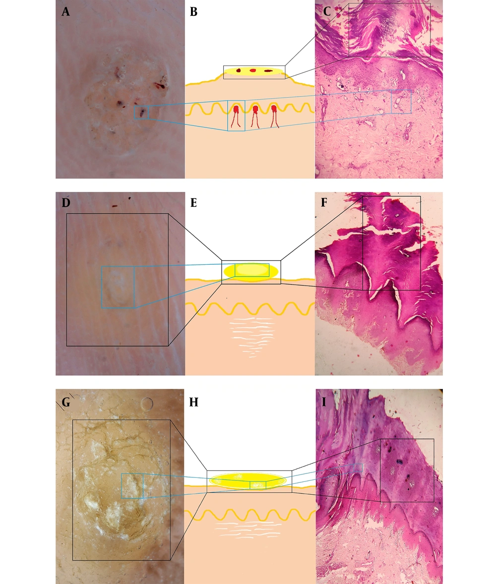
Schematic diagram showing a dermoscopic and histopathological correlation in plantar wart (A, B & C). Schematic diagram showing a dermoscopic and histopathological correlation in corn (D, E & F). Schematic diagram showing a dermoscopic and histopathological correlation in callus (G, H & I).
