1. Background
Iran is one of the seven countries in which about 90% of the worldwide cases of cutaneous leishmaniasis (CL) are seen (1, 2). Although many physical, topical, intra-lesional, and systemic treatment modalities have been suggested for CL, antimonial compounds (intralesional or systemic administration) are still considered as the standard treatment (3-9). Trichloroacetic acid (TCA) peeling has been used as a chemical peeling agent for skin rejuvenation.
TCA peeling is a papular and long-utilized procedure in dermatology and cosmetic surgery with rapid recovery and few resultant complications (10-13). TCA is also considered as a treatment option in some dermatologic conditions such as actinic keratoses, molluscum contagiosum, external genital wart, acne scarring, and photoaged skin (14-22). Our previous randomized clinical trial showed comparable efficacy of TCA and intralesional meglumine antimoniate in the treatment of papular lesions of CL (2, 3).
2. Objectives
This study was designed to compare the efficacy of the combination therapy of intralesional MA and TCA with intralesional MA alone in the treatment of CL.
3. Patients and Methods
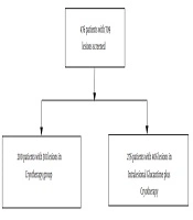
A total of 200 patients with confirmed cutaneous leishmaniasis were randomized into two groups to receive either intralesional MA alone or a combination treatment of TCA and intralesional MA. Patients with positive direct smear for CL who had six to 60 years of age without any previous history of systemic or topical therapy for CL were recruited in this study. The lesions had to be < 3 cm, less than 12 week duration, and not be located within 2 cm distance of palpebral margin. Pregnant and lactating women were excluded from the study. Random allocation software (version 1.0, May 2004, Saghaei) was used for randomization. This study was done in Skin Disease and Leishmaniasis Research center (SDLRC) and was approved by SDLRC Ethic Committee. Informed consent was taken from the patients or their parents after giving enough information. In both groups intralesional injection of MA was performed twice a week until complete resolution of the lesions or up to eight weeks Intralesional injection was done from the intact margin of the lesion in amounts enough to blanch the lesion and 1 mm rim of the surrounding normal skin. In combination therapy group, TCA 50% was applied to the lesions with a cotton swap fortnightly up to frosting the lesion. Lesions were photographed before and after completion of the treatment course. Area of the induration, erythema, and lesion were calculated using two largest perpendicular diameters. The measurements were done before treatment and at the end of eighth week by the investigators who were blinded to the type of treatment. Complete re-epithelization of the lesion and lack of induration was considered as clinical healing. Clinical response was measured at the end of treatment and was defined as complete cure (negative direct smear and clinical healing), partial cure (partial clinical improvement with decreasing erythema, induration, and lesion size), non-cure or treatment failure (no clinical change or worsening of the lesions) (5).
4. Results
Table 1 shows demographic characteristics of the patients in both groups. Type, number, and clinical response of the lesions are summarized in Table 2. There was no significant difference in demographic characteristics between two treatment groups. Ninety six patients in intralesional MA group and 91 patients in the combination group completed the study. Before treatment, mean lesion area was 359 ± 55.6 mm2 in MA treated group in contrast to 329 ± 118.7 in the combination group (MA + TCA) (P > 0.05). At the end of treatment, lesion area were 204.8 ± 165.8 mm2 and 68.3 ± 59.7 mm2 in MA monotherapy and combination therapy, respectively (P > 0.05). Complete cure was achieved in 79.2% of patients treated with MA and 85.7% of patients in the combination therapy group (P > 0.05). Kaplan-Meier test showed a significant difference in complete cure time between the two groups. In patients treated with the combination of intralesional MA and topical TCA 50%, the resolution of the lesions was accelerated in comparison to those treated with MA alone.
| Age | Number of Lesions | Sex | Number of Patients | Drug |
|---|---|---|---|---|
| (Male/Female) | ||||
| 11.3 ± 23.3 | 167 ± 1.7 | 50.46 | 96 | intralesional MA |
| 10 ± 20.6 | 152 ± 1.6 | 51.40 | 91 | intralesional MA + TCA |
| 10.7 ± 22.04 | 319 ± 1.7 | 101.86 | 187 | Total |
a Abbreviations: MA, meglumine antimoniate.
b Data are presented in Mean ± SD.
Table 2.
Type and Clinical Response of the Lesions in Two Groups Treated With Intralesional Injection of Meglumine Antimoniate Alone and Combination of Meglumine Antimoniate and TCA a
| Type of Lesions | Number of Lesions | Non-Cure | Partial Cure | Complete Cure | |||
|---|---|---|---|---|---|---|---|
| MA | TCA+ MA | MA | TCA+ MA | MA | TCA+ MA | ||
| Papule | 20 | 0 | 1 | 0 | 0 | 2 | 12 |
| Nodule | 71 | 2 | 3 | 0 | 0 | 13 | 17 |
| Plaque | 139 | 11 | 3 | 1 | 3 | 37 | 22 |
| Ulcerative nodule | 89 | 5 | 3 | 1 | 0 | 24 | 27 |
a MA, meglumine antimoniate; TCA, trichloroacetic acid.

Figure 1.
Comparison of the Place of Lesions in the Two Groups Treated With Intralesional Injection of Meglumine Antimoniate Alone and Combination of Meglumine and TCA

Figure 2.
MA, meglumine antimoniate; TCA, trichloroacetic acid.
5. Discussion
CL is an endemic disease in Iran. CL lesions have self-healing process with a permanent residual scar. Although antimonial compounds are still the first line therapy for CL, their side effects and increasing rate of resistance have motivated researchers to seek safer and alternative modalities. Topical TCA has different indications in dermatology. The topical TCA peel method is reported as a safe and effective treatment for benign pigmented lesions such as seborrheic keratosis, solar lentigines, melasma, and freckles with no significant complications (15). Application of TCA 85% in patients with external genital warts was accompanied by a high success rate at the end of the treatment period 16. El-Domyati et al. (23) reported epidermal and dermal rejuvenation in four patients with photodamaged skin treated with TCA. This change was associated with new collagen deposition and improvement of the morphologic appearance of collagen and elastic fibers (23). Yug et al. reported the treatment of three patients with acne scars by topical application of TCA 95% at six-week intervals for a total of six treatments. This procedure resulted in cosmetic and histologic improvement of the depth of acne scars with further increasing of collagen content, fragmentation of elastic fibers, and no complication. Treatment of atrophic scars with TCA was associated with activation of dermal fibroblasts and increased collagen contents (19). The same mechanism of new collagen formation and elastic fibers degradation might contribute to the accelerated healing process of CL lesions after topical application of TCA in combination therapy group. Sakai et al. assessed the changes in Langerhans cells after 40% and 60% TCA peeling and cryosurgery and reported more significant reduction in Langerhans cells in TCA treated patients raising the concern of potential carcinogenesis of long term frequent TCA peeling due to temporary impairment of skin defense system (24). The possibility for such potential side effect should be assessed in other studies with enough duration to assess this risk before adding this agent as a routine adjuvant to MA in CL patients in whom intralesional treatment is indicated. Our previous clinical trial on the efficacy of TCA on papular lesions of CL showed no significant difference while it was still comparable results with intralesional MA (25). The results of this study showed that combination therapy with intralesional MA and TCA 50% might accelerate the healing process of CL lesions with no significant difference in complete cure rate compared to the patients treated with intralesional MA alone.
Topical application of TCA 50% might have advantages when used as an adjuvant therapy to accelerate the healing process and decrease the healing time of the lesions in patients with CL. Further studies to assess the mechanism of action of TCA in the treatment of cutaneous leishmaniasis, its possible efficacy as combination therapy with other treatment modalities of CL, and assessment of its long-term usage side effects are warranted.