


1. Background
2. Objectives
3. Methods
| Radiological Parameters with Units and Case no. with Anatomical Details of Vessels Evaluated Radiologically | Pre-expansion | Post- expansion | P-Value b |
|---|---|---|---|
| 1. Vessel velocity (cm/sec) as per Colour Doppler | 0.461 | ||
| Case no. 1 | |||
| Rt. IMA | 15 | 11 | |
| Lt. IMA | 9 | 12 | |
| Case no. 2 Lt. STA | 7.7 | 21 | |
| Case no. 3 Rt. ECA | 11 | 14 | |
| 2. Lumen diameter (in mm) as per Colour Doppler | 0.500 | ||
| Case no. 1 | |||
| Rt. IMA | 2.5 | 2.3 | |
| Lt. IMA | 2.4 | 2.3 | |
| Case no. 2 Lt. STA | 0.6 | 0.8 | |
| Case no. 3 | |||
| Rt. ECA | 1.7 | 1.9 | |
| Rt. IMA | 1.2 | 1.5 | |
| 3. Lumen diameter (in mm) as per CT-angiography | 0.017 | ||
| Case no. 1 | |||
| Rt. IMA | 1.8 | 2 | |
| Lt. IMA | 1.9 | 2.2 | |
| Case no. 2 | |||
| Lt. STA (Frontal) | 1.2 | 1.3 | |
| Lt. STA (Parietal) | 1 | 1.4 | |
| Lt. STA (Occipital) | 1.5 | 1.7 | |
| Case no. 3 | |||
| Rt. ECA | 1.8 | 2 | |
| Rt. IMA | 1.5 | 1.8 | |
| 4. Density of vessels (per square cm) as per Colour Doppler | 0.065 | ||
| Case no. 1 | |||
| Rt. IMA | 0.2 | 0.4 | |
| Lt. IMA | 0.2 | 0.4 | |
| Case no. 2 Lt. STA | 1.5 | 3 | |
| Case no. 3 Rt. ECA | 0.2 | 0.3 | |
| 5. Major feeder vessels (anatomical) as per CT-angiography | Qualitative variable | ||
| Case no. 1 | Bilateral Internal Mammary Artery (IMA) | Bilateral IMA | |
| Case no. 2 | Lt. Superficial Temporal Artery (STA)- Frontal, Parietal, Occipital branches. | Lt. STA and its branches | |
| Case no. 3 | Branches of Rt. External carotid artery (ECA) and Rt. IMA | Branches of Rt. ECA and IMA | |
| 6. Waveform as per Colour Doppler | Qualitative variable | ||
| Case no. 1 | Monophasic | Biphasic | |
| Case no. 2 | Monophasic | Biphasic | |
| Case no. 3 | Monophasic | Biphasic |
a After tabulating the data in Microsoft Excel (2013), it was analysed with Stata 12. The comparison between different parameters was tested by Mann- Whitney U test, considering P-value < 0.05 to be statistically significant.
b Statistical analysis (using Mann-Whitney U test for paired non-parametric continuous data using Stata 12 software).
3.1. Surgical Technique
4. Results
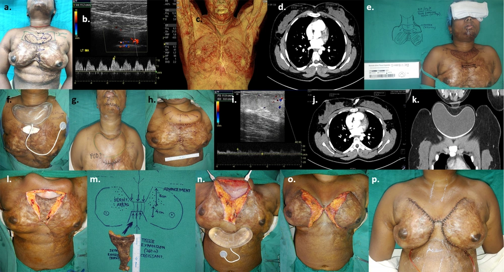
a, planning for tissue expansion of a 24-year-old female with post-burn scar on breasts & symmastia; b, pre-expansion colour doppler; c & d, pre-expansion CT angiography; e, pre-operative planning & rationale of 1 croissant shaped expander in pre-sternal area estimating area of advancement in terms of volume and dimensions of the expander (croissant 250 cc, 14.5 × 7.7 × 4.5 cm); f, expander inserted after creating the pocket; g, early post-operative image after 3 days; h, after 6 sessions of expansion (240 cc) over 2 months; i, post-expansion colour doppler; j & k, CT- angiography with 3-dimensional reconstruction post-expansion; l, removal of expander through excision of scar in the pre-sternal and inter-mammary area; m, excised scar; n, expander removed with remote port; o, expanded skin advanced inferiorly and wound closed creating adequate inter-mammary area; p, early post-operative image after 7 days.
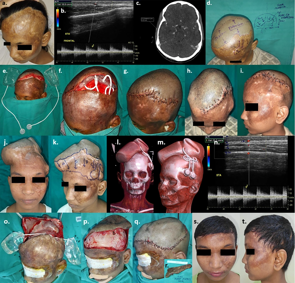
a, 19-year-old girl with post-burn scar & alopecia scalp; b, pre-expansion colour doppler; c, pre-expansion CT angiography; d, pre-operative planning of 2 expanders estimating area of advancement in terms of volume and dimensions of the expanders (cylindrical on rt. side of scalp- 250 cc, 10.2 × 5.4 × 7.2 cm and rectangular on lt. side of scalp- 300 cc, 10 × 6 × 6.3 cm); e & f, expanders inserted after creating sub-galeal pockets; g, closure of wound; h & i, early post-operative image after 1 week; j & k, After completion of expansion (240 cc on right side & 280 cc on left side) over 4 months; l & m, 3-dimensional reconstruction of CT images post-expansion; n, colour doppler post-expansion; o, to q, expander removal and definitive wound closure after excision of alopecic scar 18 × 8 cm (Figure 2. r. inset); s & t, late post-operative results after 3 months.
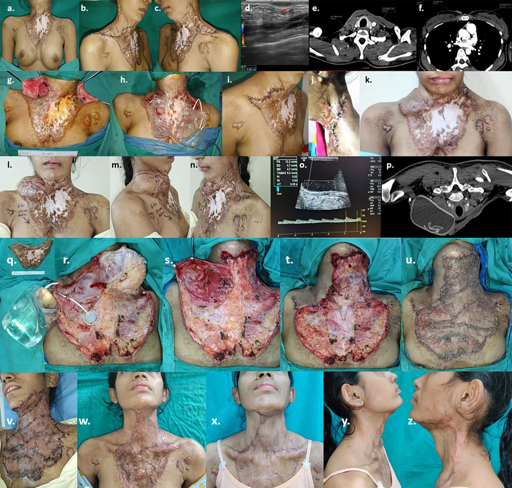
a to c, Planning for tissue expansion of a 23-year-old female with post-burn neck contracture (Grade III) and upper chest wall hypertrophic hypopigmented scar; d, pre-expansion colour doppler; e & f, pre-expansion CT angiography; g & h, insertion of 2 expanders estimating area of advancement in terms of volume and dimensions of the expanders (cylindrical on rt. supraclavicular region- 250 cc, 10.2 × 5.4 × 7.2 cm and rectangular on lt. supraclavicular region- 300 cc, 10 × 6 × 6.3 cm); i, early post-operative images after 2 weeks; j & k, left sided implant extrusion due to infection & suture dehiscence after 4 weeks following which left sided expander removed; l to n, after 7 sessions of expansion (280 cc) on the right sided expander over 4 months; o, post-expansion colour doppler; p, post-expansion CT-angiography with 3-dimensional reconstruction; q, Excision of post-burn scar and removal of expander; r to t, defect on neck & upper chest after excisional release and advancement of right sided supraclavicular expanded skin; u, defect resurfaced with intermediate thickness split skin graft; v, early post-operative image - 5th post-operative day; w, 4 weeks post-op ; x to z, 3 months post – op with adequate neck extension & cervico-mental angle.
