
1. Introduction
Penile tumors are the rarest tumors of the male urogenital tract (1), and the most common histological type is squamous cell carcinoma (SCC), which is characterized by local invasion and early lymphatic dissemination (2). Treatment is often mutilating and has a significant psychological impact, particularly when the tumor is located in the genital region. Lichen sclerosus (LS) has been identified as an important risk factor; however, it remains underdiagnosed and poorly recognized, particularly among non-dermatology physicians. We report the case of a circumcised man with long-standing LS that was neglected for several years and progressed to locally advanced penile SCC.
2. Case Presentation
The patient was a 71-year-old man who had undergone circumcision at 3 years of age. His medical history included diabetes mellitus. He had no prior history of urological disease, except for benign prostatic hypertrophy diagnosed 1 year earlier, for which his treating urologist prescribed an alpha-blocker (Tamsulosin). He reported no family history of malignancy or hereditary dermatoses.
The patient reported the onset of a pruritic hypopigmented lesion on the glans penis 4 years earlier. He had applied various ointments and topical treatments without improvement. Notably, the genital lesion did not affect daily functioning or mental health.
Because the initial findings were subtle and nonspecific, LS was not diagnosed immediately, and the condition was initially considered consistent with chronic balanitis.
The disease course was marked by lesion enlargement and the subsequent appearance of an indurated nodule overlying it. The nodule slowly increased in size, bled occasionally, deformed the glans penis, and prompted the patient to seek medical attention.
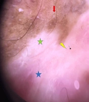
On examination, the patient had a firm, circumferential, cauliflower-like tumor involving the glans penis (Figure 1), with foul-smelling purulent discharge. Dermoscopic examination revealed white structures, black necrotic crusts, and multiple hemorrhages. The remainder of the skin examination showed a well-defined achromic and sclerotic lesion at the base of the glans, with a milky pink background, patchy white areas, comedo-like openings, and pinpoint vessels suggestive of LS (Figures 2 and 3). Lymph node examination did not reveal any palpable adenopathy, and the remainder of the physical examination was unremarkable.
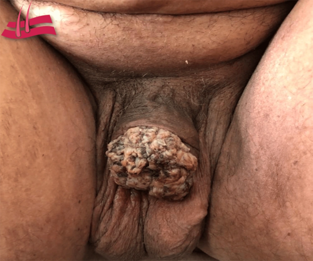
Figure 1.
Clinical image showing a vegetant lobulated penile tumor deforming the glans
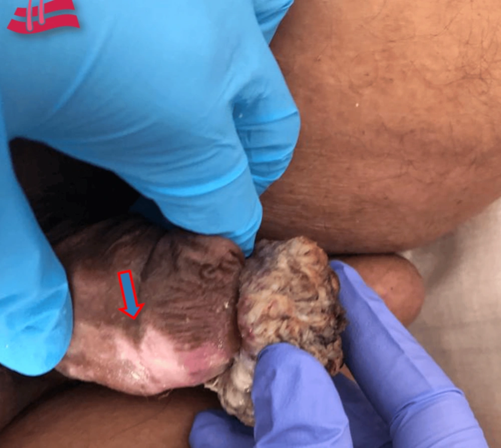
Figure 2.
Clinical image showing a well-defined pink-whitish and atrophic lesion on the base of the glans
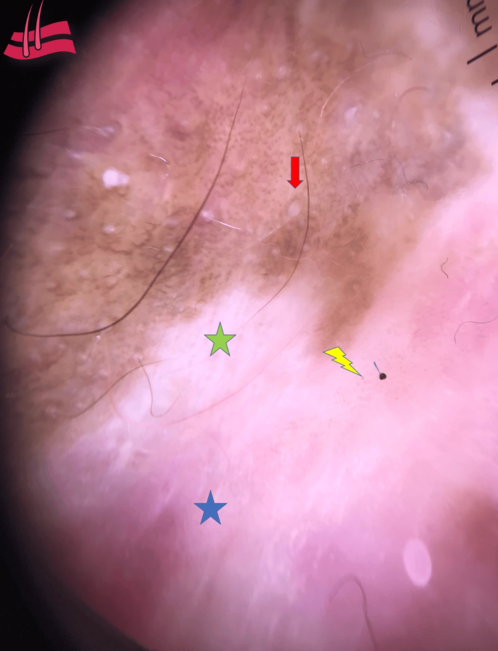
Figure 3.
Dermoscopy of the achromic lesion Dermoscopy of the achromic lesion showed Blue star: pink and whitish background Green star: Patchy white area Red arrow: Comedo-like openings Yellow bolt: pinpoint vessels
Several differential diagnoses were considered before biopsy. The chronic penile changes were compatible with inflammatory dermatoses, such as nonspecific balanitis, Zoon balanitis, and psoriasis, as well as premalignant epithelial disorders, including penile intraepithelial neoplasia, particularly erythroplasia of Queyrat. In addition, the exophytic cauliflower-like lesion prompted consideration of human papillomavirus-related condyloma, Buschke-Lowenstein tumor, verrucous carcinoma, and other verruciform or reactive proliferations. Ultimately, histopathological assessment was required to establish the final diagnosis.
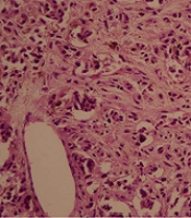
An initial biopsy of the tumor revealed moderately differentiated infiltrating SCC of the glans. Biopsy of the achromic lesion was consistent with LS, leading to the diagnosis of malignant transformation arising in LS.
The patient underwent penile magnetic resonance imaging, which revealed irregular circumferential thickening of the penile glans with low signal intensity on T1- and T2-weighted sequences and heterogeneous enhancement after gadolinium injection. The lesion infiltrated the fascia, invaded the spongy urethra, showed exaggerated contrast uptake in the urethral wall, and caused mild upstream dilation. Imaging also revealed bilateral subcentimeter inguinal lymphadenopathies.
Distant staging with abdominopelvic computed tomography did not reveal metastasis, and the tumor was classified as T2N0M0. The therapeutic approach was partial penectomy with lymph node dissection. The procedure involved excision of the distal penile segment containing the lesion, with an oncologically safe margin of approximately 1 cm. The remaining penile stump measured approximately 4 cm in length, allowing voiding in the standing position. No intraoperative complications occurred, and the patient tolerated the procedure well.
The postoperative course was uneventful, with satisfactory healing of the surgical site. The patient experienced no significant complications.
In this case, the prognosis was considered favorable because the disease was staged as T2N0M0, with no evidence of regional or distant metastasis at diagnosis. In addition, complete excision with negative margins and the absence of high-risk histopathological features, such as lymphovascular invasion or perineural invasion, supported a lower risk of recurrence.
Postoperative tolerability was evaluated during follow-up visits by clinical examination and patient-reported outcomes, with no significant complications noted.
At the 12-month follow-up, there were no signs of local recurrence or lymph node involvement on clinical examination or imaging. The patient reported improved quality of life. Psychosexual support was offered to help address the emotional and psychological impact of partial penectomy. The timeline of significant events is summarized in Table 1.
Table 1.
Timeline of Symptom Onset, Diagnosis, and Management
| Timeframe | Clinical Events |
|---|---|
| 4 years before presentation | Onset of a pruritic hypopigmented lesion on the glans penis, clinically consistent with early lichen sclerosus |
| Progressive course | Gradual enlargement of the lesion with increasing sclerosis and the appearance of an indurated nodule with intermittent bleeding |
| At dermatological evaluation | Circumferential, cauliflower-like tumor of the glans with adjacent achromic sclerotic plaques suggestive of lichen sclerosus |
| Histopathology | Tumor biopsy showed moderately differentiated invasive squamous cell carcinoma; adjacent lesion biopsy confirmed lichen sclerosus |
| Staging investigations | Penile magnetic resonance imaging demonstrated local invasion; abdominopelvic computed tomography showed no distant metastasis (T2N0M0) |
| Therapeutic intervention | Partial penectomy with inguinal lymph node dissection |
| Postoperative course | Uneventful healing without significant complications |
| 12-month follow-up | No evidence of recurrence or metastasis |
3. Discussion
Penile cancer is rare worldwide, with an incidence not exceeding 1% of cancers diagnosed in men. Its incidence is negligible in Islamic countries and Israel because of the religious practice of circumcision (3). Very limited data are available on the epidemiology of penile cancer in Morocco. A doctoral thesis conducted at the University Hospital of Fez between 2013 and 2017 included 605 patients with uro-oncological disease; only 2 patients (0.3%) had penile carcinoma, and only 1 of them had SCC. However, no risk factors were reported for this patient.
In a series of 6 cases in Rabat (1), none of the patients were reported to have LS before developing SCC, and human papillomavirus infection was the only investigated risk factor.
Penile cancer is uncommon. More than 95% of cases are SCCs. The disease is more prevalent in less industrialized regions, particularly in countries with lower hygiene standards and income levels. The highest incidence rates are reported in parts of South America, Africa, and India (4).
Because of the absence of cancer registries, data on the epidemiological profile of penile cancer are limited. However, the protective role of early circumcision has been suggested by studies showing that SCC is rare among populations in which circumcision is practiced as a religious ritual, particularly Jewish populations, in which circumcision is performed as early as the eighth day after birth (3). Muslims, however, typically undergo circumcision at a later age, between 3 and 13 years, and are consequently more affected. This difference has been attributed to the prevention of phimosis, as a retractable foreskin is suggested to facilitate retraction and proper hygiene.
Several risk factors have been identified over the years, with the primary factor being human papillomavirus infection, which is responsible for specific histological subtypes. This has led to a new World Health Organization classification that categorizes histological types according to their association with human papillomavirus infection (3).
Chronic inflammatory dermatoses, such as LS, erythroplasia of Queyrat, and Bowen disease, are considered premalignant lesions. The transformation risk for LS ranges from 4% to 8% (3). These conditions are currently grouped under the term penile intraepithelial neoplasia, analogous to cervical intraepithelial neoplasia in women. Penile intraepithelial neoplasia is divided into 2 subtypes: differentiated forms, often associated with LS, and undifferentiated forms, associated with human papillomavirus.
Other identified risk factors include phimosis, psoralen and ultraviolet A therapy in patients with psoriasis, chronic smoking, obesity, poor hygiene, and lower socioeconomic status.
In a systematic review assessing the complications of LS, SCC was identified as the most commonly reported malignancy arising in LS-affected tissues, accounting for 20% of LS-related complications in men (5). Several studies have reported a risk of malignant transformation ranging from 5% to 61% in patients with long-standing disease (6).
Prompt diagnosis and appropriate management of LS are essential not only for symptom control but also to help prevent progression to SCC (5, 6).
Treatment of premalignant penile lesions is an essential step in preventing neoplastic transformation. Several treatment options are available, including circumcision in uncircumcised individuals, followed by monitoring before considering additional treatments. For persistent lesions, treatment modalities include topical, ablative, and surgical approaches. Topical treatments include corticosteroids for LS, 5-fluorouracil, and imiquimod. Ablative treatments, such as carbon dioxide laser and neodymium-doped yttrium aluminum garnet laser, have also been used with good results for superficial lesions, but there is a risk of recurrence in cases of invasive lesions. Other methods include cryotherapy, photodynamic therapy, and surgical treatment (7, 8).
This case has several strengths that enhance its clinical relevance. Early recognition of the suspicious lesion enabled timely management. The patient underwent appropriate surgical treatment with satisfactory oncological control, and the postoperative course was favorable. In addition, regular follow-up showed good tolerability of the intervention, the absence of significant complications, and no evidence of recurrence or metastasis during the observation period.
3.1. Study Limitations
Because this report describes a single clinical case, the findings cannot be generalized to all patients with LS. The diagnosis of LS had not been confirmed before malignant transformation, which limits our understanding of the exact timeline and progression of the disease. In addition, although the patient remained recurrence-free at 12 months, longer follow-up is necessary to assess long-term outcomes.
4. Conclusions
This case illustrates the malignant transformation of long-neglected LS. Although this dermatosis is often considered benign, it should be widely recognized and monitored closely under strict medical surveillance to detect potential malignant transformation and improve prognosis with respect to survival, function, and aesthetics.