Cover image of the article: Reed Syndrome; Multiple Cutaneous and Uterine Leiomyomatosis

Image Credit:

Reed Syndrome; Multiple Cutaneous and Uterine Leiomyomatosis

Authors

Leila Yousefkhani1, Hojat EftekhariHojat Eftekhari ORCID1,*, Rana RafieiRana Rafiei ORCID1, Nahid Nikkhah2, Behnam Rafiee3
1Skin Research Center, Guilan University of Medical Sciences, Razi Hospital, Rasht, Iran
3Skin and Stem Cell Research Center, Tehran University of Medical Sciences, Tehran, Iran
2Department of Pathology, NYU Winthrop Hospital, New York, United States
*Corresponding Author: Skin Research Center, Guilan University of Medical Sciences, Razi Hospital, Sardare Jangal St., Rasht, Iran. Tel: +98-09111351439, Email: [email protected]

Journal of Skin and Stem Cell:Vol. 5, issue 1-2; e80758
Published online:Jul 21, 2018
Article type:Case Report
Received:Jun 10, 2018
Accepted:Jul 14, 2018
How to Cite:Yousefkhani L, Eftekhari H, Rafiei R, Nikkhah N, Rafiee B. Reed Syndrome; Multiple Cutaneous and Uterine Leiomyomatosis. J Skin Stem Cell. 2018;5(1-2):e80758. doi: https://doi.org/10.5812/jssc.80758

Abstract

Introduction:

Reed syndrome is a rare autosomal dominant disorder with a genetic defect in the fumarate hydratase. Patients with Reed syndrome have multiple cutaneous leiomyomas, uterine leiomyomas or leiomyosarcomas with increased risk of renal carcinoma.

Case Presentation:

The current study presented a 57-year-old female with multiple cutaneous and uterine leiomyomatosis with a positive family history. She and her family had no evidence of renal carcinoma.

Conclusions:

Multiple skin leiomyomas could be considered as a cutaneous marker for uterine leiomyomatosis and possible renal cell carcinoma in the familial presentations.

1. Introduction

Reed syndrome or familial cutaneous and uterine leiomyomatosis is a rare autosomal dominant disorder with increased risk of renal cell carcinoma (1, 2). The current study presented a female with multiple cutaneous and uterine leiomyomas.

2. Case Presentation

A 57-year-old female referred to the outpatient clinic with multiple asymptomatic to painful, erythematous, rubbery dermal nodules with smooth surfaces on her face and left arm. These lesions were 0.5 - 2 cm and developed when the patient was 25 years old (Figure 1). Size and number of the lesions gradually increased. She had a history of severe menorrhagia when she was 30 years old, which resulted in hysterectomy in her 40s. Also she had history of hypothyroidism, hypertension, hyperlipidemia, and renal stone. She was on atorvastatin, methoral, and levothyroxine therapy.
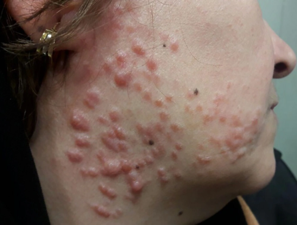
Multiple erythematous, rubbery dermal nodules with smooth surfaces on the face
Figure 1.
Multiple erythematous, rubbery dermal nodules with smooth surfaces on the face
She had three children; two daughters and one son. Her son was 37 years old and had similar lesions on similar sites in a clustered pattern from four years ago. Her older daughter was 34 and had only multiple uterine leiomyomas and severe menorrhagia. Her younger daughter was 32 and had multiple cutaneous lesions on mid-chest from six years ago with a history of severe menorrhagia. The current study patient had a cousin with similar problems. She had no history of renal carcinoma in her family.
An incisional biopsy was performed from facial skin lesions. Histopathologic evaluations showed a poorly demarcated neoplastic proliferation consisted of interlacing bundles of bland smooth muscle fibers, which had smooth muscle actin (SMA) positivity in immunohistochemistry. These fibers surrounded follicular structures and sweat glands; therefore, diagnosis of piloleiomyoma was proposed (Figure 2). Ultrasound imaging showed two simple cysts (22 and 7 mm) in the left kidney and a liver hemangioma measured 21 mm in diameter.
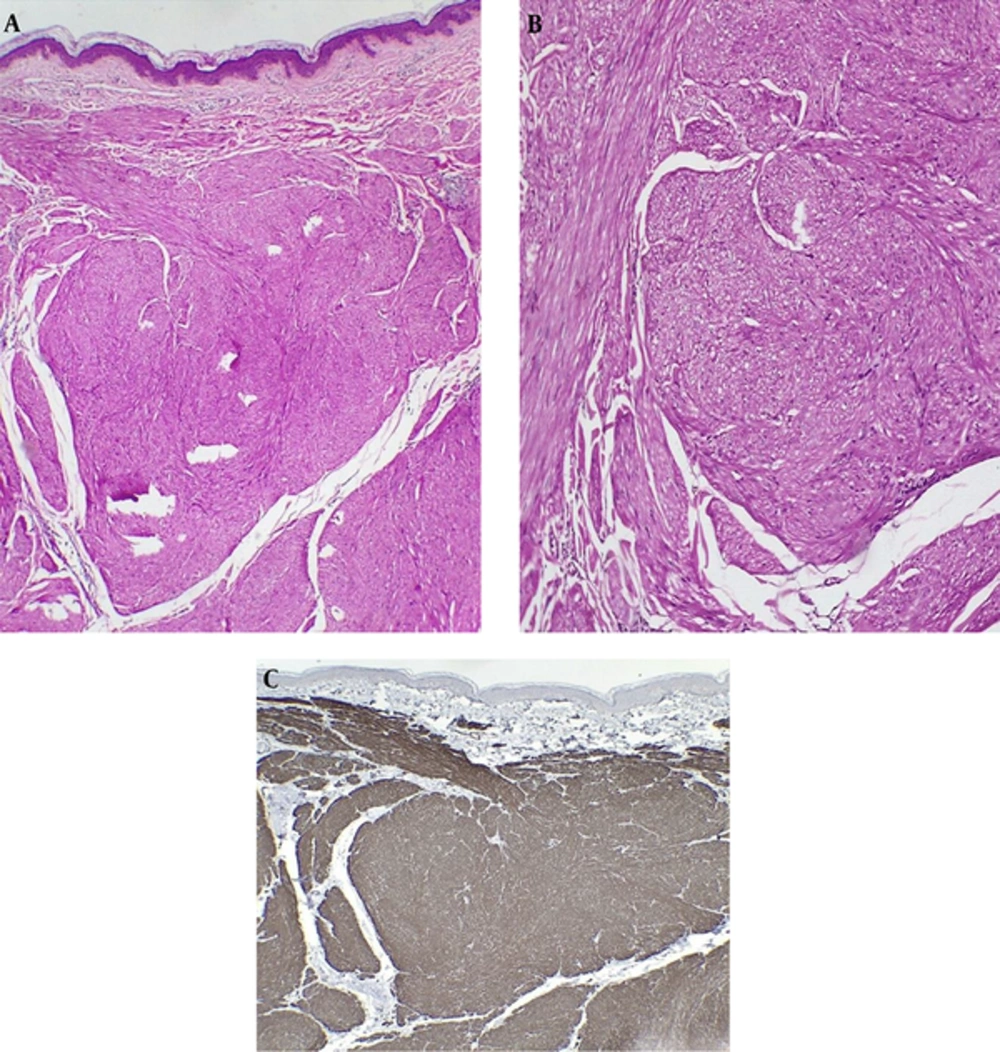
Histopathologic evaluation shows a poorly demarcated neoplastic proliferation consisted of interlacing bundles of bland smooth muscle fibers (hematoxylin and eosin staining, original magnification X40 A, X100 B), which had smooth muscle actin (SMA) positivity in immunohistochemistry (C).
Figure 2.
Histopathologic evaluation shows a poorly demarcated neoplastic proliferation consisted of interlacing bundles of bland smooth muscle fibers (hematoxylin and eosin staining, original magnification X40 A, X100 B), which had smooth muscle actin (SMA) positivity in immunohistochemistry (C).
Laboratory evaluations including complete blood count, erythrocyte sedimentation rate, and renal function tests were within normal limits. Diagnosis of Reed syndrome was made for this family. Therefore, all the affected family members were screened by renal ultrasound for possible renal cell carcinoma. Gabapentin capsule 100 mg daily was administered for the current study case to relieve occasional painful episodes of her lesions.

3. Discussion

Cutaneous leiomyoma is a benign skin tumor originated from dermal smooth muscles. Three subtypes of this tumor are described as follows: piloleiomyoma (originated from arrector pili muscles), leiomyoma of the external genitalia (originated from vulvar and mammary smooth muscles), and angioleiomyoma (originated from dermal blood vessels). The most common subtype is angioleiomyoma followed by piloleiomyoma (1, 2).
Piloleiomyoma is a skin-colored, pink or brown, asymptomatic to tender nodule usually observed on the extremities. It could be solitary or multiple with a clustered or dermatomal pattern. These lesions are usually observed in patients aged 10 to 30 years (1, 3).
Clinical differential diagnoses of leiomyoma are other skin tumors that could be painful such as spiradenoma, glomus tumor, fibrous or neural tumors (3, 4). Histopathologic evaluation differentiates leiomyoma from other tumors.
Total excision is the best method for the solitary lesion, but for multiple lesions some medications such as nifedipine, gabapentin, and doxazosin or CO2 laser irradiation could be useful (1, 3).
Reed syndrome is a syndrome with three clinical components as follows: (1) multiple cutaneous leiomyomas (mainly piloleiomyoma), (2) uterine leiomyomas or leiomyosarcomas with abdominal pain, menorrhagia, dysmenorrhea and sometimes infertility, and (3) increased risk of papillary renal cell carcinoma (3-5). It is caused by heterozygous mutation in fumarate hydratase (FH) gene, an enzyme involved in mitochondrial Krebs cycle. This gene is located on chromosome 1q42 (4-6). This mutation is related to papillary renal cell carcinoma type 2, with a high mortality in young age (4, 7).
There were signs of this syndrome in several members of this family, similar to the report by Mandal et al. (3), and similar to the current case report they did not report renal cell carcinoma. The relationship between renal cell carcinoma and multiple cutaneous and uterine leiomyomatosis is not high in studies; Alem et al. (7), reported this rate at 2% in 46 cases, Toro et al. (8), reported 6% in 35 patients, Martinez-Mir et al. (9), reported 0% in five patients and the current study observed no signs of malignancy in the case.
Screening is recommended for the patients with multiple leiomyomas including complete history and clinical examination, skin biopsy and histopathologic evaluation, periodic renal and pelvic ultrasound, complete blood count and metabolic panel, urine analysis, genetic analysis for mutation in FH gene and referring the patient to a gynecologist and nephrologist. The pelvic and renal ultrasound or computed tomography (CT) imaging should be repeated every two years. The family members should be completely examined and pelvic and renal ultrasound should be done if required (1, 4).

3.1. Conclusions

Cutaneous leiomyoma is a benign skin tumor, but multiple cutaneous leiomyomas could be considered as a sign of internal involvements such as uterine leiomyosarcoma or renal cell carcinoma, which requires more evaluations (1, 2).

References

  • 1.
    Kaddu S, Kohler S. Muscle, adipose and cartilage neoplasms. JL Bolognia, JL Jorizzo and JV Schaffer. Philadelphia: Elsevier Saunders. 2012;2:1979-92.
  • 2.
    Malhotra P, Walia H, Singh A, Ramesh V. Leiomyoma cutis: a clinicopathological series of 37 cases. Indian J Dermatol. 2010;55(4):337-41. [PubMed ID: 21430885]. [PubMed Central ID: PMC3051292]. https://doi.org/10.4103/0019-5154.74535.
  • 3.
    Mandal RK, Koley S, Banerjee S, Kabiraj SP, Ghosh SK, Kumar P. Familial leiomyomatosis cutis affecting nine family members in two successive generations including four cases of Reed's syndrome. Indian J Dermatol Venereol Leprol. 2013;79(1):83-7. [PubMed ID: 23254734]. https://doi.org/10.4103/0378-6323.104674.
  • 4.
    Emer JJ, Solomon S, Mercer SE. Reed's Syndrome: A Case of Multiple Cutaneous and Uterine Leiomyomas. J Clin Aesthet Dermatol. 2011;4(12):37-42. [PubMed ID: 22191007]. [PubMed Central ID: PMC3244358].
  • 5.
    Chan I, Wong T, Martinez-Mir A, Christiano AM, McGrath JA. Familial multiple cutaneous and uterine leiomyomas associated with papillary renal cell cancer. Clin Exp Dermatol. 2005;30(1):75-8. [PubMed ID: 15663510]. https://doi.org/10.1111/j.1365-2230.2004.01675.x.
  • 6.
    Kiuru M, Launonen V, Hietala M, Aittomaki K, Vierimaa O, Salovaara R, et al. Familial cutaneous leiomyomatosis is a two-hit condition associated with renal cell cancer of characteristic histopathology. Am J Pathol. 2001;159(3):825-9. [PubMed ID: 11549574]. [PubMed Central ID: PMC1850481]. https://doi.org/10.1016/S0002-9440(10)61757-9.
  • 7.
    Alam NA, Barclay E, Rowan AJ, Tyrer JP, Calonje E, Manek S, et al. Clinical features of multiple cutaneous and uterine leiomyomatosis: an underdiagnosed tumor syndrome. Arch Dermatol. 2005;141(2):199-206. [PubMed ID: 15724016]. https://doi.org/10.1001/archderm.141.2.199.
  • 8.
    Toro JR, Glenn G, Hou L, Duray P, Clark B, Merino M, et al. Facial papules, spontaneous pneumothorax, and renal tumors. J Am Acad Dermatol. 2003;48(1):111-4. [PubMed ID: 12522379]. https://doi.org/10.1067/mjd.2003.67.
  • 9.
    Martinez-Mir A, Glaser B, Chuang GS, Horev L, Waldman A, Engler DE, et al. Germline fumarate hydratase mutations in families with multiple cutaneous and uterine leiomyomata. J Invest Dermatol. 2003;121(4):741-4. [PubMed ID: 14632190]. https://doi.org/10.1046/j.1523-1747.2003.12499.x.

Copyright

Copyright © 2018, Journal of Skin and Stem Cell. This is an open-access article distributed under the terms of the Creative Commons Attribution-NonCommercial 4.0 International License (http://creativecommons.org/licenses/by-nc/4.0/) which permits copy and redistribute the material just in noncommercial usages, provided the original work is properly cited

Similar Articles

7
Dec
2022
Primary Malignant Melanoma of Uterine Cervix with Disseminated Involvement of Lower Genitourinary Tract

Primary Malignant Melanoma of Uterine Cervix with Disseminated Involvement of Lower Genitourinary Tract

Zahra Shiravani,
Minoo Robati,
Ali Ariafar,
Akbar Safaei,
Mojgan Hajisafari Tafti,
Shaghayegh Moradi Alamdarloo

Shiravani Z, Robati M, Ariafar A, Safaei A, Hajisafari Tafti M, et al. Primary Malignant Melanoma of Uterine Cervix with Disseminated Involvement of Lower Genitourinary Tract. Int J Cancer Manag. 2022;15(12):e114156. doi: https://doi.org/10.5812/ijcm-114156

28
Sep
2016

Nevoid Basal Cell Carcinoma Syndrome: A Case Report

Sayed Mohammad Razavi,
Amir Hossein Shakibamehr,
Saeedeh Khalesi

Razavi SM, Shakibamehr AH, Khalesi S. Nevoid Basal Cell Carcinoma Syndrome: A Case Report. Middle East J Rehabil Health Stud. 2016;3(4):e39758. doi: https://doi.org/10.17795/mejrh-39758

12
Aug
2022
A 15-Year-Old Girl with Trichorhinophalangeal Syndrome Type 1 with Non-ossifying Fibroma in Femur: A Case Report

A 15-Year-Old Girl with Trichorhinophalangeal Syndrome Type 1 with Non-ossifying Fibroma in Femur: A Case Report

Aye Miremarati,
Manijeh Tabrizi,
Setila Dalili,
Seyyedeh Azade Hoseini Nouri

Miremarati A, Tabrizi M, Dalili S, Hoseini Nouri SA. A 15-Year-Old Girl with Trichorhinophalangeal Syndrome Type 1 with Non-ossifying Fibroma in Femur: A Case Report. J Compr Ped. 2022;13(3):e129461. doi: https://doi.org/10.5812/compreped-129461

31
Jul
2017

Simultaneous Presentation of Cerebellopontine Angle Pleomorphic Xanthoastrocytoma and Malignant Melanoma in a Known Case of Neurofibromatosis 1; Probable Role of BRAF Gene: A Case Report and Review of Literature

Amir Saied Seddighi,
Afsoun Seddighi,
Sima Behrouzian,
Amir Nikouei

Seddighi AS, Seddighi A, Behrouzian S, Nikouei A. Simultaneous Presentation of Cerebellopontine Angle Pleomorphic Xanthoastrocytoma and Malignant Melanoma in a Known Case of Neurofibromatosis 1; Probable Role of BRAF Gene: A Case Report and Review of Literature. Int J Cancer Manag. 2017;10(7):e7211. doi: https://doi.org/10.5812/ijcm.7211

7
Jan
2020
Becker’s Nevus with Lichen Simplex Chronicus Limited to the Nevus Lesion: A Case Report

Becker’s Nevus with Lichen Simplex Chronicus Limited to the Nevus Lesion: A Case Report

Samer Mriani,
Bandar Tammar,
Hasan Maghrabi,
Khalid Al Aboud,
Waleed Yamani,
Zaher Al Asmary
,et al.

Mriani S, Tammar B, Maghrabi H, Al Aboud K, Yamani W, et al. Becker’s Nevus with Lichen Simplex Chronicus Limited to the Nevus Lesion: A Case Report. J Skin Stem Cell. 2019;6(2):e98571. doi: https://doi.org/10.5812/jssc.98571

More by these authors

Leila YousefkhaniPubMedScholar
Hojat EftekhariPubMedScholar
Rana RafieiPubMedScholar
Nahid NikkhahPubMedScholar
Behnam RafieePubMedScholar
Share
Cited by
Metrics