Primary Urachal Actinomycosis: Case Report and Literature Review

Author(s):
Ghodratolah MaddahGhodratolah Maddah1, Behzad Feizzdeh KerighBehzad Feizzdeh Kerigh2,*, Nema MohamadianNema Mohamadian3, Vafa BagheriVafa Bagheri4
1Endoscopic and Minimally Invasive Surgery Research Center, Mashhad University of Medical Sciences, Mashhad, IR Iran
2Department of Urology, Mashhad University of Medical Sciences, Mashhad, IR Iran
3Deprtment of Pathology, Mashhad University of Medical Sciences, Mashhad, IR Iran
4Deprtment of Surgery, Mazandaran University of Medical Sciences, Sari, IR Iran

Nephro-Urology Monthly:Vol. 5, issue 5; 997-1000
Published online:Nov 13, 2013
Article type:Case Report
Received:Mar 05, 2013
Accepted:Mar 26, 2013
How to Cite:Maddah G, Feizzdeh Kerigh B, Mohamadian N, Bagheri V. Primary Urachal Actinomycosis: Case Report and Literature Review. Nephro-Urol Mon. 2013;5(5):997-1000. doi: https://doi.org/10.5812/numonthly.10933

Abstract

1. Introduction

Actinomycosis is an infectious, chronic and granulomatous disease caused by gram-positive non-aerobic bacteria. These bacteria are te natural flora of mouth cavity especially teeth and tonsil, GI especially appendix and colon, and genital system in human that rarely causes disease. The prevalence of clinical actinomycosis is cervicofacial 50%, abdominal 20%, and thoracic 15%, respectively (1, 2).
Primary urachus actinomycosis is rarely reported in English literature and is usually mistaken with urachus malignancy (3, 4). Here, we report a primary urachus actinomycosis in a young man.

2. Case Report

A 23-years old man, the worker of granite mine referred complaining of intermittent pain around the umbilicus from several months ago. The pain was progressive and continuous from 6 months ago. Recently, the patient had umbilicus pain during voiding. The patient didn’t mention the symptoms of weight and appetite loss During examination, a solid and a little tender mass was tangible as about 12 cm expanding from umbilicus to hypogastria. Fothergil test (the mass is better tangible when the feet were above) was positive. He had no fever and the examination of other parts of his body was normal.
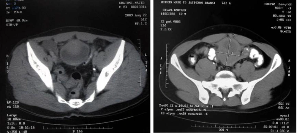
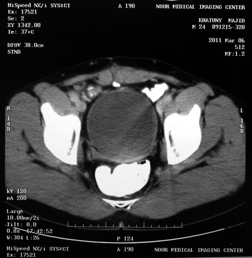
Sonography showed the heterogeneous mass with size of 124 × 61 mm expanding from the umbilicus to the bladder (Figure 1). Abdominal and pelvic CT scan showed a solid mass beginning from the umbilicus with pressure on the bladder (Figure 2). Chest X-Ray was normal. Laboratory tests of urine, urea, creatinine, and liver function tests were normal. He didn’t have leukocytosis and anemia.
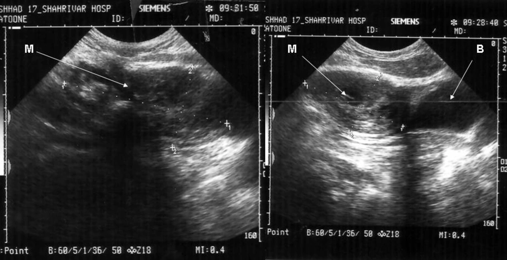
M: Mass, B: Bladder
Figure 1.

M: Mass, B: Bladder

CT Scanning Showed Solid Mass Above the Bladder
Figure 2.

CT Scanning Showed Solid Mass Above the Bladder

The patient underwent cystoscopy due to suspected urachus adenocarcinoma. Urothelium was intact but mass pressure on bladder was observed. The patient underwent laparotomy with the incision of under umbilicus middle line. The mass had adherence to bladder and omentum and posterior rectus sheath which was removed with suitable margin along with partial cystectomy. The mass was solid, irregular, and brown. Bilateral lymphadenectomy was performed due to gross lymph nodes on iliac vessels.
Microscopic evaluation of mass reported the actinomycosis without involvement of bladder urothelium (Figures 3 and 4). The lymph nodes were reactive. After diagnosis, Amoxicillin was administered for 8 weeks. The patient didn’t have any problem at 2-years follow-up (Figure 5).
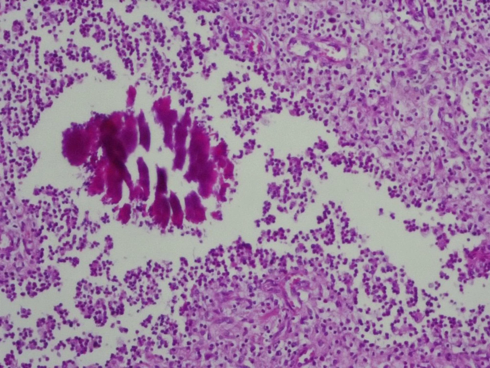
Sulphur Granules of Actinomyces With Peripheral Mixed Inflammatory Reaction. H&E Staining, 100X
Figure 3.

Sulphur Granules of Actinomyces With Peripheral Mixed Inflammatory Reaction. H&E Staining, 100X
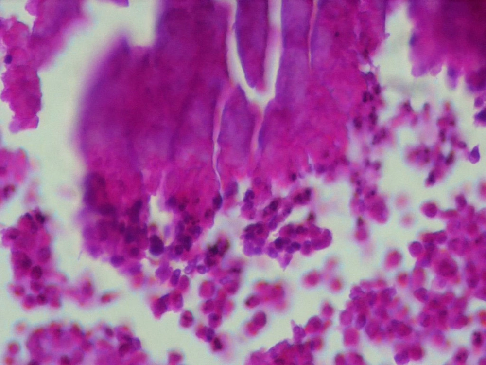
Sulphur Granules of Actinomyces With Peripheral Mixed Inflammatory Reaction. H&E Staining, 400X
Figure 4.

Sulphur Granules of Actinomyces With Peripheral Mixed Inflammatory Reaction. H&E Staining, 400X
Normal CT Scanning After Two Years Follow-up.
Figure 5.

Normal CT Scanning After Two Years Follow-up.

3. Discussion

Actinomycosis is a chronic, indolent and recurrent disease which can involve all urogenital sections such as kidney, bladder, prostate, and testicle (5). Abdominal and pelvic actinomycosis is usually caused in women due to use of intrauterine device, but other risk factors such as abdominal surgery, viscus rupture, tubo-ovarian abscess, diabetes, use of steroid and neoplasm are also were reported (6-8). It was emphasis that actinomycosis can complicate urachus through umbilicus and its abnormalities such as cyst and sinus that susceptible it to non-aerobic infection (9). Our patient didn’t have any of these risk factors.
Urachus actinomycosis is rarely reported and the patients usually refer with symptoms of tangible mass under umbilicus, abdominal pain, weight loss, appetite decrease, fever, and laboratory signs such as leukocytosis and anemia (10). Our patient also referred with abdominal and voiding pain, but didn’t have laboratory signs such as leukocytosis and anemia.
Correct diagnosis before surgery is reported in only lower than 10% of cases. Abdominal actinomycosis may be mistaken with malignancy, bowel tuberculosis, crohn disease, diverticular and rectus sheath pathologies. Malignancy and abscess are in differential diagnosis (11). In our patient, mass was removed with possible diagnosis of tumor and partial cystectomy and bilateral lymphadenectomy were performed and diagnosis was determined after surgery.
Imaging findings are non-specific in urachal actimycosis. Sonography determines the solid or cystic mass but may underestimate the expansion of inflammatory reaction. According to most reports, CT scan can better determine the status of mass than MRI (magnetic resonance imaging). The findings of PET/CT of actinomycosis are as intense hypermetabolism which is similar to malignancy (3, 12). Low signal intensity in MRI at T2-weighted sequences may suggest the actinomycosis (4). In our patient, sonography and CT-scan didn’t help to definite diagnosis and only one mass suspicious to malignancy was suggested.
In a study about pelvic actinomycosis in 33 cases, cystoscopy was performed on 12 patients and the findings of cystoscopy reported the pressure effect of external mass on bladder, bullosis edema, and vegetative proliferation in one case showing chronic inflammatory changes (8). Furthurmore, in our patient, cystoscopy was performed due to voiding pain and the possibility of urach adenocarcinoma; urothelium was intact but pressure of mass on bladder was observed.
Since the clinical laboratory and radiologic signs are not special, in most cases, the diagnosis is determined after surgery (9). If the diagnosis is performed before surgery, Penicillin is the drug of choice which should be intravenously administered and then oral Penicillin and Amoxicillin should be administered for a long time (13). However, some researchers believe that combination therapy of both surgery and antibiotic administration is the most effective modality and good results are observed in more than 90% of cases (5, 14). Also, in our patient, diagnosis was determined after removing the mass.
In most reported cases, the patients didn’t have any problem at long follow-up after surgery (3, 9); our patient also didn’t have any problem after 2-years follow-up.

Acknowledgments

Footnotes

References

  • 1.
    Kodali U, Mallavarapu R, Goldberg MJ. Abdominal actinomycosis presenting as lower gastrointestinal bleeding. Endoscopy. 2003;35(5):451-3. [PubMed ID: 12701020]. https://doi.org/10.1055/s-2003-38782.
  • 2.
    Yang SH, Li AF, Lin JK. Colonoscopy in abdominal actinomycosis. Gastrointest Endosc. 2000;51(2):236-8. [PubMed ID: 10650281].
  • 3.
    Li MF, Yang TL, Huang JS. Imaging findings of primary urachal actinomycosis. J Chin Med Assoc. 2011;74(1):44-7. [PubMed ID: 21292203]. https://doi.org/10.1016/j.jcma.2011.01.007.
  • 4.
    Yeung Y, Cheung MC, Chan GS, Cheung AN, Tam PC. Primary actinomycosis mimicking urachal carcinoma. Urology. 2001;58(3):462. [PubMed ID: 11549501].
  • 5.
    Lim KT, Moon SJ, Kwon JS, Son YW, Choi HY, Choi YY, et al. Urachal actinomycosis mimicking a urachal tumor. Korean J Urol. 2010;51(6):438-40. [PubMed ID: 20577614]. https://doi.org/10.4111/kju.2010.51.6.438.
  • 6.
    Al-Kadhi S, Venkiteswaran KP, Al-Ansari A, Shamsudini A, Al-Bozom I, Kiliyanni AS. Primary vesical actinomycosis: a case report and literature review. Int J Urol. 2007;14(10):969-71. [PubMed ID: 17880305]. https://doi.org/10.1111/j.1442-2042.2007.01843.x.
  • 7.
    Hsiao HL, Shen JT, Yeh HC, Wu WJ, Wang CJ, Huang CH. Intra- and extra-abdominal actinomycosis mimicking urachal tumor in an intrauterine device carrier: a case report. Kaohsiung J Med Sci. 2008;24(1):35-40. [PubMed ID: 18218568]. https://doi.org/10.1016/S1607-551X(08)70071-8.
  • 8.
    Marella VK, Hakimian O, Wise GJ, Silver DA. Pelvic actinomycosis. Urologic perspective. Int Braz J Urol. 2004;30(5):367-76. [PubMed ID: 15610567].
  • 9.
    Garcia Garcia JC, Nunez Fernandez MJ, Cerqueiro Gonzalez JM, Garcia Martin C, Rodriguez Garcia JC, Anibarro Garcia L, et al. [Primary actinomycosis of the abdominal wall. Description of 2 cases and review of the literature]. An Med Interna. 2001;18(2):80-3. [PubMed ID: 11322003].
  • 10.
    Cintron JR, Del Pino A, Duarte B, Wood D. Abdominal actinomycosis. Dis Colon Rectum. 1996;39(1):105-8. [PubMed ID: 8601346].
  • 11.
    Ho L, Seto J, Jadvar H. Actinomycosis mimicking anastomotic recurrent esophageal cancer on PET-CT. Clin Nucl Med. 2006;31(10):646-7. [PubMed ID: 16985378]. https://doi.org/10.1097/01.rlu.0000238193.34543.b9.
  • 12.
    Hawnaur JM, Reynolds K, McGettigan C. Magnetic resonance imaging of actinomycosis presenting as pelvic malignancy. Br J Radiol. 1999;72(862):1006-11. [PubMed ID: 10673954].
  • 13.
    Yeung VH, Wong QH, Chao NS, Leung MW, Kwok WK. Thoracic actinomycosis in an adolescent mimicking chest wall tumor or pulmonary tuberculosis. Pediatr Surg Int. 2008;24(6):751-4. [PubMed ID: 11549501].
  • 14.
    Filipovic B, Milinic N, Nikolic G, Ranthelovic T. Primary actinomycosis of the anterior abdominal wall: case report and review of the literature. J Gastroenterol Hepatol. 2005;20(4):517-20. [PubMed ID: 15836698]. https://doi.org/10.1111/j.1440-1746.2004.03564.x.

Similar Articles

12
Sep
2018
https://www.google.com/url?sa=i&rct=j&q=&esrc=s&source=imgres&cd=&cad=rja&uact=8&ved=2ahUKEwi7zfjJ2bTdAhVPLFAKHRXSDrwQjB16BAgBEAQ&url=https%3A%2F%2Fwww.atipt.com%2Fbowelbladder-dysfunction&psig=AOvVaw3qJNSUADfHVnxqe7CWZqEC&ust=1536815372945078

A Rare Case of Urachal Adenocarcinoma: Long Term Follow Up

Mohammad Reza Darabi,
Ali Reza Khoei,
Atena Aghaee,
Salman Soltani

Darabi MR, Khoei AR, Aghaee A, Soltani S. A Rare Case of Urachal Adenocarcinoma: Long Term Follow Up. Nephro-Urol Mon. 2018;10(5):e81471. doi: https://doi.org/10.5812/numonthly.81471

25
Jun
2018
Renal Actinomycosis

Renal Actinomycosis, A Rare Diagnosis Which Can Clinically Mimic a Malignancy, Case Report and Review of the Literature

Nakisa Niknejad,
Behnaz Moradi,
Nasim Niknezhad,
Masoomeh Safaei,
Fatemeh Nili

Niknejad N, Moradi B, Niknezhad N, Safaei M, Nili F. Renal Actinomycosis, A Rare Diagnosis Which Can Clinically Mimic a Malignancy, Case Report and Review of the Literature. Arch Pediatr Infect Dis. 2018;6(3):e13049. doi: https://doi.org/10.5812/pedinfect.13049

16
Oct
2023
Nephro-Urol Mon

A Rare Case of Urachal Remnant with Calculus Just Above the Bladder

Amin Mirsani,
Mohammad Amin Dana,
Seyed Hadi Sajjadi,
Raheleh Baradaran

Mirsani A, Dana MA, Sajjadi SH, Baradaran R. A Rare Case of Urachal Remnant with Calculus Just Above the Bladder. Nephro-Urol Mon. 2023;15(4):e137320. doi: https://doi.org/10.5812/numonthly-137320

30
Dec
2009

Retroperitoneal Neurilemmoma Misdiagnosed as Hepatic Tumor: A Case Report

Yan Qing Tong,
Hong Yang Guo,
Bing Han

Tong YQ, Guo HY, Han B. Retroperitoneal Neurilemmoma Misdiagnosed as Hepatic Tumor: A Case Report. I J Radiol. 2009;6(4):e78854. doi:

4
Nov
2023

Recurrent Fever and Abdominal Pain: Periodic Fever Syndrome, Inflammatory Bowel Disease or Patent Urachus; A Case Report

Khosro Rahmani,
Mohsen Jari,
Reza Shiari,
Mehrnoush Hassas Yeganeh,
Vadood Javadi

Rahmani K, Jari M, Shiari R, Hassas Yeganeh M, Javadi V. Recurrent Fever and Abdominal Pain: Periodic Fever Syndrome, Inflammatory Bowel Disease or Patent Urachus; A Case Report. Arch Pediatr Infect Dis. 2024;12(3):e139427. doi: https://doi.org/10.5812/apid-139427


Crossmark
Crossmark
Checking
Share on
Cited by
Metrics

Ordering Reprints

Articles are published under the Creative Commons license stated on each article. No permission or royalty fee is required for uses permitted by that license. CCC handles optional bulk and customized reprint orders. Any quotation covers production and delivery services only, not copyright permission. > Request Reprints from CCC 

Search Relations

Author(s):

Related Articles