Bilateral Renal Lymphangiectasia in a Thirty-two-Year-Old Woman

Author(s):
Kaushal KumarKaushal Kumar1,*, Ahsan AhmadAhsan Ahmad1, Mahendra SinghMahendra Singh1, Amit KumarAmit Kumar2, Rana Pratap SinghRana Pratap Singh1, Mumtaz HussainMumtaz Hussain1
1Department of Urology, Indira Gandhi Institute of Medical Sciences, Patna, India
2Department of Radiodiagnosis,Indira Gandhi Institute of Medical Sciences, Patna, India
*Corresponding Author: Corresponding author: Kaushal Kumar, Department of Urology, Indira Gandhi Institute of Medical Sciences, Patna, India. Tel: +91-919431457765, E-mail: Email: [email protected]

Nephro-Urology Monthly:Vol. 7, issue 1; e21736
Published online:Dec 19, 2014
Article type:Case Report
Received:Jul 03, 2014
Accepted:Aug 20, 2014
How to Cite:Kumar K, Ahmad A, Singh M, Kumar A, Pratap Singh R, et al. Bilateral Renal Lymphangiectasia in a Thirty-two-Year-Old Woman. Nephro-Urol Mon. 2015;7(1):e21736. doi: https://doi.org/10.5812/numonthly.21736

Abstract

Introduction:

Renal lymphangiectasia is a rare disorder of the lymphatic system that might be confused with various other renal cystic diseases and urinoma. Clinical presentation is non-specific and characteristic findings in radiologic imaging are the mainstay of diagnosis.

Case Presentation:

We reported a case of bilateral lymphangiectasia in a 32-year-old woman who presented with flank pain and hypertension.

Discussion:

Renal lymphangiectasia is an uncommon benign condition. Most of the cases improve with conservative treatment. Surgery is rarely required, except in cases such as those with persistent pain refractory to medication.

1. Introduction

Renal lymphangiectasia is a rare renal condition characterized by dilatation of perirenal, per pelvic, and intrarenal lymphatic vessels (1, 2). This condition can be found in children and adults of both sexes and can be unilateral or bilateral (3-5). Isolated case reports are the only available source of information about this condition. Familial, developmental, and acquired causes have been suggested. Clinical signs and symptoms are non-specific and characteristic findings on ultrasonography (USG), computerized tomography (CT), and magnetic resonance imaging (MRI) arethe mainstay of diagnosis.

2. Case Presentation

A 32-year-old womanpresented with recent history of intermittent low-grade bilateral flank pain. She had no history of any medical illness and family history was not significant. Physical examination findings were normal. Her blood pressure was 160/90 mm Hg. Urinalysis results were normal. Laboratory analysis revealed normal renal function. Ultrasonography revealed bilateral perirenal and parapelvic septate collections with increased echo texture and enlarged kidneys. Subsequent CT scan of abdomen showed per pelvic collections of various densities with indentationon renal capsule. Collecting systemis splayed without dilatation. Excretory phase had normal excretion of contrast without any extravasation, ruling out any communication with pelvicalyceal system. Analysis of USG-guided aspirated fluid showed few lymphocytes with abundant protein and high levels of renin. No organism was isolated from aspirated fluid. A diagnosis of bilateral renal lymphangiectasia was made. Patient was treated with angiotensin-converting enzyme (ACE) inhibitors, 50 mg of Diclofenac SOS, and salt restriction. On follow-up, her blood pressure came down to 130/80 mm Hg. She was advised for long-term follow-up. Pain gradually subsided without any surgical intervention (Figures 1-4).
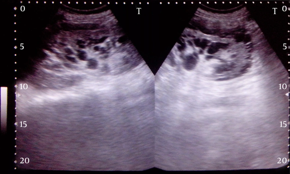
Ultrasonographic Image Showing Perirenal and Per Pelvic Collections With Septa
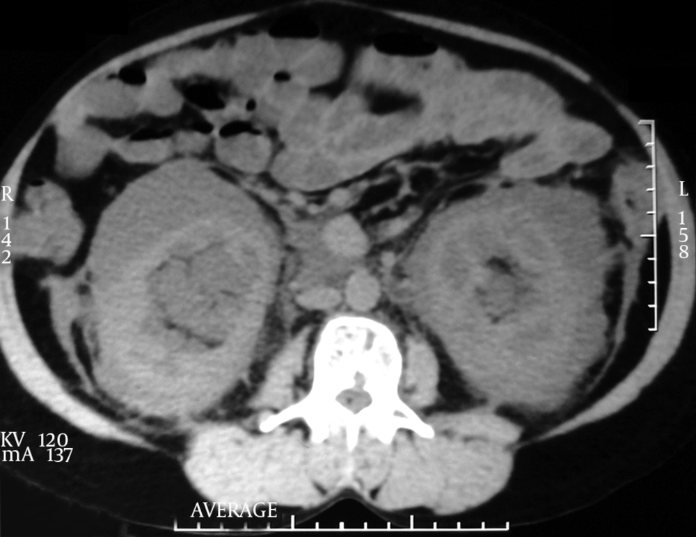
Figure 1.

Ultrasonographic Image Showing Perirenal and Per Pelvic Collections With Septa

Computed Tomography Image Showing Symmetric, Hypodense Perirenal Collections
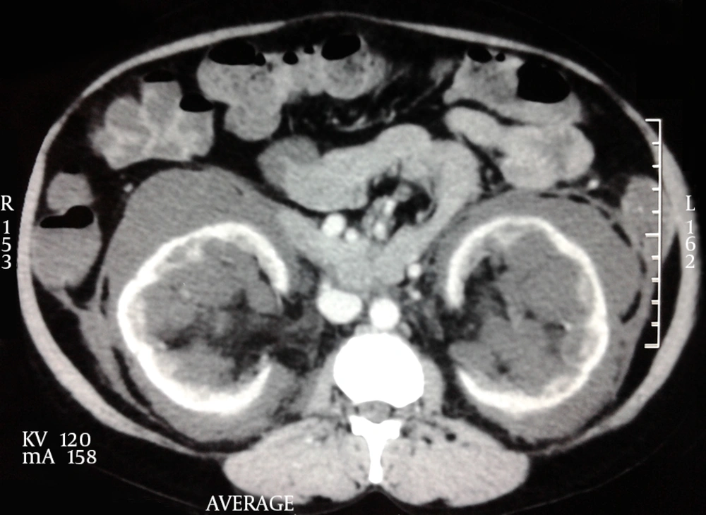
Figure 2.

Computed Tomography Image Showing Symmetric, Hypodense Perirenal Collections

Contrast-Enhanced Computed Tomography Image Showing Normal Cortical Enhancement With Non-enhancing Perirenal and Peripelvic Collections
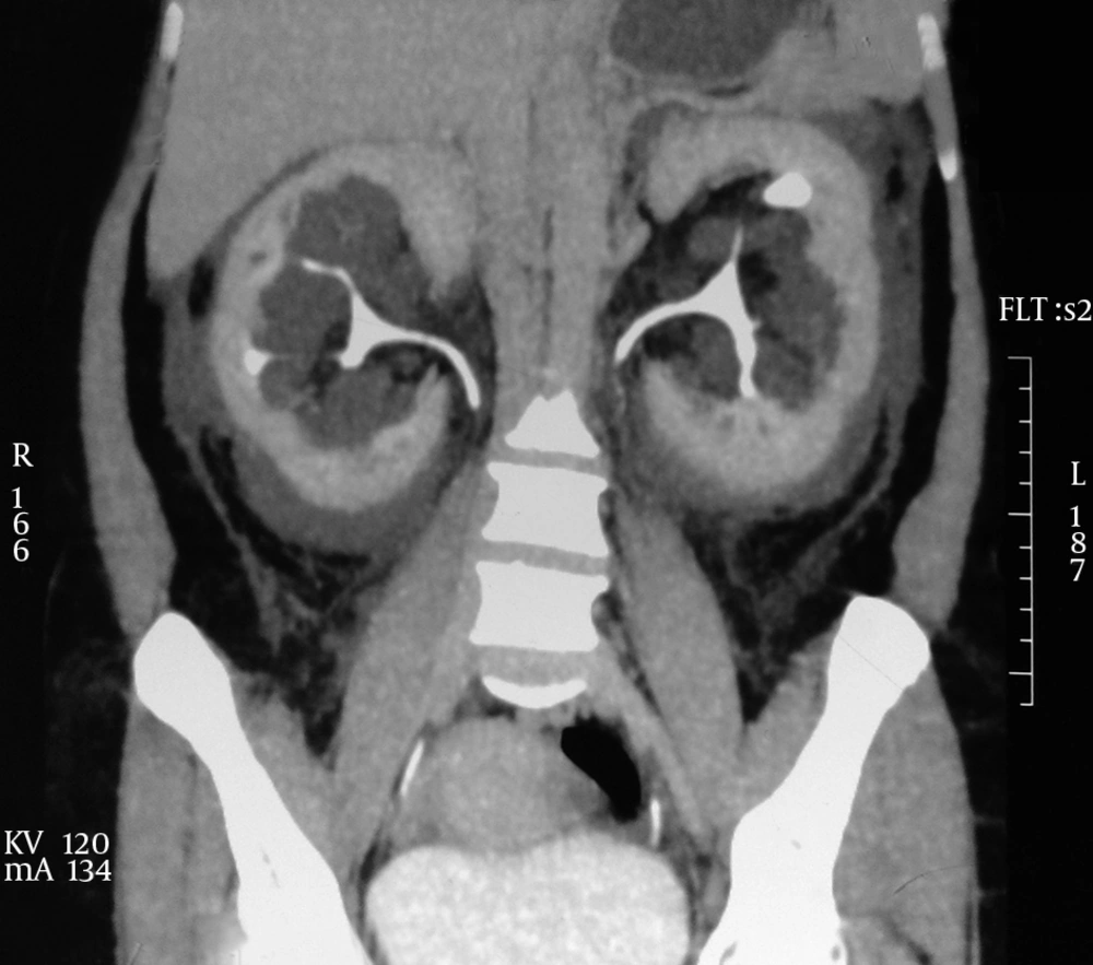
Figure 3.

Contrast-Enhanced Computed Tomography Image Showing Normal Cortical Enhancement With Non-enhancing Perirenal and Peripelvic Collections

Computed Tomographic Urogram Showing Splayed and Nondilated Pelvicalyceal System
Figure 4.

Computed Tomographic Urogram Showing Splayed and Nondilated Pelvicalyceal System

3. Discussion

Renal lymphangiectasia is an uncommon benign condition associated with disturbances in lymphatic drainage of perirenal and hilar areas causing cystic dilatation of perirenal and peripelvic lymphatics (6, 7). Although the exact pathophysiology is unclear, both developmental malformation and acquired obstructive inflammatory causes has been suggested. Familial association has been also found in a few cases. Clinical manifestation is non-specific. Patients might present with flank pain, abdominal mass, lower extremity edema, gross hematuria, ascites, or hypertension (7). Renal function is usually well preserved. The cause of hypertension is presumed to be compression of renal parenchyma by the subcapsular collection resulting in excessive renin release. Differential diagnosis of this condition includes polycystic kidney disease, multicystic dysplastic kidneys, multilocular cystic nephroma, and urinoma depending upon clinical presentation. Diagnosis can be made on the basis of characteristic findings on USG, CT-scan, and MRI. Needle aspiration of chylous fluid from the perinephric fluid collections confirms the diagnosis.
Asymptomatic cases do not require treatment. Percutaneous aspiration of perinephric collection has been done to relieve pain not responding to analgesics (8). Marsupialization has been also reported to make communication with peritoneal cavity to drain fluid (9). Hypertension is controlled with ACE inhibitors. A diuretic is used to control the ascites. Rarely, renal lymphangiectasia is associated with renal vein thrombosis and needs nephrectomy (10).


Acknowledgments

References

  • 1.
    Ramseyer LT. Case 34: renal lymphangiectasia. Radiology. 2001;219(2):442-4. [PubMed ID: 11323470]. https://doi.org/10.1148/radiology.219.2.r01ma17442.
  • 2.
    Llorente JG, Garcia AD, Sacristan JS, Chicharro GN. Renal lymphangiectasia: radiologic diagnosis and evolution. Abdom Imaging. 2002;27(6):637-9. [PubMed ID: 12395251]. https://doi.org/10.1007/s00261-001-0147-z.
  • 3.
    Merguerian PA, Sargent SK, Dunn JL. Unilateral lymphangiectasis of the kidney: an unusual cause of renal enlargement in an infant. J Urol. 1995;153(2):447-9. [PubMed ID: 7815616]. https://doi.org/10.1097/00005392-199502000-00053.
  • 4.
    Gupta R, Sharma R, Gamanagatti S, Dogra PN, Kumar A. Unilateral renal lymphangiectasia: imaging appearance on sonography, CT and MRI. Int Urol Nephrol. 2007;39(2):361-4. [PubMed ID: 17171417]. https://doi.org/10.1007/s11255-006-9039-z.
  • 5.
    Varela JR, Bargiela A, Requejo I, Fernandez R, Darriba M, Pombo F. Bilateral renal lymphangiomatosis: US and CT findings. Eur Radiol. 1998;8(2):230-1. [PubMed ID: 9477271]. https://doi.org/10.1007/s003300050368.
  • 6.
    Kutcher R, Mahadevia P, Nussbaum MK, Rosenblatt R, Freed S. Renal peripelvic multicystic lymphangiectasia. Urology. 1987;30(2):177-9. [PubMed ID: 3617306].
  • 7.
    Ashraf K, Raza SS, Ashraf O, Memon W, Memon A, Zubairi TA. Renal lymphangiectasia. Br J Radiol. 2007;80(954):e117-8. [PubMed ID: 17684071]. https://doi.org/10.1259/bjr/16931054.
  • 8.
    Pianezza ML, Mokhtassi A, Wu L, D'A Honey RJ. Case report: renal lymphangiectasia. Can J Urol. 2006;13(4):3204-7. [PubMed ID: 16952331].
  • 9.
    Schwarz A, Lenz T, Klaen R, Offermann G, Fiedler U, Nussberger J. Hygroma renale: pararenal lymphatic cysts associated with renin-dependent hypertension (Page kidney). Case report on bilateral cysts and successful therapy by marsupialization. J Urol. 1993;150(3):953-7. [PubMed ID: 8345618].
  • 10.
    Riehl J, Schmitt H, Schafer L, Schneider B, Sieberth HG. Retroperitoneal lymphangiectasia associated with bilateral renal vein thrombosis. Nephrol Dial Transplant. 1997;12(8):1701-3. [PubMed ID: 9269653].

Copyright

Copyright © 2014, Nephrology and Urology Research Center. This is an open-access article distributed under the terms of the Creative Commons Attribution-NonCommercial 4.0 International License (http://creativecommons.org/licenses/by-nc/4.0/) which permits copy and redistribute the material just in noncommercial usages, provided the original work is properly cited.

Similar Articles

23
May
2015

Adrenal Cystic Lymphangioma Presented With Hypertension: A Case Report

Bita Geramizadeh,
Shahrzad Yazdanpanah,
Heshmatollah Salahi,
Mahsa Marzban

Geramizadeh B, Yazdanpanah S, Salahi H, Marzban M. Adrenal Cystic Lymphangioma Presented With Hypertension: A Case Report. Nephro-Urol Mon. 2015;7(3):e27049. doi: https://doi.org/10.5812/numonthly.7(3)2015.27049

7
Jul
2024
Nephro-Urol Mon

Low Intraoperative Bleeding During Laparoscopic Nephrolithotomy: A Case Report

Ahmad Sulaiman Lubis,
Ahmad Zulfan Hendri,
Dhirajaya Dharma Kadar,
Pradipta Haydan Aldiosta,
Wildan Chanieful Wajiih,
Fadhli Rizal Makarim

Sulaiman Lubis A, Zulfan Hendri A, Dharma Kadar D, Haydan Aldiosta P, Chanieful Wajiih W, et al. Low Intraoperative Bleeding During Laparoscopic Nephrolithotomy: A Case Report. Nephro-Urol Mon. 2024;16(2):e146785. doi: https://doi.org/10.5812/numonthly-146785

27
Dec
2012

Ureterocele Associated with Renal Agenesia Presented as a Pelvic Mass in an Adult

Mohammad Ghasem Mohseni,
Seyed Reza Hosseini,
Alborz Salavati,
Shahrzad Dadgari

Mohseni MG, Hosseini SR, Salavati A, Dadgari S. Ureterocele Associated with Renal Agenesia Presented as a Pelvic Mass in an Adult. I J Radiol. 2013;10(1):45-47. doi: https://doi.org/10.5812/iranjradiol.10262

30
Dec
2013

Ureteral Stump Metastasis of Renal Cell Carcinoma: Report of a Case

Ladan Ajori,
Babak Javanmard,
Aida Moeini,
Hooman Bahrami‐Motlagh,
Mohammad Mohsen Mazloomfard

Ajori L, Javanmard B, Moeini A, Bahrami‐Motlagh H, Mazloomfard MM. Ureteral Stump Metastasis of Renal Cell Carcinoma: Report of a Case. Int J Cancer Manag. 2013;6(Supplement):e80477. doi:

23
May
2015

Unusual Presentation of Renal Cell Carcinoma in Crossed Ectopic Kidney

Mohammad Reza Nowroozi,
Hamidreza Ghorbani,
Erfan Amini,
Amir Arbab,
Alireza Ghadian

Nowroozi MR, Ghorbani H, Amini E, Arbab A, Ghadian A. Unusual Presentation of Renal Cell Carcinoma in Crossed Ectopic Kidney. Nephro-Urol Mon. 2015;7(3):e26760. doi: https://doi.org/10.5812/numonthly.7(3)2015.26760

Download PDF100.20 KB

Crossmark

Crossmark

Checking

Share on
Cited by
Metrics

Ordering Reprints

Articles are published under the Creative Commons license stated on each article. No permission or royalty fee is required for uses permitted by that license. CCC handles optional bulk and customized reprint orders. Any quotation covers production and delivery services only, not copyright permission. > Request Reprints from CCC 

Search Relations

Author(s):

Related Articles