

1. Background
After the introduction of percutaneous renal access, there has been remarkable improvements in the equipment and technique of percutaneous nephrolithotomy (PCNL) after 3 decades, which has confirmed this procedure as the method of choice for management of large and multiple renal stones (1). Recently, with advances in endoscopic equipment, more experience, and also advanced devices for destruction of stones, success rate of PCNL has increased to more than 85%. Nowadays, percutaneous nephrolithotomy is the modality of choice for large and challenging renal calculi. The key point of any successful and safe PCNL technique is perfect access to the collecting system, through the papilla of the calyx (2).
Traditionally, PCNL has been performed under fluoroscopic control. This modality has the potential risk of radiation for the surgical team and patients. Although the usage of protective apron can limit this hazard, yet, they are bothersome and do not emphasize complete protection (3). Recently, ultrasonography, as an alternative modality, has been suggested instead or accompanied by fluoroscopy for puncture the pelvicaliceal system during PCNL. Diminishing the radiation exposure, no need for contrast dye, localization of radiolucent calculi, and the probability of continuous real time puncture are some considerable advantages of ultrasonography guided PCNL. Also, visceral tissues, such as intestines, liver, spleen, and lungs are observable under three-dimensional ultrasonography guide (4). The PCNL has traditionally been done in prone position, yet this position is not optimal for all patients, particularly the morbidly obese or those with respiratory compromise. This insight, and the interest for easier and more comfortable access to the entire urinary tract led to the introduction of alternative patient positions for PCNL. Flank position is more familiar for urologists, and can be used safely in patients with severe obesity and kyphoscoliosis. Repositioning of the patient is easier than prone position (5). Herein, this study assessed the feasibility of flank position ultrasound guided percutaneous nephrolithotomy for management of renal stones.
2. Methods
This cross-sectional study was done at the urology department of Shahid rahnemoon hospital, Yazd, Iran. Between September 2012 and June 2016, 250 cases (134 males and 116 females) with renal stone underwent ultrasonography-guided percutaneous nephrolithotomy under lateral decubitus position. Patients with renal or upper ureteral stones larger than 2.5 cm in diameter were included in this study. Exclusion criteria were complex kidney anomalies, uncontrolled coagulopathies, and active urinary tract infection. Procedures were performed by a single surgeon, under general anesthesia and in flank position. First, after induction of general anesthesia, a 4 to 6 Fr open ended ureteric stent cystoscopically was placed in lithotomy position and fixed to the 10 to 16 Fr Foley’s catheter. Therefore, the position of patients was changed to the flank with one bolster under the rested flank. Identification of the targeted calyx was performed with 3.5 MHz ultrasound. If needed, about 10 to 50 mL of sterile saline was used through the ureteric catheter for creation of artificial hydrodistention of the pyelocaliceal system. An 18 G diamond tip needle (cook urological) was applied for percutaneous access into the targeted calyx. After passage of a stiff guide wire, the tract was dilated under real time ultrasonographic control, with facial dilatator, and finally a working sheath was inserted in the collecting system. Visualization and stone removal was performed with 26 Fr Rigid nephroscope. Swiss Pneumatic Lithoclast was applied for stone fragmentation. Intra operative ultrasonography was performed to determine significant fragments. Finally, 14 to 18 Fr nephrostomy tube was placed. For uncomplicated patients, ureteral catheter and nephrostomy tube were removed after 24 to 72 hours. Uncomplicated cases were discharged with proper antibiotics on postoperative days or if necessary, a DJ stent was inserted for 4 to 6 weeks. KUB and ultrasound, and rarely abdominopelvic computerized tomography (CT) scan were performed 1 to 3 months post operatively for evaluation of stone free rate. Residual fragments greater than 5 mm were considered significant. Auxiliary procedures, such as ESWL, TUL or re-PCNL, were selected for complete treatment. The demographic variables and the intra and postoperative surgical and follow up outcomes were recorded and evaluated.
3. Results
Out of 250 patients, 134 (53.6%) were male and 116 (46.4%) female. The mean age of the patients was 42 ± 13.4 (10 to 62) years. Twelve of the patients were children under 12 years old. Mean stone size was 4.2 ± 1.1 (2.5 to 5.3) cm. Stones were located on the right side in 131 (52.4%) and on the left side in 119 (47.6 %) cases. Multiple or challenging renal stones existed in 155 (62%) of the cases (Figure 1). Mild, moderate, and severe hydronephrosis were detected in 58 (23.2%), 42 (16.8%), and 18 (7.2%) patients. The history of previously open stone surgery was obtained in 24 (9.6%) cases. Successful access to the PC system was obtained in 95% of patients. Mean access and operative times were 15.5 ± 2.3 and 68 ± 14.5 minutes, respectively. Four of the patients were pregnant, and stones were successfully managed in this group. The lower, middle, and upper pole calyces were selected for puncture in 83 (33.2%), 143 (57.2%), and 24 (9.6%) of the patients, respectively. The mean hospital stay was 3 ± 0.6 (2 to 6) days. The primary complete stone free rate at the time of hospital discharge was 68%, and after 6 weeks, it reached 75%, without intervention. With auxiliary procedures (URS and ESWL), ultimate stone free rate increased to 88% (Figures 2 and 3). The mean hemoglobin reduction was 1.9 ± 0.9 gr/dL. Blood transfusion more than 2 points of blood was needed for 6 (2.4%) patients. Significant prolonged or delay hemorrhage, requiring angioembolization or surgical exploration, was not shown in any of the cases. According to Clavien Dindo classification, no serious complications were encountered. One patient was complicated by moderate degree of pneumothorax and managed with chest tube insertion. Mild degree of postoperative fever (less than 38.50 C) was recorded in 25 (10%) patients, which was obviated with routine antibiotics and antipyretics. Prolonged urinary leakage more than 10 days occurred in 9 (3.6%) patients that resolved with DJ stent insertion for 4 weeks. None of the patients were complicated by visceral injury.
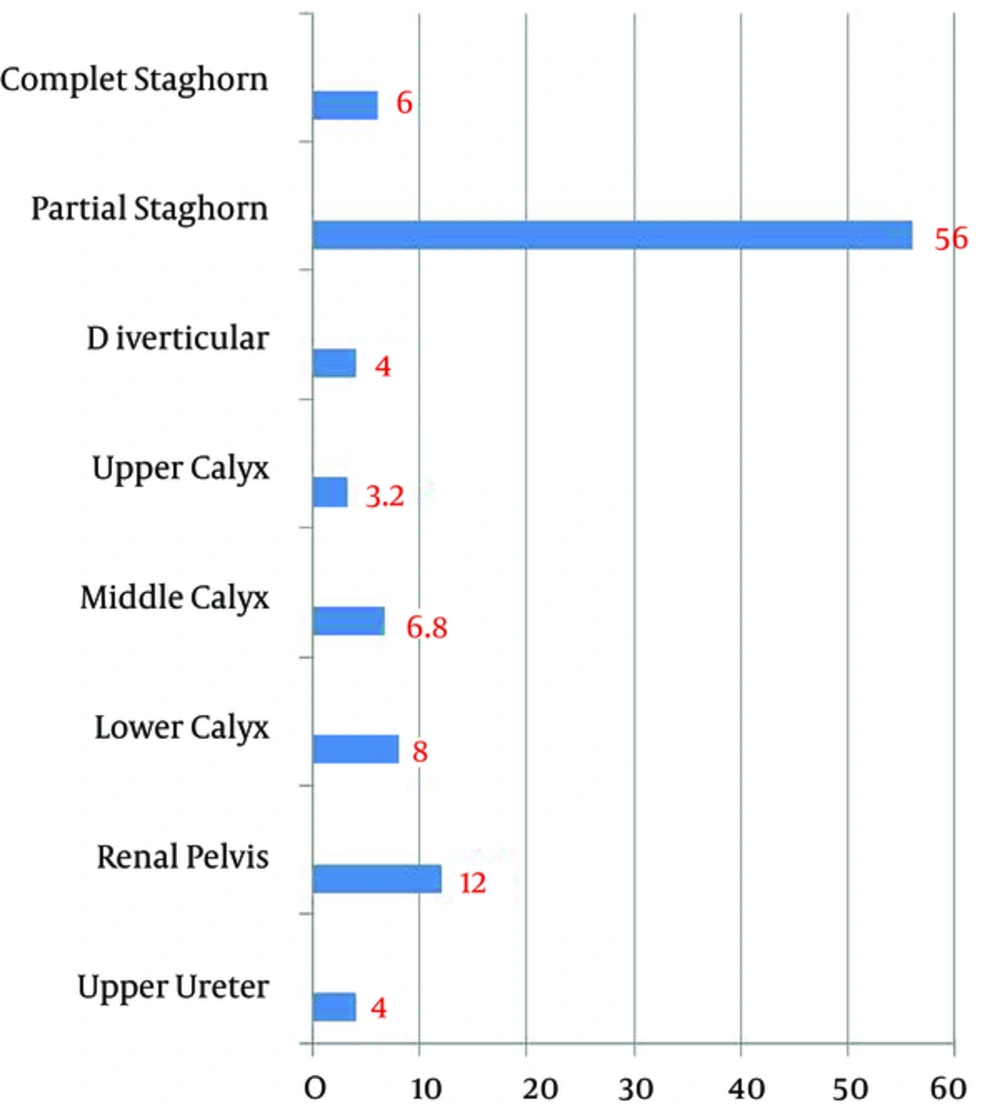
Figure 1.
Stone Location Before Percutaneous Nephrolithotomy Percentage
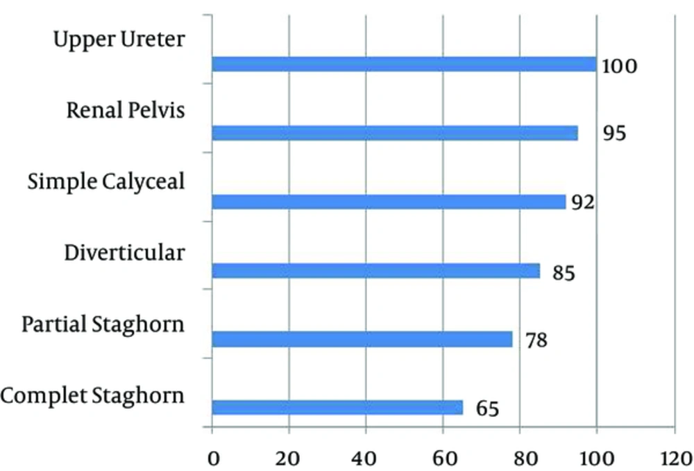
Figure 2.
Percentage of Stone Free Rate After Six Weeks Without Auxiliary Procedures
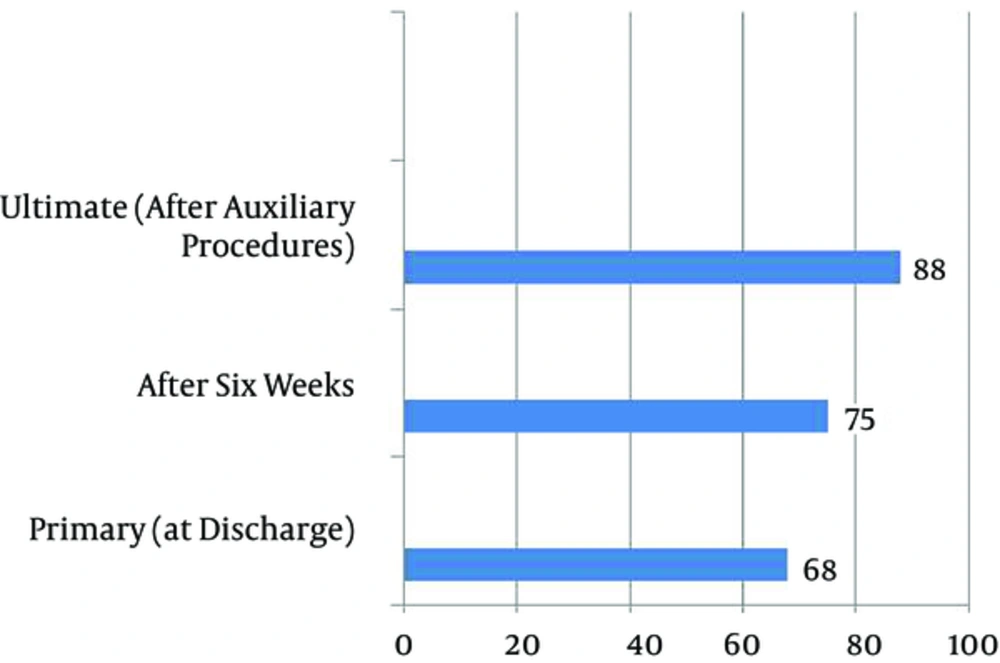
Figure 3.
% of Stone Free Rates
4. Discussion
Recently, percutaneous nephrolithotomy has become the method of choice for management of multiple or large kidney stones. The proper patient selection, perfect equipment and technique, and accurate follow-up are three considerable points for acceptable results and avoiding significant complications in this modality (6). Key prerequisite for success of PCNL is appropriate renal access. Access can be obtained under X-ray fluoroscopic or ultrasonographic control. Because of the remarkable vascularity situation of the kidney, the access tract should directly passage through the calyceal papilla. Puncture from the posterior calyx minimizes vascular injury (7). Because of the teratogenic effect of radiation, fluoroscopy is contraindicated for pregnant patients. Ultrasound-guided PCNL under flank position is preferred for this group (8). In our study, 4 pregnant patients with moderate to severe degree of hydronephrosis and large renal pelvis stones underwent successful PCNL, without significant mother or fetus problems. Heroic use of fluoroscopy, expose of the patient and operative theater to cumulative hazard effect or radiation (9). Bone marrow, gonads, thyroid gland, and eye lens are the most radiosensitive areas (10). In the high-volume centers it is very important to reduce radiation time on the base of ‘ALARA’ (as-low-as-reasonably-achievable) (11). Ultrasonography obviates the need of contrast media because of the usage of hydrodistention. Desai (12) evaluated the efficacy of ultrasonographic access of kidney and pointed some benefits such as radiation avoidance, diminishing visceral organ trauma, and significant reno-vascular injury. Shortest access tract to the pyelocalyceal can be achieved under ultrasonography guidance. Because of the susceptibility of the reproductive system in pediatrics, US-guided access is of particular help in this group (13). Twelve of our patients were children under 12 years old. The PCNL was performed in this group with acceptable results. Hosseini et al. (14) assessed the feasibility of ultrasonography-guided percutaneous nephrolithotomy in 47 cases. Total stone-free rate was reported as 93.61%. They concluded that PCNL using US is a good alternative to the fluoroscopic modality. Basiri et al. (15) evaluated the results of flank position PCNL under ultrasonographic guidance in 30 patients. The stone-free rate was 88.9% and 75.0% for simple and multiple renal calculi, respectively. They claimed the satisfactory outcomes of this technique. In the current study, the primary complete stone free rate at the time of hospital discharge was 68%, and after 6 weeks, it reached 75%, without intervention. With auxiliary procedures (URS and ESWL), ultimate stone free rate was raised to 88%. Intra and postoperative bleeding was pointed as one of the most important complications of PCNL. Significant bleeding required transfusion, ranging from 1% to 15% depending on the stone burden and the experience of the surgeon. Six of the patients (2.4%) developed intraoperative bleeding and needed more than 2 pints of blood transfusion. Karami et al. (5), in a randomized prospective trial, reported the feasibility of flank position ultrasonography guided PCNL of 40 patients. Renal access was achieved successfully in all patients. The total stone clearance rate was 85%. The main dilemma of this modality encountered access to a non-dilated pyelocalyceal system. However, this problem can be resolved by retrograde injection of 30 to 50 mL of sterile saline (16). Also real time dilation of access tract under ultrasonography control needs to more learning curve than fluoroscopic modality (17). The PCNL has traditionally been done in prone position. This position is not recommended for situations such as morbid obesity, respiratory compromise, late pregnancy, kyphoscoliosis, and severe vertebral deformities. For these conditions, flank position is one of the most comfortable and preferable options (18).
4.1. Conclusion
Overall, US-guided percutaneous nephrolithotomy is a valuable alternative to the fluoroscopic modality with comparable results. Preventing radiation hazards, diminishing the damage to adjacent organs, and feasibility of usage under all positions are some of the remarkable advantages of this modality. Some limitations may be encountered for this modality. Also, flank position is a more comfortable selection for patients, especially in limiting conditions and when urologists and anesthesiologists are more familiar with this technique.


