
1. Background
Coronary artery disease (CAD) remains one of the leading causes of illness and death worldwide, placing a significant burden on healthcare systems across the globe. Well-known risk factors such as diabetes mellitus (DM), hypertension (HTN), smoking, and elevated low-density lipoprotein (LDL) cholesterol are key contributors to the development of CAD (1). Yet, despite their established role, these traditional risk factors often fall short in accurately predicting which individuals will go on to develop CAD, particularly in its early stages (2). As a result, early detection and effective risk stratification remain difficult, especially in asymptomatic individuals or those being evaluated before surgery.
Coronary angiography is currently the gold standard for diagnosing and assessing the severity of coronary artery stenosis and atherosclerosis (3). By directly visualizing the coronary arteries, this technique provides a precise evaluation of disease extent. However, angiography is an invasive procedure that carries certain risks, including vascular complications, contrast-induced nephropathy, and hemodynamic instability — risks that are especially concerning in patients with unstable clinical conditions. These limitations underscore the need for reliable, non-invasive, and easily repeatable diagnostic tools that can either complement or, in selected cases, serve as an alternative to invasive coronary evaluation.
In this context, carotid artery intima-media thickness (IMT) has emerged as a promising marker of systemic atherosclerosis and cardiovascular risk (4, 5). Measured non-invasively by high-resolution B-mode ultrasonography, IMT represents the combined thickness of the intimal and medial layers of the arterial wall, offering a quantifiable indicator of subclinical atherosclerosis. In particular, the IMT of the common carotid artery (CCA) has been shown to correlate strongly with established cardiovascular risk factors such as age, gender, smoking, diabetes, HTN, and dyslipidemia (6-8). Importantly, IMT is capable of detecting early atherosclerotic changes before the onset of clinical symptoms or significant coronary obstruction, providing an opportunity for timely intervention and preventive measures.
Growing evidence also suggests that carotid IMT serves not only as a general marker of risk but also as a predictor of the severity and extent of CAD itself. Numerous studies have found that patients with angiographically confirmed CAD tend to have significantly higher carotid IMT values than healthy controls (9). This association supports the idea that carotid IMT could function as a non-invasive proxy for coronary atherosclerosis and offer valuable insight into overall vascular disease (VD) burden.
2. Objectives
Against this backdrop, the present study set out to examine the relationship between carotid artery IMT and coronary artery stenosis in patients undergoing coronary angiography. A better understanding of this association could enhance non-invasive cardiovascular risk assessment and potentially reduce the need for invasive diagnostic procedures. Moreover, validating carotid IMT as a predictor of CAD severity may improve clinical decision-making and help guide more targeted preventive strategies for individuals at elevated risk.
3. Methods
3.1. Study Design and Population
This cross-sectional observational study included 230 consecutive patients (112 males and 118 females), with a mean age of 58.2 ± 10.7 years (range: 35 - 95 years), who were referred for coronary angiography due to suspected CAD. The study was conducted between January 2020 and August 2021. All patients were admitted to our hospital’s cardiology ward, and written informed consent was obtained from each participant prior to enrollment.
3.2. Coronary Angiography
Coronary angiography was performed using the Seldinger technique via femoral artery access. All procedures were conducted in a dedicated angiography suite using a Siemens (Munich, Germany) system equipped with Quantor version 2.0 coronary quantitative analysis software. Each angiogram included a detailed assessment of all major coronary arteries. Significant CAD was defined as the presence of at least one coronary artery lesion causing ≥ 50% luminal stenosis. Patients with lesions causing < 50% stenosis were classified as having minimal atherosclerosis and were not included in the CAD-positive group.
3.3. Carotid Intima-Media Thickness Assessment
Each patient underwent bilateral carotid ultrasound using a high-resolution B-mode, color Doppler, and pulsed Doppler ultrasound system (ALOKA SSD 5500, Japan) with a 7.5 MHz linear array transducer. All scans were performed by a single experienced sonographer who was blinded to the patients’ clinical and angiographic data.
The IMT measurements were taken at multiple sites, including the distal and proximal walls of the CCAs, the carotid bifurcation, and the internal carotid arteries. For each segment, the thickest IMT value was recorded, and the mean of the maximal values across all segments was used as the patient’s representative IMT value. An IMT ≤ 1.0 mm was considered normal, while focal IMT > 1.3 mm was classified as plaque. Stenosis exceeding 70% was categorized as acute vertebral or carotid atherosclerosis. In patients with ultrasound findings suggestive of significant stenosis, the degree of narrowing was confirmed by correlating results with coronary angiography.
3.4. Data Collection
Demographic data (age, sex), clinical information, and comorbid conditions — including HTN, DM, dyslipidemia, smoking status, and substance addiction — were collected through structured patient interviews and medical record review. The IMT values were compared across different CAD severity categories: Normal angiography, one-vessel disease, two-vessel disease, and three-vessel disease.
3.5. Ethical Approval
The study was approved by the Ethics Committee of Zahedan University of Medical Sciences (IR.ZAUMS.REC.1399.268).
3.6. Statistical Analysis
All statistical analyses were performed using SPSS version 24.0. Continuous variables were reported as mean ± standard deviation (SD), and categorical variables as percentages. Differences in mean IMT across CAD severity groups were assessed using one-way analysis of variance (ANOVA). Multivariate logistic regression was used to identify independent predictors of coronary stenosis, with results reported as odds ratios (ORs) and 95% confidence intervals (CIs). To further explore the relationship between IMT and VD, IMT values were also analyzed by quartiles. Correlations were evaluated using Pearson’s or Spearman’s coefficients, as appropriate. A P-value < 0.05 was considered statistically significant.
4. Results
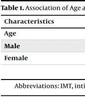
A total of 230 patients undergoing coronary angiography were included in the analysis. The study population consisted of 112 males (48.7%) and 118 females (51.3%), with a mean age of 58.2 ± 10.7 years. The most common comorbidity was HTN, present in 58.3% of patients, followed by DM and hyperlipidemia, each affecting 36.5% of the cohort. In addition, 23.5% of patients were current smokers, and 18.7% had a history of substance addiction (Tables 1 and 2).
Table 1.
Association of Age and Sex with Intima-Media Thickness
| Characteristics | No. (%) | Values (Mean IMT ± SD) | P-Value |
|---|---|---|---|
| Age | - | 58.2 ± 10.7 | 0.032 |
| Male | 112 (48.6) | 0.75 ± 0.14 | 0.031 |
| Female | 118 (51.4) | 0.71 ± 0.12 | 0.037 |
Abbreviations: IMT, intima-media thickness; SD, standard deviation.
Table 2.
Association of Comorbidities with Coronary Stenosis (Logistic Regression Analysis)
| Characteristics | No. (%) | Values (Beta Coefficient ± SE) | P-Value |
|---|---|---|---|
| HTN | 134 (58.3) | 0.91 ± 0.36 | 0.010 |
| DM | 84 (36.5) | 0.31 ± 0.40 | 0.400 |
| Dyslipidemia | 84 (36.5) | -0.66 ± 0.41 | 0.110 |
| Smoking | 54 (23.5) | 0.79 ± 0.45 | 0.080 |
| Addiction | 43 (18.7) | 0.02 ± 0.41 | 0.020 |
| IMT (mean ± SD) | - | 10.08 ± 1.54 | < 0.001 |
Abbreviations: IMT, intima-media thickness; DM, diabetes mellitus; HTN, hypertension; SD, standard deviation.
Among the six comorbidities assessed — HTN, DM, dyslipidemia, smoking, addiction, and elevated carotid IMT — only HTN and IMT showed statistically significant associations with the presence of significant coronary artery stenosis (Table 2). Patients with HTN had a 2.46-fold higher risk of developing significant coronary stenosis (OR = 2.46, 95% CI: X-Y, P < 0.05). Each 1 mm increase in carotid IMT was associated with a 2.20-fold increase in the risk of coronary artery stenosis (OR = 2.20, 95% CI: X-Y, P < 0.01).
The overall mean carotid IMT for the study cohort was 0.73 ± 0.13 mm. The IMT values were modestly but significantly correlated with age (Pearson’s R = 0.143, P = 0.03). Interestingly, female patients exhibited significantly higher mean IMT values compared to males (P = 0.03). There was a strong and consistent relationship between carotid IMT and the severity of CAD. Patients with more advanced CAD — particularly those with three-vessel disease — demonstrated significantly higher mean IMT values compared to patients with less extensive or no coronary involvement (ANOVA P < 0.001). This positive association between IMT and CAD severity was further supported by Spearman’s correlation analysis (R = 0.48, P < 0.001).
Substance addiction also emerged as a factor associated with increased IMT. Patients with a history of addiction exhibited significantly higher mean IMT values compared to those without addiction (P = 0.02; Table 3). When patients were grouped by IMT quartiles, a clear and consistent trend emerged: Higher IMT quartiles were associated with a greater prevalence and severity of CAD. Patients in the highest IMT quartile showed the highest frequency of multi-vessel disease, reinforcing the potential role of carotid IMT as a predictive marker for CAD.
Table 3.
Relationship Between Intima-Media Thickness and Severity of Coronary Artery Stenosis
| Coronary Stenosis Severity | Values (Mean IMT ± SD) | P-Value |
|---|---|---|
| Vessel disease present | 0.78 ± 0.13 | < 0.001 |
| No vessel disease | 0.65 ± 0.09 | < 0.001 |
Abbreviations: IMT, intima-media thickness; SD, standard deviation.
5. Discussion
Atherosclerosis is now widely recognized as a systemic disease that begins early in life, often in the first few decades, and progresses silently until clinical manifestations occur (10). Both the coronary and carotid arteries are among the primary vascular sites affected by atherosclerotic changes. The close relationship between atherosclerosis in these two vascular beds has been well documented, reflecting the systemic nature of the disease process (1, 12). In this context, carotid IMT has emerged as a valuable surrogate marker of atherosclerosis, providing a quantifiable, non-invasive measurement that correlates with overall VD burden.
Numerous studies have shown that carotid IMT increases in response to several traditional cardiovascular risk factors, including HTN, DM, dyslipidemia, sex differences, and population-specific variables (6, 13-15). These same risk factors contribute to the pathogenesis of CAD, reinforcing the concept of IMT as a systemic indicator of atherosclerotic burden.
While coronary angiography remains the gold standard for diagnosing and assessing coronary atherosclerosis, its invasive nature carries inherent risks, including vascular injury, contrast reactions, and hemodynamic instability (3). As a result, there is growing clinical interest in non-invasive alternatives such as carotid IMT measurement, which is a well-established marker of cardiovascular risk and has been strongly linked to adverse outcomes, including stroke and coronary events (16, 17).
Multiple studies have demonstrated that increased carotid IMT is associated with a higher risk of CAD. For example, IMT values greater than 1 mm have been linked to a twofold increase in CAD risk in men and a fivefold increase in women (18). In a study by Kablak-Ziembicka et al., IMT increases with advancing CAD; patients with mean IMT over 1.15 mm have a 94% likelihood of having CAD, and the coexistence of CAD with severe stenosis of aortic arch arteries is relatively high and was found in 16.6% of patients with three-vessel CAD (18). Similarly, Zaman et al. reported that to diagnose stenosis less than 50%, the sensitivity, specificity, positive predictive value (PPV), negative predictive value (NPV), and accuracy were 83.9%, 95.6%, 61.0%, 98.6%, and 94.7%, respectively. For stenosis greater than 50%, the sensitivity, specificity, PPV, NPV, and accuracy were 84.2%, 99.3%, 86.5%, 99.1%, and 98.5%, respectively. For stenosis exceeding 75%, the values were 100.0%, 99.9%, 97.0%, 100.0%, and 99.9% (19). It has also been confirmed that there is consistent sensitivity but variable specificity across different populations (20, 21). In contrast, another study reported lower sensitivity (31.9%) but high specificity (90.5%) using an IMT cutoff of 1 mm, suggesting that while IMT may not detect all cases, a positive result is highly specific (4). Another study found similar trends, reporting sensitivity and specificity of approximately 50% and 96%, respectively, for IMT values above 0.9 mm (21). Another study presented more moderate findings, indicating that IMT’s diagnostic performance can vary depending on patient characteristics and measurement methodology (22).
In the present study, we found that each 1 mm increase in carotid IMT was associated with a 2.20-fold increase in the risk of coronary artery stenosis, supporting the predictive value of this marker. These findings align with those of Thangaprajan et al., who reported that B-mode ultrasound measurements of IMT and plaque thickness correlated well with coronary CT angiography findings in patients presenting with acute coronary syndrome (ACS) symptoms (23). Similarly, Thangaprajan et al. demonstrated that increasing IMT values correlated with both the extent of coronary atherosclerotic lesions and the severity of CAD, further validating the utility of carotid ultrasound in assessing coronary disease burden (23). Epidemiological studies have also consistently shown that individuals with IMT ≥ 1 mm face significantly elevated risks of future cardiovascular events (24), underscoring the clinical relevance of IMT measurement in risk stratification.
Despite these promising results, our study has several important limitations. First, the relatively small sample size may limit the generalizability of our findings and reduce statistical power, particularly for subgroup analyses. Larger, multi-center studies are needed to validate our results in more diverse populations. Second, the cross-sectional design precludes establishing causal or temporal relationships between IMT progression and CAD severity. Longitudinal studies would offer deeper insight into the progression of IMT over time and its prognostic implications. Third, variations in the incidence and severity of ACS events based on the time of day and day of the week — factors not controlled for in our study — may have introduced additional variability into our findings. Additionally, although the sonographer was blinded to clinical and angiographic data, some degree of inter- and intra-observer variability in IMT measurement remains possible. Finally, while IMT is a valuable surrogate marker of atherosclerosis, it does not capture all relevant features of coronary disease, such as plaque composition and stability that influence clinical outcomes.
5.1. Conclusions
The study demonstrates a significant association between carotid artery IMT and both the presence and severity of coronary artery stenosis in patients undergoing coronary angiography. The HTN and elevated IMT emerged as independent predictors of CAD, with higher IMT values correlating positively with the extent of coronary vessel involvement. Furthermore, substance addiction was linked to increased IMT values, suggesting an additional contributory role in vascular changes. Given its non-invasive, accessible nature, carotid IMT measurement offers a promising tool for cardiovascular risk stratification and may help identify patients at heightened risk for significant CAD. Future prospective studies are warranted to further validate carotid IMT as a reliable surrogate marker of coronary atherosclerosis and to explore its potential role in guiding clinical management and preventive strategies.