
1. Background
After orthodontic treatment, retainers are necessary to prevent unwanted tooth movement. Retention remains a fundamental stage of orthodontic therapy. During this period, teeth must be maintained in their new positions to allow the surrounding bone and soft tissues to adapt and stabilize (1, 2). Retention after orthodontic treatment deserves the same level of attention as the active phase. Studies have shown that relapse rates are highest during the first and second years after treatment. Various factors contribute to relapse, including the type of malocclusion, forces exerted by soft tissues, growth, and function (3).
Fixed retainers are typically used when there is a risk of arch instability and long-term retention is desired. These retainers are bonded in place and are generally positioned lingually, which has the advantage of not relying on patient compliance (4). Consequently, an increasing number of orthodontists use fixed retainers. Various composites, including restorative and orthodontic composites, have been used to fabricate fixed retainers (5). Flowable composites, which have a higher resin content than microfilled composites, are now commonly used for fabricating lingual retainers (5, 6). However, fixed retainers also have disadvantages, including the need for long-term monitoring to prevent periodontal problems, low bond strength to tooth structure that may increase the risk of debonding, difficulty maintaining oral hygiene, and increased plaque accumulation and caries risk (4, 7).
Preventing caries is a major challenge in patients with fixed retainers. Fixed retainers complicate oral hygiene and increase microbial plaque accumulation (3). After orthodontic appliances are bonded, patients show increased numbers of Streptococcus mutans and lactobacilli in dental plaque and saliva. Studies have demonstrated that these bacterial counts return to normal after appliance removal. Streptococcus mutans plays a crucial role in the initiation of carious lesions. Orthodontic adhesives can also predispose enamel to demineralization because their rough surfaces provide a favorable environment for the rapid adhesion and growth of oral microorganisms (8).
Therefore, new materials and methods with anticaries properties are being developed to reduce the incidence of caries. In recent years, nanomaterials have attracted considerable attention and have been introduced into the biomedical field. Nanomaterials are characterized by several unique physicochemical properties, including extremely small size, a high surface area-to-volume ratio, the ability to penetrate cells and organelles, and enhanced chemical reactivity. Therefore, they have potential as antimicrobial materials (9, 10). Nanomaterials made from metals and metal oxides, such as silver, gold, copper, iron, titanium dioxide, and zinc oxide, have been shown to have antimicrobial properties (11).
Copper is a widely studied element in medical and dental research because of its antimicrobial properties (12). In dentistry, copper nanoparticles can be added to dental cements, restorative materials, adhesives, resins, orthodontic wires and brackets, implant surface coatings, and other materials (13). Studies have shown that brackets coated with copper oxide nanoparticles have greater antimicrobial effects against S. mutans than control materials (14, 15). Copper oxide can exert antimicrobial effects against a broad range of pathogenic bacteria. In addition, because copper oxide nanoparticles are relatively inexpensive and chemically stable, they may represent a suitable material for dental applications (15-17).
2. Objectives
Reducing caries risk while maintaining adequate bond strength between the tooth and the wire is essential for successful orthodontic treatment. This investigation addressed a clinically relevant challenge: reducing the caries risk associated with fixed retainer placement without compromising the retainer’s primary function. Therefore, this study evaluated the effects of incorporating copper oxide nanoparticles into an orthodontic adhesive on antimicrobial properties and debonding strength.
3. Methods
3.1. Preparation of the Nanocomposite
Copper oxide nanoparticles with an average particle size of 25 nm were purchased from ARMINANO (Iran). To prepare the 2 wt% nanocomposite, 0.02 g of CuO NP powder was weighed using a digital analytical balance and manually mixed with 0.98 g of flowable composite resin (Filtek Flow, 3M ESPE, USA) under semi-dark conditions to minimize premature photopolymerization. Similarly, to prepare the 4 wt% nanocomposite, 0.04 g of CuO NPs was incorporated into 0.96 g of the same flowable composite resin using the same procedure (Figure 1).
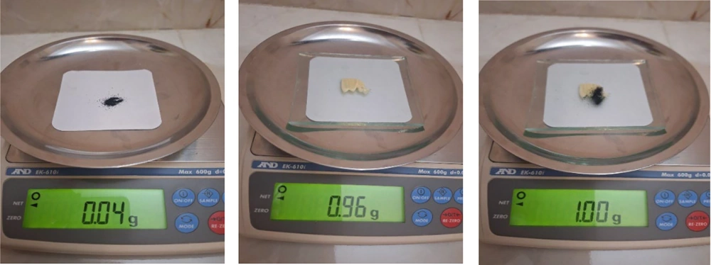
Figure 1.
Preparation of 1 g of nanocomposite with a concentration of 4%
The nanoparticles were gradually added to the composite resin and manually mixed with a sterile spatula for approximately 5 minutes until a visually homogeneous mixture was obtained. Mixing was performed under semi-dark conditions to minimize premature activation of the resin material. During mixing, efforts were made to minimize nanoparticle aggregation. Before specimen preparation, no visible phase separation or obvious nanoparticle agglomeration was observed; however, no microscopic evaluation was performed to quantitatively confirm nanoparticle dispersion. All specimens were light-cured using the same LED curing unit under standardized conditions according to the manufacturer’s instructions. The wavelength and light intensity were kept constant for all groups. After preparation, specimens were stored under identical conditions until testing (18).
3.2. Bond Strength Testing
A total of 60 extracted healthy premolars without caries or cracks were collected. The teeth were stored in normal saline solution until testing. Three experimental groups were defined based on the concentration of CuO NPs incorporated into the flowable composite resin: Group 1 (control) received 0% CuO NPs, Group 2 received 2% CuO NPs, and Group 3 received 4% CuO NPs. The teeth were randomly assigned to the 3 groups (n = 20 teeth per group). Before bonding, the tooth surfaces were cleaned using pumice-free prophylaxis. For shear bond strength testing, the teeth were mounted in pairs; thus, every 2 teeth from the same group constituted 1 testing assembly (experimental unit). Consequently, each group of 20 teeth yielded 10 paired specimens for bond strength testing and statistical analysis. For each testing assembly, the teeth were mounted in self-curing acrylic up to the cementoenamel junction area.
The lingual tooth surfaces were etched for 20 seconds with 37% phosphoric acid (H-One ULTRA), rinsed thoroughly, and gently air-dried. A bonding agent (Transbond XT, 3M Unitek) was applied in 2 stages using a microbrush; after the first application, the bonding agent was gently air-dried. After the second application, it was light-cured for 20 seconds (Woodpecker, LED, F China, 2 mW/cm², 1600 nm, 480 - 420 nm).
Subsequently, twisted stainless steel orthodontic wires (Dentaurum, Germany), measuring 10 mm in length and 0.44 mm in diameter, were placed on the lingual surfaces of the teeth, centered mesiodistally, and positioned 3 mm from the incisal edge. Two loops were created at both ends of the wire to improve retention. Transbond XT composite without CuO NPs (0%) was applied in the control group, while composites containing 2% and 4% nanoparticles were applied to the remaining groups on both the wire and tooth surfaces and cured for 40 seconds (Figure 2).
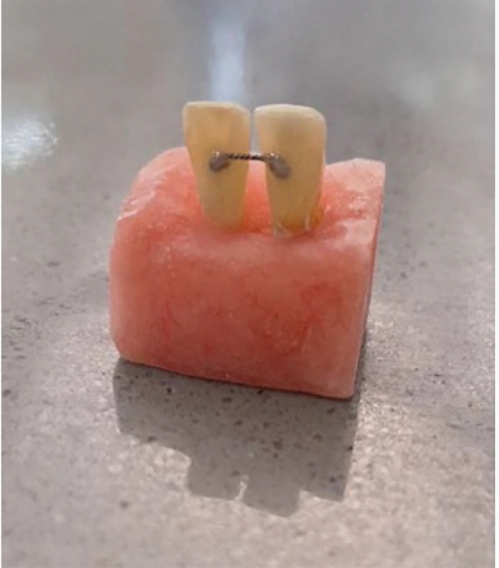
Figure 2.
Connection of orthodontic wire to mounted teeth.
The teeth were then thermocycled (Vafaei Industrial, C-300, Iran) to simulate aging processes in the oral environment. The teeth underwent 1000 thermal cycles (5°C - 55°C, 13 hours), with 15-second baths and 10-second rests.
Bond strength between the wire and tooth was measured using a universal testing machine (Z020, Zwick GmbH & Co. KG, Ulm, Germany). A blade 0.8 mm thick was positioned perpendicular to the bond interface and advanced at a rate of 1 mm/min until debonding was observed in at least 1 of the teeth.
No debonding, specimen loss, or failed measurements occurred before analysis. All 30 paired assemblies were successfully tested and included in the results reported in Table 1.
Table 1.
Summary of SBS Measurements Across Groups
| Groups | Samples No. | paired specimens | Mpa | P-Value | |||
|---|---|---|---|---|---|---|---|
| Mean ± SD | 95% CI for Mean | Min | Max | ||||
| Composite with Copper Oxide Nanoparticles 2% | 20 | 10 | 21.06 ± 7.32 | (17.63, 24.49) | 7.24 | 34.90 | 0.013 |
| Composite with Copper Oxide Nanoparticles 4% | 20 | 10 | 19.66 ± 7.24 | (16.27, 23.05) | 5.57 | 29.48 | |
| Control | 20 | 10 | 32.23 ± 13 | (26.15, 38.31) | 17.95 | 51.05 | |
3.3. Antimicrobial Testing
The agar DAD method was used to evaluate antimicrobial activity. Although direct contact tests and biofilm-based methods can provide a more clinically relevant assessment of the antimicrobial activity of orthodontic composites containing nanoparticles with limited ion release, the DAD method is widely used as a primary screening test for antimicrobial properties in dental material studies. The absence of an inhibition zone with this method does not necessarily indicate a complete lack of antimicrobial activity. Therefore, the limitations of this method were considered when interpreting the study results (19).
To measure antimicrobial properties, composite disks containing nanoparticles were prepared in triplicate for each group, for a total of 9 samples. This test was repeated 3 times for each nanocomposite concentration, resulting in a total of 27 samples.
3.4. Preparation of Composite Disks
Cylindrical metal molds measuring 6 mm in diameter and 2 mm in thickness were used as templates for preparing composite disks. After the molds were placed on glass slabs, 3 composite formulations were poured into them: a 2% CuO NP composite for the first group, a 4% formulation for the second group, and a nanoparticle-free (0%) composite as the control for the third group. To create a smooth, pore-free surface, another glass slab was gently pressed onto the molds, and each sample was cured using a light-curing device from both sides for 40 seconds each, for a total curing time of 80 seconds. After curing, the composite specimens were removed from the molds using gentle manual pressure. The samples were then placed under a UV laminar flow hood (JTLV C2, Jal Tajhiz Production, Iran) for 30 minutes for sterilization.
3.5. Disk Agar Diffusion Test
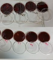
This test was designed to evaluate the antimicrobial properties of composite disks containing CuO NPs by assessing nanoparticle diffusion from the disks. For this purpose, a suspension of S. mutans ATCC 35668 equivalent to a 0.5 McFarland standard (1.5 × 10⁸ CFU/mL) was inoculated onto Brain Heart Infusion agar plates (Merck, Germany) using a sterile swab. The composite disks were then placed on the agar surface, 2 cm apart and 2 cm from the edge of the plate. After the plates were incubated for 48 hours, the diameter of the inhibition zone was measured using a ruler (Figure 3).
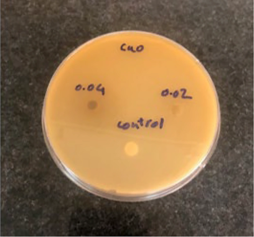
Figure 3.
Composite disks in BHI agar plates.
3.6. Statistical Analysis
Statistical analysis was performed using SPSS version 26. Descriptive data are presented as means and standard deviations. Normality was confirmed using the Kolmogorov-Smirnov test (P > 0.05 for all groups), and homogeneity of variances was confirmed using Levene’s test (F(2, 57) = 2.41, P = 0.099). One-way analysis of variance followed by the Tukey HSD post hoc test was used to compare shear bond strength among the 3 groups. Statistical significance was set at P = 0.05.
4. Results
4.1. Bond Strength Test
The highest mean shear bond strength (SBS) was observed in the control group (13 ± 32.23 MPa), whereas the lowest SBS was observed in the group with 4% copper oxide nanoparticles (7.24 ± 19.66 MPa). Table 1 presents the mean bond strength across the 3 groups. The comparison of SBS among the 3 experimental groups is shown in Figure 4.
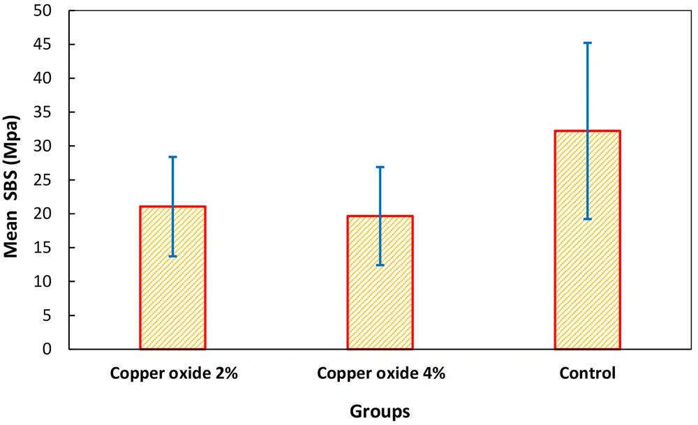
Figure 4.
Comparison of SBS among the three experimental groups.
Tukey HSD post hoc pairwise comparisons are presented in Table 2. Both the 2% CuO and 4% CuO groups showed significantly lower shear bond strength than the control group. No statistically significant difference was observed between the 2 nanoparticle concentration groups. These results indicate that the addition of CuO NPs at either concentration significantly decreased bond strength compared with the control group, with no difference between the 2% and 4% concentrations.
Table 2.
Pairwise Comparison of SBS in the Studied Groups
| Pairwise Comparisons | Mean Difference SBS, MPa | 95% CI for Difference | P Value | Significance |
|---|---|---|---|---|
| 2% CuO vs 4% CuO | +1.4 | -4.66, 7.46 | 0.9 | ns |
| 2% CuO vs Control | -11.17 | -17.23, -5.11 | 0.011 | * |
| 4% CuO vs Control | -12.57 | -18.63, -6.51 | 0.003 | * |
4.2. Disk Agar Diffusion Test
The DAD test showed no inhibition zone in any sample, regardless of the concentration used.
5. Discussion
The prolonged presence of fixed retainers in the mouth can make oral hygiene more difficult for patients, potentially leading to caries development (1). Several methods have been used for caries prevention, including fluoride and chlorhexidine in toothpaste and mouthwashes. Incorporating nanoparticles into composite resins is an effective approach to caries prevention because nanoparticles exhibit strong antibacterial properties due to their small size (20). However, in addition to their antimicrobial activity, the effects of these nanoparticles on the physical and mechanical properties of orthodontic composites require evaluation.
The high surface-to-volume ratio of copper nanoparticles has been shown to enhance antimicrobial efficacy and suppress biofilm formation. The antimicrobial effect of copper nanoparticles primarily involves the generation of reactive oxygen species and bacterial DNA damage (21). In this study, CuO NPs were added to orthodontic composite retainers to evaluate their antimicrobial effects and their impact on bond strength between teeth and orthodontic wires. Thermocycling was performed according to previous studies to simulate the oral environment.
All groups exceeded the clinically acceptable SBS threshold of 5 - 8 MPa (22, 23). The lowest mean value (19.66 MPa for 4% CuO) was more than double this threshold, and even the lower bound (12.42 MPa) remained above 8 MPa. Thus, despite the statistically significant reduction (P = 0.013), the bond strength of all groups remained clinically acceptable for retainer use.
Although the bond strength values observed in this study remained within clinically acceptable thresholds (≥6 - 8 MPa), the 4% CuO NP concentration should be used with caution. Additional thermomechanical testing is warranted to evaluate long-term performance under simulated intraoral conditions.
The findings of this study are consistent with those of Sodagar et al. (24), who investigated the effect of propolis nanoparticles at concentrations of 1%, 2%, 5%, and 10% on shear bond strength in orthodontic composites and found that bond strength decreased significantly.
In another study by Sodagar et al. (25), which examined shear bond strength in Transbond XT composites containing 1%, 5%, and 10% titanium dioxide nanoparticles, similar results were obtained. That study also showed that increasing nanoparticle concentrations reduced bond strength. Mirhashemi et al. (26) reported findings similar to those of the present study. They examined the effects of 1%, 5%, and 10% zinc oxide/chitosan nanoparticles on shear bond strength in orthodontic composites and found that shear bond strength decreased significantly, with the greatest reduction observed at the 10% concentration. In the study by Akhavan et al. (27), which examined the effect of silver/hydroxyapatite nanoparticle composites on the bond strength of orthodontic composites, the findings were also similar to those of the present study. In that study, bond strength decreased significantly at concentrations of 5% and 10%.
By contrast, the findings of Felemban et al. (28) were inconsistent with the present study. Their study evaluated the effects of adding zirconium dioxide-titanium dioxide composite nanoparticles at concentrations of 0.5% and 1% to adhesives used in orthodontic brackets and showed increased bond strength after nanoparticle incorporation. This difference may be due to the different types of nanoparticles used, as well as the type of brackets examined. Similarly, the findings of Argueta-Figueroa et al. (29) were contrary to those of the present study. In their investigation of orthodontic adhesives containing copper nanoparticles at concentrations of 0.01%, 0.0075%, and 0.005%, they found that shear bond strength increased after nanoparticle incorporation. This difference may be attributed to the very low nanoparticle concentrations used, as studies indicate that nanoparticles at very low concentrations may yield better bond strength outcomes.
The present study also assessed the antimicrobial properties of Transbond XT composite containing 2% and 4% CuO NPs against S. mutans. The antimicrobial effect generated by ion release within the composite resin was determined using the DAD test. This test is important because carious lesions typically develop around orthodontic wires; therefore, an ideal adhesive material for orthodontic applications should have strong antibacterial properties and the ability to diffuse into the surrounding environment. However, the present results do not demonstrate clinical efficacy in caries prevention because biofilm formation, ion release kinetics, and long-term durability were not assessed.
The results showed that neither the 2% nor the 4% CuO NP concentration produced zones of inhibition against S. mutans in composite disks. This finding suggests that CuO NPs did not diffuse in the solid medium of the disks. The findings of this study were consistent with those reported by Mirhashemi et al. (19). In that study, the authors examined the antimicrobial action of composites containing silver nanoparticles at concentrations of 1% and 2%. Similar to the present results, they found no inhibition zones in composites containing silver nanoparticles. In another study conducted by Sodagar et al. (24), which investigated the antimicrobial effects of curcumin nanoparticles at concentrations of 1%, 5%, and 10%, no inhibition zones were formed at any concentration, which is consistent with the findings of the present study. Argueta-Figueroa et al. (29) also reported similar findings in their study of the antibacterial properties of orthodontic adhesives containing copper nanoparticles at concentrations of 0.01%, 0.0075%, and 0.005%, concluding that no inhibition zones were observed.
However, the results of Sodagar et al. (30) contradicted those of the present study. Their study on the antimicrobial effects of silver/hydroxyapatite nanoparticles at concentrations of 5% and 10% demonstrated zones of inhibition against 3 bacteria: S. mutans, Streptococcus sanguinis, and Lactobacillus acidophilus. This discrepancy may be due to differences in nanoparticle type, as silver/hydroxyapatite nanoparticles have shown superior antimicrobial effects in various studies. Moreover, in another study by Sodagar et al. (24), which examined the antimicrobial properties of propolis nanoparticles at concentrations of 2%, 5%, and 10% in orthodontic composites, the results were contrary to those of the present study. Their findings showed zones of inhibition against S. mutans and S. sanguinis at concentrations of 2%, 5%, and 10%, possibly because of differences in nanoparticle type.
Additional tests, such as biofilm inhibition assays and eluted component tests, have also been used to assess antimicrobial effects. Copper oxide nanoparticles have been shown in numerous studies to have antimicrobial properties when tested using different methods. Of particular note, the DAD test reveals zones of inhibition at relatively low concentrations. The reason for this difference may be that CuO NPs at concentrations of 2% and 4% lack the ability to diffuse in solid media, although they may show antimicrobial effects in other tests.
The potential for gradual release of CuO nanoparticles from the resin matrix over time, as well as possible surface oxidation of the nanoparticles in the oral environment, may affect antibacterial activity and biocompatibility. In addition, possible interactions of released nanoparticles or ions with salivary components, including proteins, mucins, and electrolytes, could modulate antimicrobial efficacy. Therefore, the clinical safety and stability of the developed nanocomposite should be evaluated in future long-term studies.
5.1. Conclusions
Within the limitations of this in vitro study, incorporation of 2% and 4% CuO NPs into a flowable composite resin significantly reduced shear bond strength compared with the control, although the values remained within clinically acceptable ranges. Using the DAD method against S. mutans ATCC 35668, no inhibition zones were observed, suggesting a lack of nanoparticle diffusion in solid media. It should be noted that the absence of an inhibition zone in this assay does not conclusively demonstrate a complete absence of antimicrobial activity. The present findings support only in vitro conclusions; clinical caries prevention, ion release, cytotoxicity, and long-term durability were not evaluated. Further studies are needed to assess the clinical feasibility of this approach for fixed retainer applications.
5.2. Limitations
This study had several limitations. First, it was conducted under controlled laboratory conditions, which cannot fully replicate the complex oral environment. Factors such as saliva, thermal cycling, variable occlusal forces, and long-term biofilm dynamics were not simulated, which limits direct clinical extrapolation of the findings.
Second, antimicrobial evaluation was limited to S. mutans, a primary but not exclusive contributor to caries. The effect on multispecies biofilms, which more accurately represent clinical plaque and include fungi such as Candida albicans, remains unknown.
Third, the bond strength and antimicrobial tests represented immediate or short-term outcomes. The long-term durability of the bond under simulated oral stress and the potential for nanoparticle release or sustained antimicrobial effects over months or years were not assessed.
5.3. Suggestions
Based on the findings of this study, the following directions are proposed for future research.
First, lower nanoparticle concentrations should be investigated. Given the observed concentration-dependent decline in bond strength and the need to preserve mechanical integrity, future formulations should evaluate sub-1% or trace-level loadings (eg, 0.5% and 1%) of CuO NPs. This approach may help identify a therapeutic threshold that provides detectable antimicrobial benefit while minimizing the negative impact on the adhesive properties of the composite.
Second, advanced antimicrobial testing methods should be used. The DAD test is unsuitable for assessing nondiffusible, contact-based antimicrobial surfaces. Future studies should use direct-surface antimicrobial assays.