


1. Background
Coronaviruses disease (COVID-19) originated from China, Wuhan, and soon spread all over the words (1-4). Rotaviruses are enveloped RNA viruses belonging to the Coronaviridae family, which are widely found in humans and mammals (5). Although most coronavirus infections are mild, many can cause infections in different parts of the animal's body as well as in the human respiratory system (6). The virus is one of the major causes of respiratory infections in humans. The extent of transmission of the virus is unclear, but available evidence suggests human-to-human transmission. Acute coronavirus respiratory syndrome (SARS-CoV) and Middle Eastern coronavirus respiratory syndrome (MERS-CoV) are two pathogens from the coronavirus family that caused great epidemics during the past two decades, which infected more than 10 000 people and claimed many lives (4, 5, 7, 8). The mortality rates of SARS and MERS are estimated as 10% and 37%, respectively (9, 10). According to experts, the coronavirus may be the only part of the iceberg floating in the water that has not yet been revealed to us by sharp angles. In December 2019, a series of viral pneumonia were reported in Wuhan, Hubei Province, China (11, 12). The analysis of samples taken from the lower part of the lungs of infected patients showed a new coronavirus, which was then named COVID-19 (13). According to published statistics, most patients with COVID-19 present mild symptoms and good advertising. Rarely do patients have extensive pneumonia, pulmonary edema, acute respiratory failure, or organ failure (14). The widespread outbreak of pneumonia in Wuhan (China) spread rapidly to other countries, including Iran, Italy, the United States, and eventually turned into a pandemic (15). Iran is one of the countries that has confronted the epidemic after Japan and China, according to the World Health Organization (WHO). Guilan province was one of the first provinces of Iran that Covid-19 cases were identified. Unfortunately, the extent of the disease, its management, and long-term complications are not yet fully understood.
2. Objectives
Regarding the importance of management and control of Covid-19 patients and the necessity of publishing evidence in this field, the present study aimed to investigate the CT-scan findings of patients with a definitive diagnosis of COVID-19 in Guilan, Iran.
3. Methods
3.1. Study Population
In this retrospective, observational study, CT-scan findings of 1000 patients with a definitive diagnosis of COVID-19 admitted to nine central hospitals of COVID-19 in Guilan province of Iran are investigated. The electronic medical records of patients were assessed and analyzed. Data were collected from 20 April to 30 July 2020. In this study, medical records of 1000 patients were collected, including 620 males and 380 females; aged 28-62 years, with a mean age of 55 ± 1.2. The diagnosis of patients was according to positive results of the real-time reverse transcriptase-polymerase chain reaction (rRT-PCR) of respiratory secretions from nasopharyngeal or oropharyngeal samples. Demographic characteristics and CT-scan findings of patients were collected. All patients were under treatment with anti-viral drugs and other treatments.
3.2. CT Scan Characteristics
In this study, the high-resolution CT scan was performed for 1000 patients with SOMATOM Spirit, SOMATOM Perspective, or SOMATOM Definition AS+ scanners and with these parameters: tube voltage 120 kV and detector collimation widths 63 × 0·6 mm, 127 × 0·7 mm, 65 × 0·7 mm, and 66 × 0·7 mm. CT images were reconstructed via a slice thickness of 1·5 mm and an interval of 1·5 mm. All scans were performed from level of upper thoracic to costophrenic angle in patients with supine position and breath-hold.
3.3. CT Images Analysis
All CT scans were reviewed by two university professors (specialists) of pulmonary diseases (A.F, A.T) and two senior radiologists with at least ten years of experience in cardiothoracic imaging, and any controversy was resolved through discussion. CT-scans were assessed for the following characteristics: (1) presence of ground-glass opacities (GGO); (2) crazy-paving pattern; (3) consolidation; (4) number of infected lobes of lungs; (5) presence of any underlying lung disease; (6) level of lungs involvement; and (7) location of involvement. The level of lung involvement was measured according to no involvement (0%), small (1-25%), mild (26-50%), moderate (51-75%), and severe (76-100%). The overall lung involvement was calculated by summing up the scores of five lobes (ranging from 0-20).
3.4. Statistical Analysis
Data were analyzed using SPSS (version 21; SPSS Inc., Chicago, Illinois, United States). The findings are described using descriptive statistics, including mean, standard deviation, and frequency.
3.5. Ethical Statement
This research has been approved by the Ethics Committee of the Guilan University of Medical Sciences (IRB approval: IR.GUMS.REC.1398.541). We observed principles mentioned by the 1964 Helsinki Declaration and its subsequent amendments.
4. Results
4.1. Clinical Features
In this study, 1000 medical records of patients with a definitive diagnosis of Covid-19 admitted to selected COVID-19 hospitals in Guilan are investigated (620 males and 380 females). The youngest and oldest participants were 28 to 62 years, respectively, with a mean age of 55 ± 1.2. The most common reason for infection with COVID-19 close contact with infected people (67%), followed by attending crowded places (23%) and contact with domestic animals (10%) (Table 1).
| Variables | No. (%) |
|---|---|
| Age (y) | 55 ± 1.2 |
| Gender | |
| Male | 620 (60) |
| Female | 380 (40) |
| Symptoms | |
| Fever | 900 (90) |
| Cough | 950 (95) |
| Fatigue | 890 (89) |
| Dyspnea | 990 (99) |
| Sputum production | 665 (66.5) |
| Headache | 135 (13.5) |
| Rhinorrhea | 835 (83.5) |
| Vomiting | 550 (55) |
| Diarrhea | 400 (40) |
| Exposure history | |
| Close contact with infected people | 545 (54.5) |
| Attend in crowded places | 385 (38.5) |
| Contact with domestic animals | 30 (3) |
| Comorbidity | |
| Diabetes | 550 (55) |
| Hypertension | 250 (25) |
| Chronic lung disease | 450 (45) |
| Cancer | 440 (44) |
| Cardiovascular disease | 385 (38.5) |
| Cerebrovascular disease | 165 (16.5) |
| The period between the onset of initial symptoms and the first scan (d) | 3 ± 1.3 |
| Total hospital stay (d) | 22 ± 1.1 |
| Time between the onset of initial symptoms and discharge (d) | 20 ± 1.5 |
| Total number of samples | 1000 |
Characteristics of Patients Infected with COVID-19a
According to degree of infection, 105 patients (10.5%) were mild, 650 (65%) common, 240 (24%) severe, and 5 patients (0.5%) critical. Based on the lung CT-scan findings, no pulmonary involvement was observed in 60 patients (57.1%) with mild Covid-19, and the most common manifestations was bilateral lung infection (n = 50 or 47.6%). In the common type, the most common manifestation was GOO (n = 640 or 98.4%), followed by bilateral lung infection (n = 610 or 93.8%) and thickening of the lung (n = 310 or 47.6%). In severe type, the most common manifestation was GOO (n = 240 or 100%), bilateral lung infection (n = 240 or 100%), and thickening of the lung (n = 240 or 100%). In the critical type, all types of lung lesions, including bilateral lung infection, crazy paving pattern, consolidation, GOO, thickening of the lung, pleural effusion, and unilateral, were observed (Table 2).
| Kind and Place of Lesion | Degree of Infection | |||
|---|---|---|---|---|
| Mild Type (n = 105) | Common Type (n = 650) | Severe Type (n = 240) | Critical Type (n = 5) | |
| Bilateral lung infection | 50 (47.6) | 610 (93.8) | 240 (100) | 5 (100) |
| Unilateral lung infection | 10 (9.5) | 280 (43) | 0 | 0 |
| Consolidation | 0 | 240 (36.9) | 200 (83.3) | 5 (100) |
| Crazy paving pattern | 0 | 280 (43) | 180 (75) | 5 (100) |
| Pleural Effusion | 0 | 0 | 0 | 5 (100) |
| Ground-glass Opacities | 0 | 640 (98.4) | 240 (100) | 5 (100) |
| Thickening of Lung | 20 (19) | 310 (47.6) | 240 (100) | 5 (100) |
| No lung involvement | 60 (57.1) | 0 | 0 | 0 |
The Type of Pulmonary Lesions and Degree of Infection in Patients with COVID-19 [No. (%)]
4.2. CT Manifestations According to Days of Hospitalization
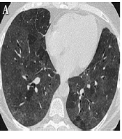
In the present study, we classified patients’ lung involvement findings into 4 stages according to quarters of hospitalization duration, ranging from 0 to 30 days: stage 1 or primary (0 to 7 days): ground-glass opacities (GGO) (n = 890 or 89%) (Figure 1A), stage 2 or progressive (8 to 15 days): increased crazy-paving pattern (n = 445 or 44.5%) (Figure 1B), stage 3 or peak involvement (16 to 22 days): consolidation (n = 89 or 89%) (Figure 1C), and stage 4 or decreased pulmonary involvement severity (greater than 23 Day): gradual resolution of consolidation (n = 89 or 89%).
; B: 77-year-old woman, day 11 after symptom onset: bilateral, peripheral ground-glass opacity associated with Crazy-paving pattern C: 67-year-old woman, day 21 after symptom onset: bilateral and peripheral predominant Consolidation.")
Transverse section of chest CT scan results of patients infected with COVID-19 in three stages. A: 58-year-old man, day 4 after symptom onset with ground glass opacities (GGO); B: 77-year-old woman, day 11 after symptom onset: bilateral, peripheral ground-glass opacity associated with Crazy-paving pattern C: 67-year-old woman, day 21 after symptom onset: bilateral and peripheral predominant Consolidation.
4.2.1. CT Manifestations by the Severity of the Disease
The severity of the disease was considered as overall lung involvement, which was determined by the number of lobes involved and using a scoring system of 0 to 5 for each infected lobe (5 lobes, the lowest and highest scores were 0 and 25, respectively). Of 1000 patients, 350 (35%) had 0-5 score, 250 (25%) had 6-10 score, 260 (26%) had 11-15 score, 65 (6.5%) had 16-20, and 75 (7.5%) had 21-25 score (Table 3).
| Number of Infected Lobes in Patients According to CT Scan Findings | Score of Lung Involvement | ||||
|---|---|---|---|---|---|
| 0-5 | 6-10 | 11-15 | 16-20 | 21-25 | |
| Right upper lobe | 170 (48.5) | 45 (18) | 30 (11.5) | 4 (6.1) | 4 (5.3) |
| Right middle lobe | 75 (21.4) | 7 (2.8) | 8 (3.07) | 4 (6.1) | 13 (17.3) |
| Right lower lobe | 75 (21.4) | 7 (2.8) | 185 (71.1) | 42 (64.6) | 38 (50.6) |
| Left upper lobe | 10 (2.8) | 177 (70.8) | 5 (1.9) | 5 (7.6) | 13 (17.3) |
| Left middle lobe | 12 (3.4) | 8 (3.2) | 7 (2.6) | 5 (7.6) | 4 (5.3) |
| Left lower lobe | 8 (2.2) | 6 (2.4) | 25 (9.6) | 5 (7.6) | 3 (4) |
| Total | 350 (35) | 250 (25) | 260 (26) | 65 (6.5) | 75 (7.5) |
The Severity of Patient's Lung Involvement in CT Scan [No. (%)]
4.2.2. CT Manifestations According to the Stages of the Lung Involvement
Patients were classified into 4 stages according to quarters of hospitalization duration, ranging from 0 to 30 days: stage 1 or primary (0 to 7 days): ground-glass opacities (GGO) (n = 890 or 89%), stage 2 or progressive (8 to 15 days): increased crazy-paving pattern (n = 445 or 44.5%), stage 3 or peak involvement (16 to 22 days): consolidation (n = 890 or 89%), and stage 4 or decreased pulmonary involvement severity (greater than 23 Day): the gradual resolution of consolidation (n = 890 or 89%).
5. Discussion
Currently, the use of RT-PCR from the nasopharynx and oropharynx samples is the method of choice for the definitive diagnosis of Covid-19 patients. However, this method requires a longer time and has a false negative rate of more than 5%. In this regard, the chest CT-scan can be a highly useful method for the early diagnosis of patients (16, 17). By using chest CT-scan findings patients can be easily classified based on the severity of their lung involvement, which in turn facilitates treatment (18). In the present study, the most common finding of pulmonary involvement in the chest CT-scan was the presence of ground-glass opacities in patients, both moderately and severely, followed by pulmonary consolidation and Crazy paving pattern. In the present study, all patients underwent more than 3 chest CT-scans, which provided sufficient and reliable data for assessing patient’s lung involvement. According to the severity of the disease, most of the patients were in the common group, followed by the severe group. Pulmonary consolidation was found mostly in common, severe, and critical types of lung involvement. At the present time, the pathological causes of these infections are not clear and the following hypothesis has not been approved by pathologists. Overall, we think that any changes in lung interstitium can be due to infiltration of inflammatory cells, edema, and interstitial thickening of lungs, and changes in pulmonary parenchyma may result in edema, alveolar hemorrhage, and hyaline membrane of lung formation. The CT-scan findings showed that pleural effusion is very scarce, only was observed in 5 cases, which all of them were in critical stages of the disease. This finding shows that only patients in advanced stages of the infection present pleural effusion, with a poor prognosis. The findings of the present study are in line with Liu et al., who reported that atelectasis and pleural effusion was a very scarce finding according to the chest CT-scans (18). In our study, the period between the onset of initial symptoms and the first CT-scan and diagnosis of Covid-19 infection was 3 days (3 ± 1.3).
Unlike other studies, in the present study, we investigated the chest CT-scan profile of Covid-19 patients based on the severity of the disease, which seems to be a better indicator of the association between the severity of the disease and the chest CT-scan findings. In the present study, we focused on infection duration in order to recognize imaging patterns. However, for many patients, the severity of the disease is not consistent with the disease course. In a study that investigated 121 symptomatic patients with COVID-19 and their CT findings concerning the association between the time since the onset of symptoms and the initial chest CT scan, Bernheim et al. found that patients in the early stage of the disease (0-2 days) had few signs of GGO and consolidation compared to those in the intermediate (3-5 days) and late (6-12 days) stages of the disease (19). The present study had a number of limitations, including using a retrospective design, and the timing of chest CT-scans was not the same for all patients. The latter could be a factor in creating a bias in describing image characteristics. In addition, no pathological test was performed for the affected patients, so it was not possible to investigate the association between the CT findings and the results of pathological tests. In general, this study demonstrated that chest CT-scan is one of the most effective, fast, and efficient diagnostic methods for identifying Covid-19 patients. According to the chest CT-scan findings, GOO and consolidation were the most common findings in Covid-19 patients in the present study.
5.1. Conclusion
In conclusion, this study demonstrated that Covid-19 infection tends to manifest on chest CT scans as consolidation, bilateral lung infection, crazy paving pattern, GOO, and thickening of the lung. Also, in mild, common, and severe cases of Covid-19, chest CT findings are usually as no lung involvement, GOO, and bilateral lung infection, respectively. In critical infections, a mixture of involvement as bilateral lung infection, GOO, consolidation, crazy paving pattern, pleural effusion, and thickening of the lung was found. Underlying diseases and comorbidities, especially diabetes, as well as old age and male sex might be associated with an increased risk of poor prognosis for Covid-19 patients.
