


1. Background
Space exploration has made significant advancements in recent years, leading to an increased interest in understanding the effects of space traveling on the human body. For the spaceship not to go out of its orbit, its angular acceleration must be equal to the Earth's gravity; therefore, the spacecraft circles the Earth several times every day, which leads to multiple sunrises and sunsets in short time intervals (1). Since living organisms on the surface of the Earth have adapted to a daily rhythm with a sequence of 24 hours of light and darkness (2), astronauts experience rapid light and darkness rhythms, which can disrupt the circadian rhythm in spacecraft. This issue can profoundly affect astronauts' physical and mental health (3).
Disruption of the circadian rhythm, caused by the frequent and rapid alternation of light and darkness during spaceflight, has been linked to increased levels of pro-inflammatory cytokines, such as interleukin-6 and tumor necrosis factor-alpha (TNF-α) (4). Moreover, circadian disruption can induce oxidative stress (5), which might adversely affect brain cells and alter biomarkers, such as cholinesterase (AChE) (6) which hydrolyzes acetylcholine (ACh).
Acetylcholine plays an important role in the prefrontal cortex and cerebellum (7-9). Cholinesterase activity follows a circadian cycle and, similar to other elements of the cholinergic system, increases during the day and decreases at night (10, 11).
2. Objectives
By elucidating how circadian disruption affects cholinesterase activity in these important brain regions, the present study seeks to contribute valuable insights into the broader understanding of the physiological and molecular mechanisms underlying the consequences of space travel on the brain. Such knowledge is essential for developing effective countermeasures to mitigate the potential adverse effects and ensure the well-being of astronauts during extended missions in space.
3. Methods
3.1. Subject
Male Wistar rats were used for this study. The rats were housed under standard laboratory conditions, with controlled temperature (22 ± 2°C) and humidity (55 ± 5%). They were provided food and water ad libitum throughout the experiment.
3.2. Experimental Design
The rats were divided into two groups, with eight rats in each group. Group 1 rats were kept on a 12-hour light/12-hour dark cycle (control group) for 14 days. Group 2 rats were exposed to a 45-minute light/45-minute dark cycle for 14 days.
3.3. Tissue Collection and Processing
After 14 days of the respective interventions, the rats were fasted overnight and then anesthetized via the intraperitoneal injection of ketamine and xylazine (100 mg/kg and 2.5 mg/kg, respectively). The cerebellum and prefrontal cortex were harvested from each rat. The samples were washed in ice-cold normal saline solution and weighed. They were then homogenized in 20 mM phosphate buffer (pH = 7.4) at a tissue/buffer ratio of 1:10 (w/v) using 1 ml of buffer per 100 mg of tissue. The homogenates were centrifuged at 4000 g at 4°C for 10 minutes, and the resulting supernatants were aliquoted and stored at -80°C until further analysis.
3.4. Cholinesterase Activity Analysis
Cholinesterase activity was measured using a photometric method, following the manufacturer's instructions (Biorex, Shiraz, Iran). Each sample was measured at least twice, and if the coefficient of variation (CV) transgressed 15%, it was repeated until the CV was less than 15%. The mean values of the duplications were used for statistical analysis.
3.5. Statistical Analysis
The data are presented as mean ± standard error of the mean (SEM) and analyzed using unpaired student’s t-test. Statistical analysis was performed using SPSS software. A P-value of less than 0.05 was considered statistically significant.
4. Results
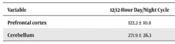
The amounts of cholinesterase activity in the cerebellum and prefrontal cortex of the rats exposed to a light/dark cycle of 45/45 minutes was significantly lower than in the rats exposed to a 12/12 hours light/dark cycle (Table 1).
5. Discussion
Spaceships must orbit the Earth at a speed whose angular acceleration is equal to the Earth’s gravity in order not to go out of orbit. Therefore, spacecraft, such as the International Space Station, which rotates at an altitude of approximately 408 km above the Earth's surface, orbits the Earth once every 90 minutes. This means that it orbits the Earth approximately 16 times per day, and, therefore, astronauts experience approximately 16 sunrises and sunsets per day, each lasting about 45 minutes (1). Therefore, astronauts experience a rapid day/night cycle, which disrupts the normal circadian rhythm from 12/12 hours to 45/45 minutes, which can have profound effects on the astronaut's physical and mental health. This study aimed to investigate the impact of this stressor on cholinesterase activity, specifically in the prefrontal cortex and cerebellum of rats. Therefore, we exposed rats to spacecraft conditions (i.e., 45/45-minute light/dark cycle) for 14 days. The findings of this study indicated that the disruption of the light/dark cycle had a significant impact on the amount of cholinesterase activity in the cerebellum and prefrontal cortex of rats.
A previous study has shown that changing the circadian cycle from 12/12 hours to 45/45 minutes increases the level of retinoic acid in the serum and hippocampus of rats (12). It seems necessary to conduct further studies on the effects of circadian disruption on the brain, such as the alteration of cholinesterase.
Cholinergic signaling, specifically cholinesterase, is simultaneously involved in central cognitive processes such as learning, memory, and stress responses, mediating neuromuscular and anti‐inflammatory responses (12). The cholinergic system has been shown to have a critical role in the cerebellum and prefrontal cortex (8, 9). Acetylcholine signaling suppresses inflammation in the brain (13). Previous research demonstrates that inflammation stimulates the release of ACh (14). Acetylcholine can reduce inflammation by decreasing the release of pro-inflammatory cytokines, such as TNF-α (15), and directly inhibits inflammation (16). Cholinesterase, which regulates the concentration of ACh, is reduced in many inflammatory diseases (17-21). It is hypothesized that the reduction in cholinesterase activity might lead to an increase in ACh levels during inflammation, subsequently modulating the inflammatory response.
Considering that circadian disruption can induce inflammation, it is plausible to suggest that one of the potential causes of the decrease in cholinesterase activity observed in the present study is the occurrence of inflammation in such a situation. Moreover, previous studies have indicated that circadian disruption can contribute to the occurrence of inflammation (4, 22, 23).
Inflammation in the upper parts of the body, especially in the brain, can influence the functions of the cerebellum and prefrontal cortex, which play pivotal roles in various human activities; therefore, understanding the impacts of space traveling and its underlying mechanisms on these brain regions is of great importance. Further investigations are needed to fully elucidate the underlying mechanisms through which disrupted circadian rhythm induces inflammation and affects cholinesterase activity. Understanding these mechanisms will be crucial for developing effective strategies to mitigate the adverse effects of space travel on the brain, particularly cognitive functions controlled by the cerebellum and prefrontal cortex.
This study provides valuable insights into the impact of changes in the light/dark cycle on cholinesterase activity in the cerebellum and prefrontal cortex. The observed decrease in cholinesterase activity suggests a potential link between these interventions, inflammation, and modulation of the cholinergic system. Further research is warranted to validate these findings and explore the precise mechanisms involved, with the ultimate aim of safeguarding the brain function of astronauts during space missions.
5.1. Conclusions
It seems that cholinesterase activity in rats' cerebellum and prefrontal cortex reduces following exposure to rapid light/dark rhythm.
