1. Background
Foot-and-Mouth disease (FMD) is an infectious and sometimes fatal viral disease (1). This disease influences cloven-hoofed animals, such as domestic and wild bovids (2). The FMD has severe implications for animal farming. since FMD is highly infectious, it can spread by polluted animals, aerosols, contact with contaminated farming equipment, vehicles, clothing, or feed, and through domestic and wild predators (3).
The virus responsible for FMD is a picornavirus, a member of the genus Aphthovirus. Seven serotypes (4) were observed for this virus including A, C, O, Asia 1, SAT1, SAT2 and SAT3 (5).
The FMD results in serious economic losses when outbreaks of the disease occur. Like other picornaviruses, FMD virus has a single-stranded positive RNA molecule (6), encapsulated in an icosahedral capsid made of 60 copies each of four proteins: VP1, VP2, VP3 and VP4 (7). The virus exhibits great antigenic variability extensively characterized in tissue culture and in the field (8-11). Of the four structural proteins, only VP1 has been shown to elicit neutralizing antibodies in animals (12-15). The main antigenic site on FMD virus has been identified around amino acid residues 140 to 160 of VP1, with the extreme carboxy terminus (residues 200 to 213) of that protein likely contributing to the antigenicity (16-19).
Humans are very rarely infected with FMD through contact with polluted animals. The virus that causes FMD cannot spread to humans via consumption of infected meat because it is sensitive to stomach acid. The last confirmed human case was found in 1966 (4, 20) in the United Kingdom. Moreover, only a few other cases have been recorded in countries of continental Europe, Africa, and South America. The FMD in humans has symptoms including malaise, fever, vomiting, red ulcerative lesions of the oral tissues, and sometimes, vesicular lesions of the skin. It was reported that FMD killed two children supposedly due to infected milk in England in 1884 (20).
Since FMD is on top of the world organization for animal health (OIE) List, early detection has great importance. Complement fixation test (CFT) was widely used for detection of the FMD virus, yet was replaced by the enzyme linked immunosorbent assay (ELISA) due to the sensitivity and assessment of a lot of samples achieved by this new technique. At present, real time-polymerase chain reaction (RT-PCR) along with ELISA and cell culture is used for early detection and determination of type of FMD virus in the world reference laboratory (21). In this study, we aimed to introduce the sucrose gradient procedure as a convenient method for purification of antigens.
2. Objectives
In this study, we aimed to introduce the sucrose gradient procedure as a convenient method for purification of 146s antigens of FMD virus serotype A.
3. Methods
3.1. Virus Source
Foot-and-Mouth disease virus serotype A was supplied by the Research and Production of Foot-and-mouth disease vaccine center of Razi institute. This virus was inactivated by ethylene amine double method.
3.2. Purification of 146santigen of Foot-and-Mouth Disease Virus Serotype Aby Using the Sucrose Gradient Procedure
The inactivated virus of Foot-and-Mouth disease was frozen and thawed. The solution containing the virus was centrifuged twice at 1100 g. The solution was poured into a dialysis bag (Specterapor, Germany), and then the bag was placed in a container containing polyethylene glycol in a refrigerator. Degreasing using trichloroethylene, centrifugation at 2300g for 20 minutes (VS-1000Bm, Korea) and at 6000 g for four hours (VS-30000i, the united states), respectively, were performed. The pellet was washed by Tris 0.05 M and discarded, and then the pellet was resuspended with Tris 0.1 M. First, sucrose was provided at concentrations of 20%, 25%, 30%, 35%, 40%, 45% and 50%. For creation of sucrose gradient, using a 5-mL Hamilton syringe, concentrations of 20%, 25%, 30%, 35%, 40%, 45% and 50% were poured into a Beckman tube. Then, 1 mL of the prepared virus solution, using Hamilton syringe, was poured on the sucrose gradient. The Beckman tube was placed in an ultracentrifuge at 10000 g for three hours (VS-30000i, the united states). The pellet was washed several times by Tris 0.05 M.
3.3. Measurement of the Purified Protein
First, the spectrophotometer (Shimadzu, the united states) was zeroed by Tris 0.05 M and then absorbance of the solution, containing the 146s antigen at wavelengths of 240-259 nm and 280 nm, was read. Moreover, nanodrop was used for determination of protein concentration
3.4. Determination of Purity of the 146s Antigen
One of the significant techniques for purity evaluation of a protein sample is SDS-PAGE electrophoresis. An electric field is applied across the gel and proteins migrate across the gel away from the kathode and towards the anode. Each protein, depending on its size, moves differently through the gel matrix. The gel is usually run for a few hours. Then, smaller proteins travel further down the gel while larger ones remain closer to the point of origin. For observation of the 146s antigen band on SDS-PAGE electrophoresis, the resulting solutions of sucrose gradient were used.
3.5. Confirmation of Presence of the 146s Antigen by Using Dot Blot
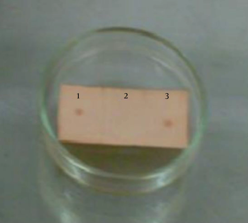
Moreover, presence of the 146s antigen was confirmed by dot blot. Two-μL of the purified 146s antigen and 2-μL of the Phosphate Buffered Saline (PBS) (negative control) were spotted on a piece of nitrocellulose membrane with a distance of 1 cm from each other. The membrane was placed in a plastic container and sequentially incubated at 37°C for 15 minutes, followed by blocking in Bovine Serum Albumin (BSA) 1% buffer, incubation at 37°C for 45 minutes, rinsing with buffer (PBS containing tween 20 (5%)), incubation at 37°C for 45 minutes with specific antibody solution against 146s (1:10 dilution), rinsing with buffer (PBS containing tween 20 (5%)), adding anti-rabbit antibody (1:1000 dilution), incubation at 37°C for 45 minutes, washing with PBS containing tween 20 (5%), placing in DAB solution and peroxidase, respectively. Finally, detection by eye was performed.
4. Results
4.1. Purification by Sucrose Gradient Procedure
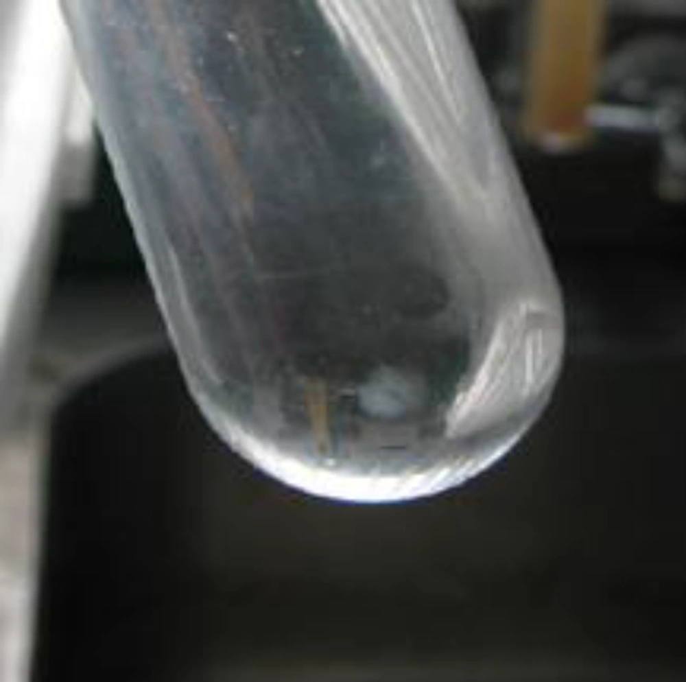
The pellet of the146s antigen of serotype A was developed at a concentration of sucrose 50% (Figure 1).
4.2. Measurement and Confirmation of the 146s Antigen
The absorbance rate of Foot-and-Mouth disease virus serotypes A at the wavelengths of 240, 259 and 280 nm was 1.238, 1.573 and 1.157, respectively. Moreover, the amount of 146s antigen at the same wavelengths was 163.416, 207.636 and 152.724 μg/mL, respectively. The amount of purified protein by nanodrop (ND-1000, the united states) was 0.275 mg/mL.
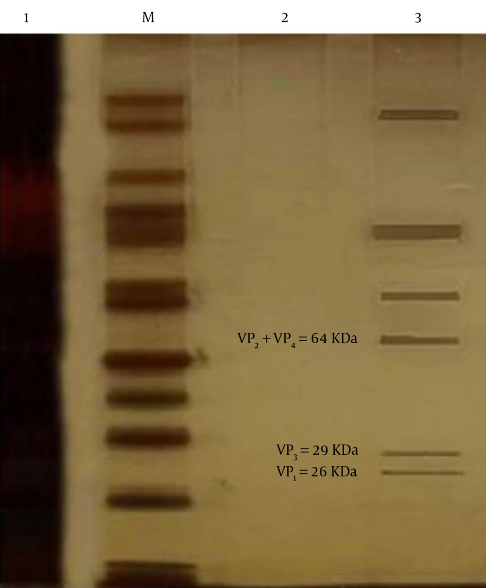
With respect to protein size, resolving of gel in SDS-PAGE was 15%. We used a protein marker (Fermentas, Germany) for determination of protein size in the sample. The 146s antigen was observed at 26, 29 and 64 KDa (Figure 2).
In the dot blot assay, brown spots represent reaction between the 146s antigen and standard specific antibody. Intensity of the brown stain is dependent on the amount of the antigen.
5. Discussion
Foot-and-Mouth Disease (FMD) is the most infectious viral disease that involves cloven-hoofed livestock species. The FMD is an economically destructive disease of livestock and has remarkable global socioeconomic outcomes. Even though its vaccines were available since the early 19th century, the eradication of FMD from parts of the world remained uncertain. The disease still involves millions of animals around the world and remains the main hurdle to the commerce of animals and animal products (22). The FMD virus relates to the Picornaviridae family and chiefly infects cloven-hoofed animals. Its genome is a single-stranded RNA that is surrounded by a protein coat consisting of four structure proteins including; VP1, VP2, VP3 and VP4 (23). The FMD virus is separated into seven immunologically distinctive serotypes: A, O, C, Asia I, and South African Territories (SAT1, SAT 2, and SAT3) (20). These seven serotypes distribute no cross-immunity. This means that, animals that have formerly been contaminated with one serotype remain vulnerable to the six other serotypes. Occurring in Europe, South America and Asia, FMDV serotype O is the most common between the seven serotypes, worldwide. The FMD infectivity may be mild or subclinical in related species, further complicating clinical diagnosis (24, 25). A definite diagnosis of FMD is promising only with laboratory tests. The aim of this study was to purify 146s antigen of FMD virus serotype A.
In the current study, the whole 146s particle was used. VP4 is internal and the other particles are on the surface of the capsid. The VP1, 2 and 3 particles are antigenic indexes on the surface of the virus, but only VP1 is decomposed and can stimulate antibody neutralization. In the present study, 20% - 50% of sucrose gradients were used for purification of 146s antigen of FMD virus. Consistent with our study, many researches applied sucrose gradients for purification of FMD virus.
In the study of Booth et al., (1987), which was done for the determination of response dose ratio of micro-neutralization tests on foot-and-mouth disease, FMD virus was harvested in BHK21 tissue culture. It was then inactivated at 4°C for 48 hour with 0.05% acetyl ethyleneimine. Next, the reaction was stopped by addition of excess sodium thiosulphate. The crude fluids were concentrated by precipitation with saturated ammonium sulfate and pelleted in the ultracentrifuge. The resuspended pellets, were finally purified by zonal centrifugation in 15-45% sucrose gradients for 180 minutes at 30000 rpm (26).
In the study of Cartwright et al., (1980) the correlation of serology and immunology between 12s and 146s particles of FMD virus was investigated. In their study, 146s particle was purified by 15% - 45 % sucrose gradients (27).
In the study of King et al., (1980) in order to assess the mutant FMD virus, it was purified by 15% - 45% sucrose gradients (28).
Francis et al., (1982) at the beginning of their study applied 15% - 25% sucrose gradients for purification of virus antigens. They studied the quantitative difference between antigens of FMD virus and synthetic polypeptide (29).
Collen et al. (1984) used 146s antigen serotype O induced antibody production in mouse spleen cells. They used 5% - 30% sucrose gradients for purification of 146s particles (30).
Shirai et al., (1989) purified 146s antigen with 10% - 40% sucrose gradients (31).
Aggarwal and Barett (2002) purified FMD virus by 15-30% sucrose gradients. They studied about recognition of antigenic regions on the surface of FMD virus (32).
In the study of Xia Feng et al., (2016) a suitable method to identify and quantify the 146s antigen of a serotype O FMD vaccine, a Double-Antibody Sandwich (DAS) ELISA was compared with a SDG analysis. The DAS ELISA was highly linked with the SDG method (R2 = 0.9215, P < 0.01). In contrast to the SDG method, the DAS ELISA was quick, vigorous, repeatable and very sensitive, with the lowest quantification limit of 0.06 μg/mL (33).
As several studies showed that this method is favorable; however, a new method with more specificity and more accuracy is needed.


