


1. Background
2. Objectives
3. Methods
3.1. Expression and Purification of Recombinant Human HMGB1(rhHMGB1), rhHMGB1 A Box and rhHMGB1 B Box
| Names | Sequences |
|---|---|
| hHMGB1 | 5′-CCCGAATTCATGGGCAAAGGAGATCCTAA-3′ |
| 5′-CCCCTCGAGTTATTCATCATCATCATCTTC-3′ | |
| hHMGB1 A-box | 5′-CCCGAATTCCCGAGAGGCAAAATGTCATC-3′ |
| 5′-CCCCTCGAGGATATAGGTTTTCATTTCTC-3′ | |
| hHMGB1 B-box | 5′-CCCGAATTCCCCAAGAGGCCTCCTTCGGC-3′ |
| 5′-CCCCTCGAGTCGATATGCAGCAATATCCT-3′ |
3.2. SELEX Procedure, Cloning, DNA Sequencing and Secondary Structure Prediction
| Names | Sequences |
|---|---|
| ssDNA library | 5′-GGACAAGAATCACCGCTC-N40-CGTACAGGAGGCATACAG-3′ |
| Forward primer | 5′-GGACAAGAATCACCGCTC-3′ |
| Reverse primer | 5′-CTGTATGCCTCCTGTACG-3′ |
3.3. Aptamer Binding Assay
3.4. Determination of Equilibrium Dissociation Constants (Kd)
3.5. ELASA
3.6. Cell Culture Experiments
3.7. Dot-Blot, South-Western Blot and Western Blot
3.8. Aptamer-Based Histochemical Staining
3.9. Immunofluorescence
3.10. qRT-PCR
| Names | Sequences |
|---|---|
| TNF-α | 5′-CGAGTGACAAGCCTGTAGCC-3′ |
| 5′-CCAGCTGGTTATCTCTCAGC-3′ | |
| IL-1β | 5′-TGTACCTGTCCTGCGTGTTG-3′ |
| 5′-GAAGACGGGCATGTTTTCTG-3′ | |
| IL-6 | 5′-AGAGCTGTGCAGATGAGTAC-3′ |
| 5′-GTCATGTCCTGCAGCCACTG-3′ | |
| TLR9 | 5′-CCTGTAGCTGCTGTCCAGTC-3′ |
| 5′-GCACAGACTTCAGGAACAGC-3′ | |
| β-actin | 5′- GAAGTGTGACGTGGACATCC-3′ |
| 5′- GATCCACACGGAGTACTTG-3′ |
3.11. Statistical Analysis
4. Results
4.1. Expression and Purification of Recombinant Human HMGB1(rhHMGB1), rhHMGB1 A Box, and rhHMGB1 B Box
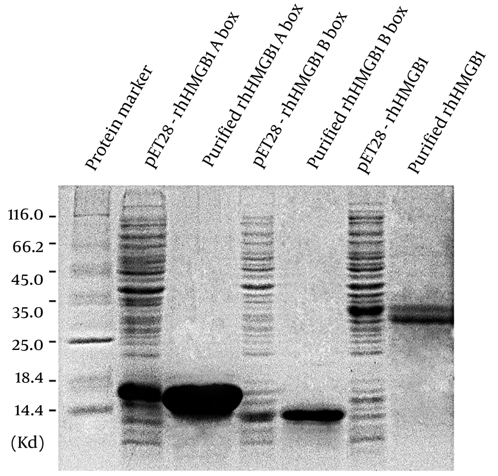
Expression and purified of rhHMGB1, rhHMGB1 A box and rhHMGB1 B box. Proteins were resolved by SDS-PAGE followed by Coomassie brilliant blue stain.
4.2. Selection and Characterization of the DNA Aptamer Against HMGB1
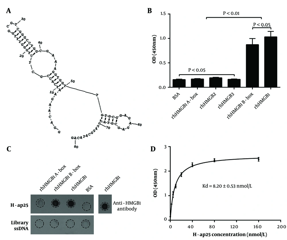
Characterization of H-ap25. The predicted secondary structure of H-ap25 presented a stable structure composed of a hairpin (9 - 50) and a stem-loops (52 - 69) linked by a T (A). H-ap25 specifically bound to rhHMGB1 and rhHMGB1 B box but not rhHMGB1 A box, as indicated by aptamer binding assay (B) and Dot-blot (C). In addition, H-ap25 does not bind to HMGB2 and HMGB3, which have high homologiesy to with HMGB1 (B). Equilibrium dissociation constant (Kd) of H-ap25 was 8.20 ± 0.53 nmol/L (D). The results are presented as the mean ± standard deviation (SD) of triplicate reactions.
4.3. Application of H-ap25 in South-Western Blot, ELASA and Immunohistochemical Staining
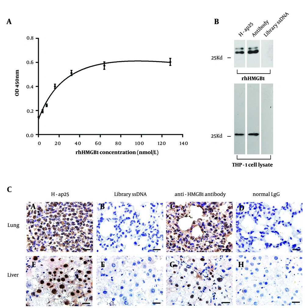
Application of H-ap25 in the detection of HMGB1. Sandwich ELASA with H-ap25- biotin exhibited a nice linear relationship between the concentration of rhHMGB1 and the optical density. The results are presented as the mean ± standard deviation (SD) of triplicate reactions (A). H-ap25-biotin specifically recognized denatured rhHMGB1 and cellular HMGB1 in South-Western blot (B). Aptamer-based histochemical staining showed specific nuclear staining both in mouse liver and lung tissues, comparable to the results of immunohistochemistry with anti-HMGB1 antibody (C). Scale bars = 20μm.
4.4. Enhancement of H-ap25 to HMGB1 on Pro-Inflammatory Through Activing TLR9/NF-κB Pathway
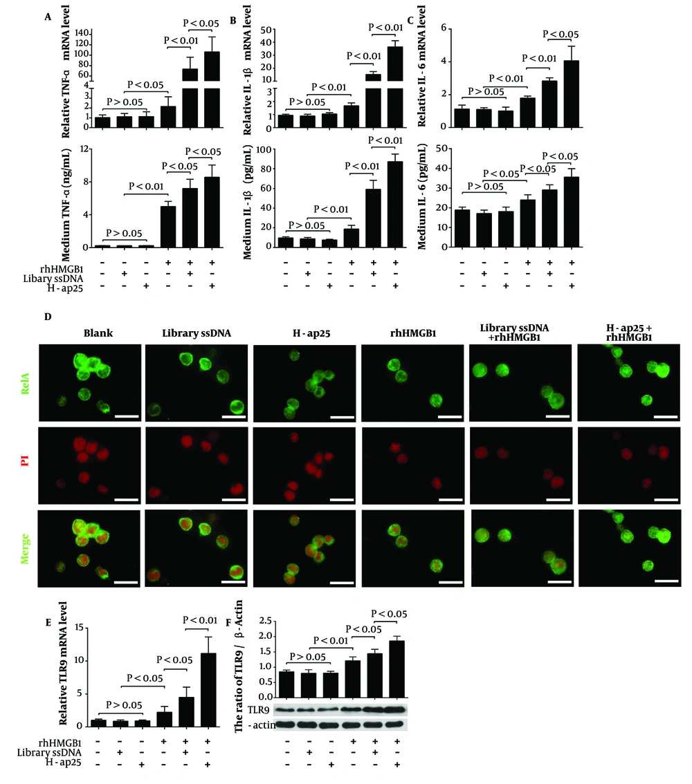
H-ap25 enhanced the pro-inflammatory response of HMGB1 in THP-1 cells. Both qRT-PCR (upper panel) and ELISA (lower panel) showed that rhHMGB1 up-regulated TNF-α (A), IL- 1β (B) and IL-6 (C) expression in THP-1 cells. These effects were markedly enhanced by both H-ap25 and library ssDNA whereas H-ap25 exhibited a stronger action than library ssDNA. Immunofluorescence demonstrated that the combination of Hap25 and rhHMGB1 exhibited a stronger effect of inducing p65 nuclear translocation than either rhHMGB1 or library ssDNA + rhHMGB1 (D). Scale bars = 40 μm. qRT-PCR (E) and Western blot (F) showed that the combination of H-ap25 and rhHMGB1 was most effective on up-regulation of TLR9 in THP-1 cell than either rhHMGB1 alone or rhHMGB1 + library ssDNA. The results of qRT-PCR and Western blot are presented as the mean ± standard deviation (SD) of four separate experiments.



