Methylglyoxal, a highly potent dicarbonyl compound, is the most important precursor of AGEs. One of the primary sources of MG and AGEs is food, particularly Western diets, and alcoholic beverages; however, they can also be produced endogenously as a by-product of the glycolysis pathway (
46). Previous studies have shown that MG and AGEs contribute to cognitive impairments (
27,
47,
48). The role of AG in improving diabetes complications caused by increased serum AGE levels has been demonstrated in various studies (
49-
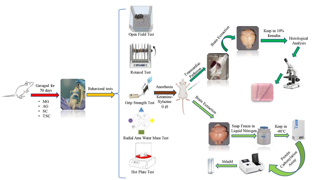
51). This study aimed to evaluate the neurobehavioral toxicity of MG in male Wistar rats. Additionally, we investigated the neuroprotective effects of AG, SC, and TSC, due to their structural similarity, on MG neurotoxicity using a battery of behavioral tests, followed by biochemical and histopathological assessments.
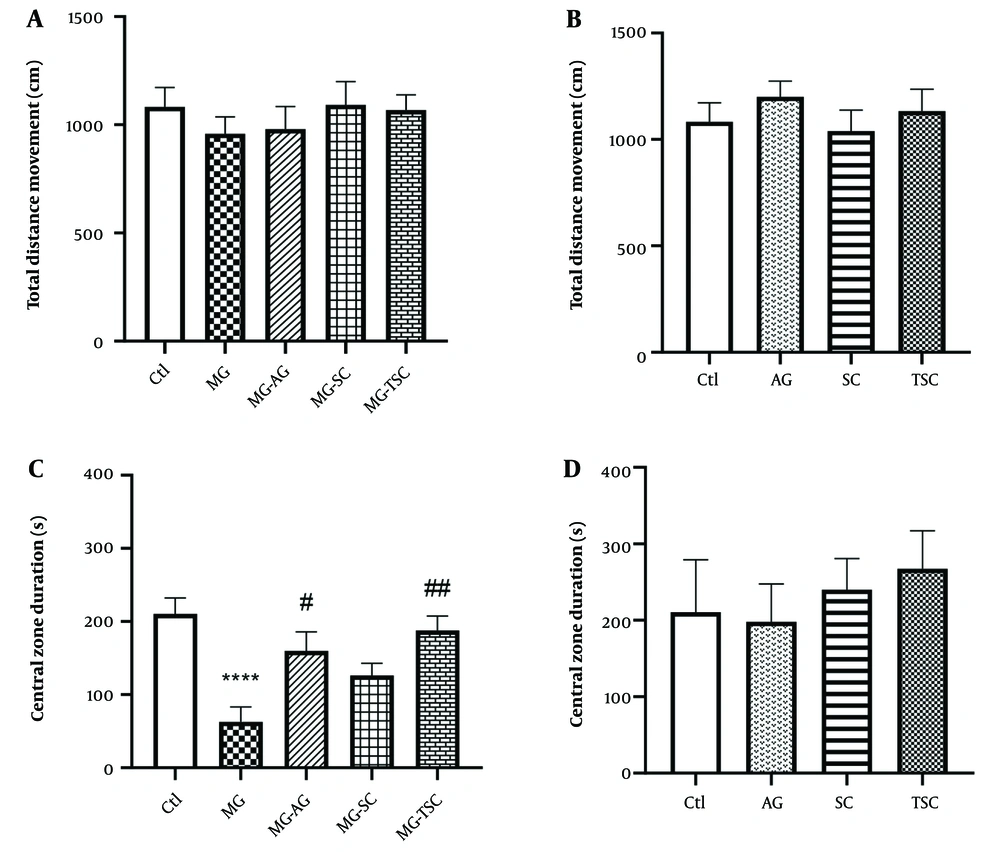
Based on the results, long-term treatment (70 days) with MG induced anxiety-like behaviors by reducing the time spent in the central zone of the open field apparatus. Furthermore, anxiety-like behavior was significantly reduced in the MG-AG and MG-TSC groups compared to the MG group. These findings suggest that AG and TSC may have an anxiolytic effect on MG-induced neurotoxicity. Patil et al. treated Sprague-Dawley rats with oral MG (50 mg/kg/day) or in combination with AG (1 g/L/day, through drinking water) for 45 days. Their results from the elevated plus maze test showed that anxiety developed in rats after MG treatment, as evidenced by the reduction in the number of entries and distance traveled in the open arms. They reported that AG administration for 45 days reduced anxiety behavior compared to the MG group (
51).
Methylglyoxal can induce oxidative stress by increasing AGE formation (
52), inactivating antioxidant enzymes (
53), and activating inducible nitric oxide synthase (iNOS) (
54). It has been shown that AG can reduce anxiety by interfering with the iNOS-cGMP pathway. iNOS can influence anxiety through nitric oxide production, leading to an increase in cyclic guanosine monophosphate (cGMP). This secondary messenger enhances neuronal communication, contributing to increased anxiety. Additionally, AG is able to inhibit guanylyl cyclase, an essential enzyme involved in cGMP production, thereby reducing nitric oxide synthesis and cGMP formation, which results in an anxiolytic effect (
55,
56).
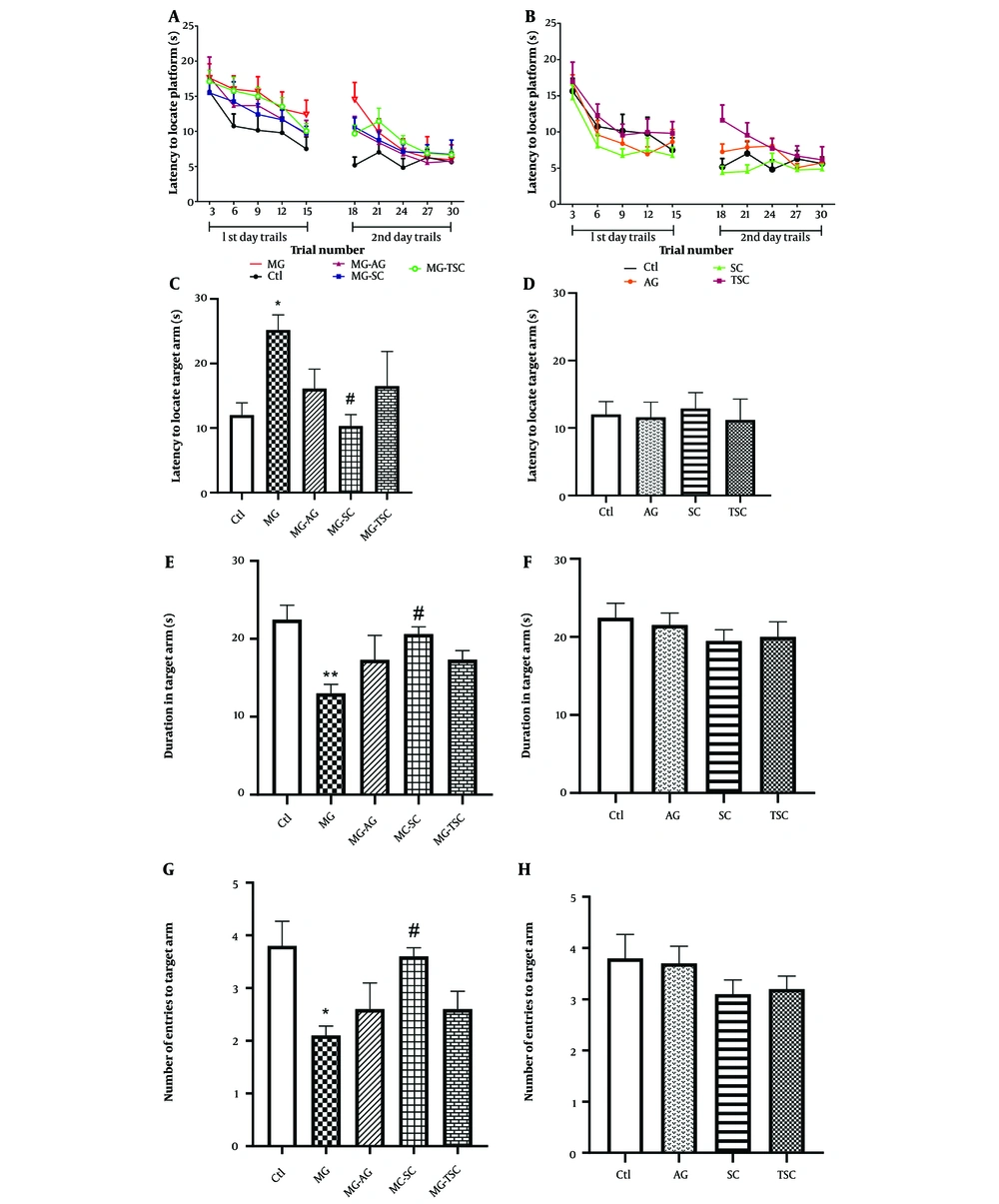
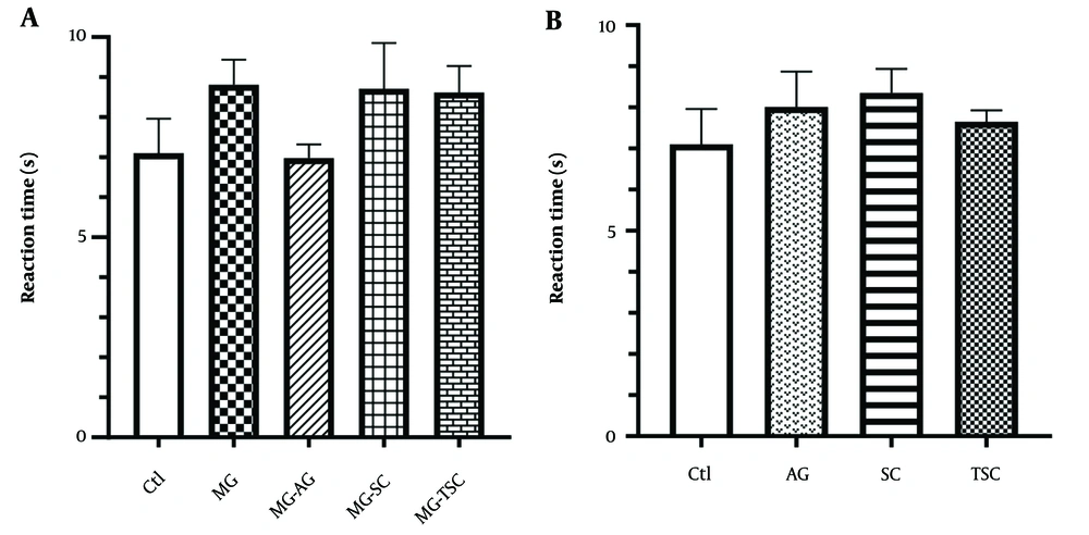
We investigated learning and memory impairment using the radial arm water maze test. In the MG group, there was an increase in the latency to locate the target arm on the probe day. Additionally, the number of entries into the target arm and the duration spent in the target arm were both reduced in the MG group.
Furthermore, the latency to locate the target arm was decreased in the MG-SC group, and the number of entries to the target arm and the duration spent in the target arm showed an increase compared to the MG group. These findings suggest that SC may prevent memory impairment induced by MG treatment. Previous research has indicated that MG, as a major precursor of AGEs, is linked to cognitive decline (
20,
57,
58). Methylglyoxal reacts with the free amine groups of amino acids (
59), leading to AGE formation and oxidative damage to proteins (
60). Long-term oral MG treatment elevates serum MG levels (
61). Methylglyoxal detoxification primarily occurs through glyoxalase 1 (GLO1) and glyoxalase 2 (GLO2). Glyoxalase 1 converts methylglyoxal-glutathione hemithioacetal into S-(D)-lactoylglutathione, which is subsequently hydrolyzed by GLO2 into D-lactate and glutathione (
62).
Pucci et al. found that MG treatment decreases GLO1 activity, leading to MG accumulation in the hippocampus (
61). As a result, RAGE expression increases, and the AGE-RAGE axis is activated (
7). The AGE-RAGE axis plays a crucial role in cognitive impairment by activating intracellular signaling pathways, such as nuclear factor-κB (NF-κB), which lead to the production of pro-inflammatory cytokines like IL-1 and IL-6 (
63,
64). Additionally, it increases reactive oxygen species (ROS) production, causing inflammation in neural tissues (
65-
67).
High intracerebroventricular (ICV) injection of MG (3 µmol/µL/day) administered over six days led to a decrease in the recognition index in the object recognition test but had no effect on learning and memory assessed by the open field and Y-maze tests (
67). In another study, a high ICV injection of MG (3 µM/µL) impaired cognitive functions, including both short- and long-term memory, as well as short-term spatial memory, as evaluated by the novel object recognition and Y-maze tests (
29). Treatment of mice with MG (100 mg/kg, orally) for four weeks also caused working memory impairment as shown by the Y-maze test (
61). Long-term treatment with MG (0.5% in drinking water) in Sprague-Dawley rats did not cause significant cognitive impairment in spatial and working memory, though the serum MG level increased (
68). This body of evidence demonstrates that whether or not cognitive impairment occurs depends on the specific MG study protocol employed.
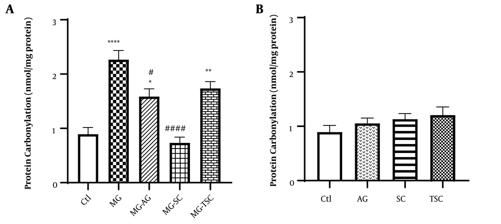
In another part of our study, we measured the protein-carbonyl content in the brain tissues of the studied animals. The binding of reactive aldehydes to amino acid side chains, known as protein carbonylation, is one of the major irreversible oxidative protein alteration markers related to various chronic diseases such as AD and Parkinson's disease (
69-
72). Carbonyl stress accumulates reactive carbonyl species that interact with nucleophilic substrates, inducing biomolecular malfunction and altering crucial cellular proteins (
24,
73). This leads to inflammation and autoimmune reactions (
74). Based on the results, MG treatment for 70 days increased protein-carbonyl content in the brain tissues of the studied animals. We observed a significant increase in protein carbonylation in the MG, MG-AG, and MG-TSC groups compared to the control group. Furthermore, the MG-SC and MG-AG groups showed a decrease in protein-carbonyl content compared to the MG group. Preventing carbonyl stress is a new therapeutic approach for reducing AGEs formation by inhibiting the formation of Schiff bases and Amadori adducts (
75,
76). Colzani et al. (2016) evaluated and compared the activity of AG toward four reactive carbonyl species using high-resolution mass spectrometry (HRMS) in vitro, and reported that AG's quenching reactivity depends on the type of reactive carbonyl species (RCS). AG inhibits protein carbonylation by binding to carbonyl or dicarbonyl RCS, including MG and glyoxal (
77). These findings suggest that SC and AG may serve as practical agents for reducing MG-induced protein carbonylation, possibly by trapping dicarbonyl groups. Further research is needed in this area.
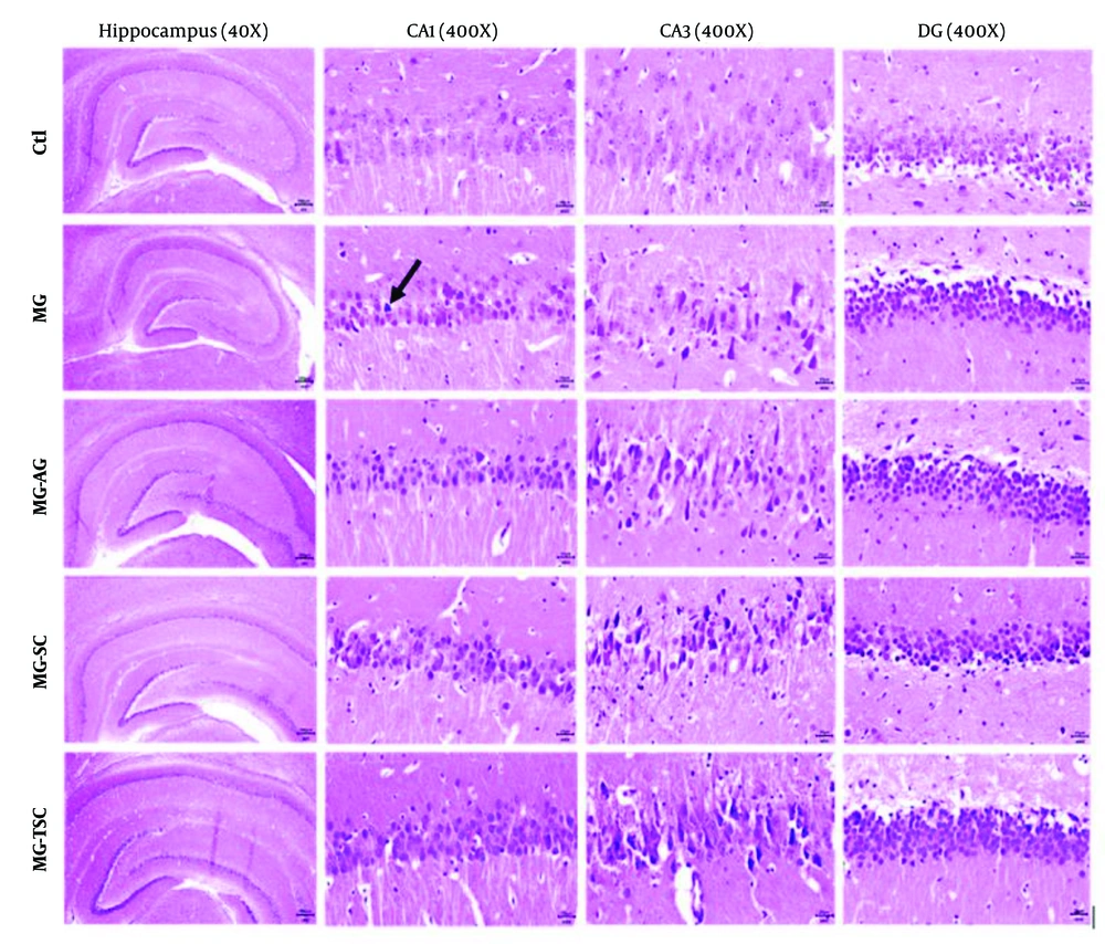
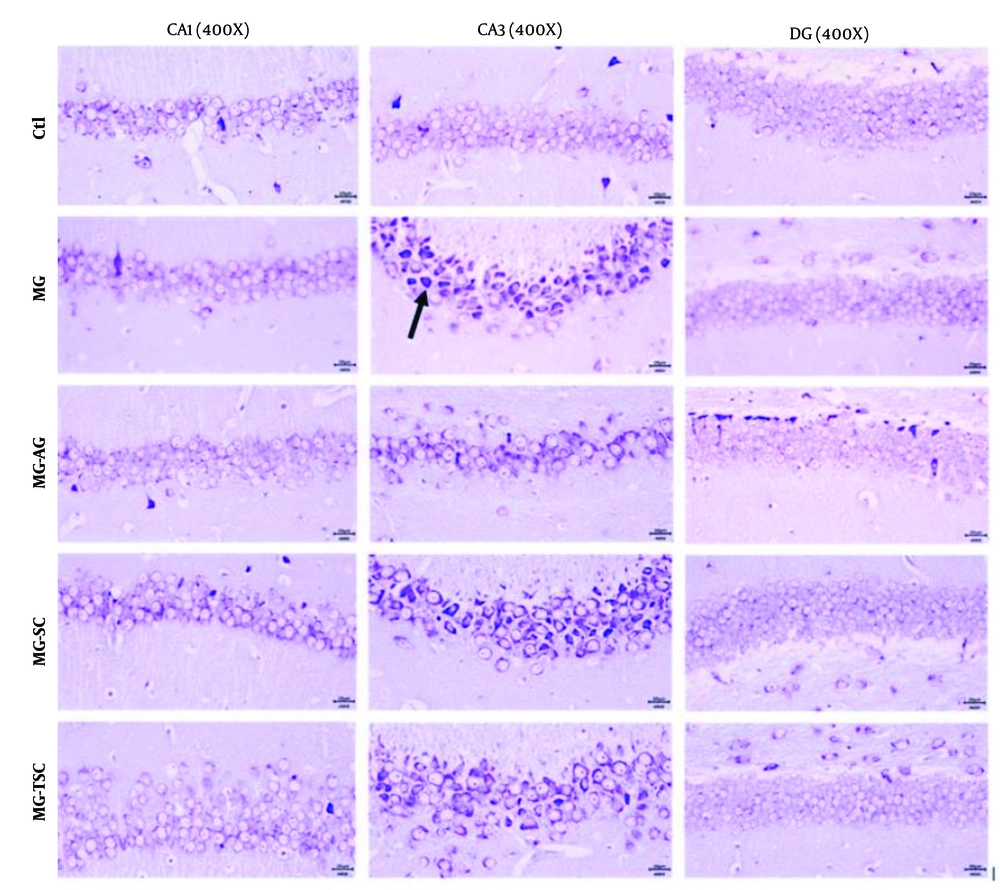
It has been demonstrated that cognitive functions such as memory, learning, and anxiety are closely related to hippocampal function (
51). In this study, we examined the effects of MG, AG, SC, and TSC treatment on the CA1, CA3, and DG regions of the hippocampus using hematoxylin-eosin (H&E) staining and cresyl violet staining. According to the histological results, neuronal necrosis was most prominent in the CA3 region across all treatment groups. The MG group exhibited extensive neuronal loss and nuclear pyknosis, particularly in the CA1 and CA3 regions. In contrast, the other groups (MG-AG, MG-SC, and MG-TSC) demonstrated improved neuronal survival and reduced necrosis, with the MG-TSC group showing the best outcomes, particularly in the CA1 and DG areas. It has been shown that the central nervous system is sensitive to the toxic effects of MG (
30). In one study, oral administration of MG (50 mg/kg/day) for 45 days led to mild neuronal degeneration in the CA1 region of the hippocampus, while co-treatment with AG (1 g/L/day) significantly reduced degenerative changes compared to the MG group (
51). Another study exposed Sprague-Dawley male rats to MG (0.5% in drinking water) for 12 months, and their histological results indicated no significant differences in cellular morphology or apoptosis compared to the control group (
68).
Overall, our findings support the idea that long-term MG exposure can mediate behavioral, biochemical, and histopathological changes in rat brains, affecting memory, anxiety, protein-carbonyl content, and cellular degeneration. Additionally, our results suggest that AG, SC, and TSC hold promise for further investigation as potential neuroprotective agents against MG-induced neurobehavioral toxicity.
5.1. Conclusions
Methylglyoxal is known as a highly reactive by-product of glucose metabolism and serves as a major precursor of AGEs, playing a critical role in the development of neurodegenerative diseases, including AD and Parkinson's disease. In addition to its endogenous production, MG is also introduced into the body through exogenous sources such as food and cigarette smoke. This study aimed to examine the neurobehavioral toxicity of MG, as well as evaluate and compare the neuroprotective effects of AG, SC, and TSC on MG-induced neurotoxicity using neurobehavioral, biochemical, and histopathological assessments.
We observed that oral administration of MG impaired memory and increased anxiety-like behaviors in animals. Additionally, MG treatment led to increased protein carbonylation in brain tissue, a marker of oxidative stress. SC was effective in preventing memory impairment, while both SC and AG reduced protein carbonylation in the brain tissue. Aminoguanidine and TSC also decreased anxiety-like behaviors. Furthermore, MG exposure caused neurodegenerative changes in the hippocampus, specifically in the CA1 and CA3 regions and AG, SC, and TSC improved neuronal survival particularly in the CA1 and DG areas.
According to the results, further research is needed to elucidate the precise mechanisms by which AG, SC, and TSC exert their neuroprotective effects against MG-induced neurotoxicity.