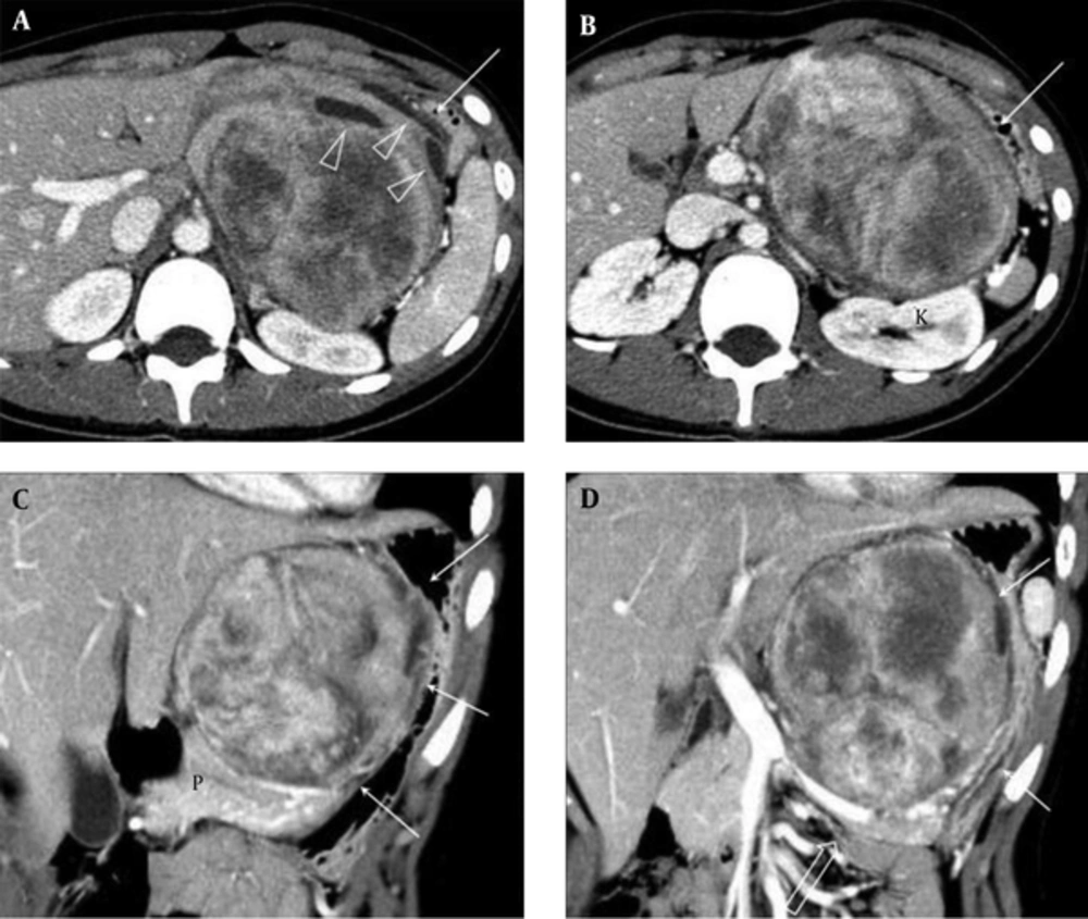
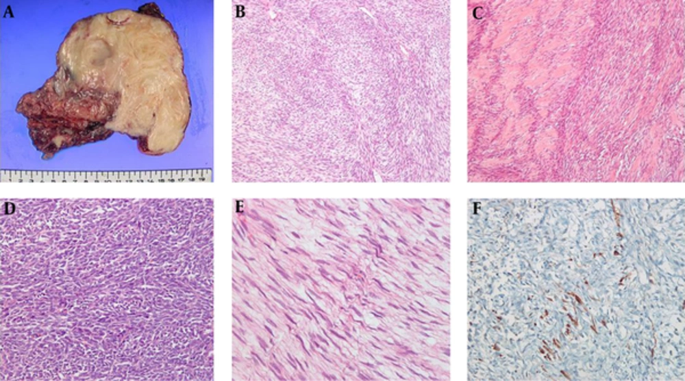
An MPNST arising in the abdomen is very rare, and to the best of our knowledge, only a few cases of retroperitoneal MPNSTs have been reported (
7,
8). Various neoplastic diseases develop in the lesser sac. They usually mimic gastric or pancreatic tumors. As the tumor of our case was large, its origin was difficult to define; either the stomach or lesser omentum was a possibility. A massive, well-circumscribed mass exhibiting heterogeneous enhancement and some cystic changes was apparent on enhanced CT scans. Similarly, a malignant gastric GIST usually presents as a large, well-circumscribed heterogeneous mass with a central region of low density (
9). Based on the CT findings and location of the mass, we assumed that the tumor was a GIST arising from the lesser curvature of the stomach or an extra-GIST of the lesser omentum. The absence of any NF1 signs contributed to no suspicion of MPNST. Therefore, we did not conduct a preoperative biopsy; the fragility of GISTs and the resultant risks of hemorrhage and dissemination preclude preoperative biopsy (
10). The mass was pathologically identified as an MPNST after surgery.
Retroperitoneal MPNST has been reported to develop in the paraspinal region, close to a nerve trunk (
11). The CT features of an MPNST include a large heterogeneously enhancing mass with an irregular or infiltrative margin, invasion of adjacent organs or soft tissues, and bone destruction (
11,
12). The CT findings of our case were different except for the large heterogeneously enhancing mass. Both the unusual tumor location and CT findings of our present case of MPNST provide valuable information for future clinical diagnoses.
MPNST is typically associated with a poor outcome compared with those of other soft tissue sarcomas. The recurrence rate is as high as 40%, and the most common metastatic sites are the lungs and the bone (
3). The 5-year survival rate ranges from 30 to 50% (
5). The negative prognostic factors for MPNST are similar to those for other soft tissue sarcomas and include the tumor site (head, neck, and trunk vs. the extremities), a large tumor size, high-grade, and positive surgical margins (
1). Some reports suggested that NF1 patients with MPNST experience poorer survival than do sporadic MPNST patients (
13) although this has not been a consistent finding (
14). Tumor size, tumor site, and microscopically incomplete resection explain the poor outcome of our present patient. Her younger age may also have influenced her unfavorable outcome (
13).
Complete surgical resection is the mainstay of MPNST treatment. Incomplete surgical resection increases the risk of MPNST-specific death nearly six-fold (
4). The tumor location in our present case adversely affected creating an adequate surgical margin (
15). Adjuvant radiotherapy may improve local tumor control; however, any evidence that survival is prolonged by radiotherapy is limited (
16-
18). If the preoperative diagnosis had been correct (identification of MPNST with a biopsy), neoadjuvant radiotherapy may have reduced the tumor volume, rendering complete excision possible (
19). Role of chemotherapy for MPNST was proved more in pediatric patients than in adults. The overall response rate to primary chemotherapy was reported as 45% in a group of pediatric MPNST patients (
6). First-line chemotherapy typically consists of a combination of ifosfamide and doxorubicin (
1). In our present case, the MPNST recurred and progressed systematically despite the use of these chemotherapeutic agents. Research on the molecular pathway involved in MPNST pathogenesis and the development of effective target agents would aid in treatment of this very aggressive malignancy.
In conclusion, MPNSTs remain diagnostically and therapeutically challenging. This case report improves our understanding of this extremely rare but highly malignant disease.