1. Introduction
Transjugular intrahepatic portosystemic shunt (TIPS) is an effective way to decompress portal hypertension. Due to the high rate of stenosis and occlusion of the TIPS stent, patients are recommended to receive routine follow-up imaging surveillances, such as Doppler ultrasonography, every three to six months. TIPS dysfunction is an important issue with no clear recommendation regarding the use of prophylactic medications to prevent the formation of thrombus or stenosis. Accumulating histologic evidences suggest that the transection of bile ducts during the TIPS procedure and the formation of biliary fistula are major etiologies of the shunt occlusion (1, 2). However, only very few cases of TIPS-associated biliary fistula, which were identifiable with imaging modalities, have been reported. We describe a case of TIPS occlusion complicated with biliary fistula, which was revised successfully by the re-implantation of a stent-graft.
2. Case Presentation
A 43-year-old male patient had undergone the TIPS procedure during May 2012 at the Seoul St Mary’s hospital for the treatment of recurrent variceal bleeding and F3 esophageal varices. Transjugular intrahepatic portosystemic shunt was created with an 8 mm × 7.5 cm polytetrafluoroethylene (PTFE)-covered stent (Seal bifurcated stent graft extension; S&G Biotech, Seongnam-si, Korea) per the standard protocol, between the right portal vein and the right hepatic vein. The stent-graft had bare segments; 5 mm at the hepatic vein side and 2 cm at the portal vein side. During the eight months after the TIPS placement, variceal bleeding did not occur. However, in January 2013, the patient’s follow-up liver ultrasound examination showed a cut-off in the blood flow within the TIPS stent, and the liver computed tomography (CT) exam revealed a thrombotic occlusion within the TIPS stent with the thrombus extending to the right portal vein. Esophagogastroduodenoscopy also showed the progression of esophageal varices. Therefore, the patient was admitted for TIPS revision. On arrival, the patient was asymptomatic and his mental status was alert. His body temperature was 36.5°C, blood pressure 120/70 mm Hg, pulse rate 68/minute and respiratory rate 20/minute. The Child-Pugh score was five (class A). Serum aspartate aminotransferase (AST) was 30 U/L, alanine aminotransferase (ALT) 20 U/L, total bilirubin 0.59 mg/dL, direct bilirubin 0.24 mg/dL, alkaline phosphatase 179 IU/L, total protein 6.8 g/dL, albumin 3.7 g/dL, prothrombin time 14.4 seconds (INR 1.34), white blood cell count (WBC) 1160/uL, hemoglobin 11.5 g/dL, and platelets 46000/uL.
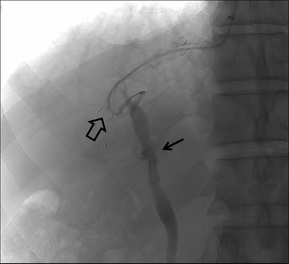
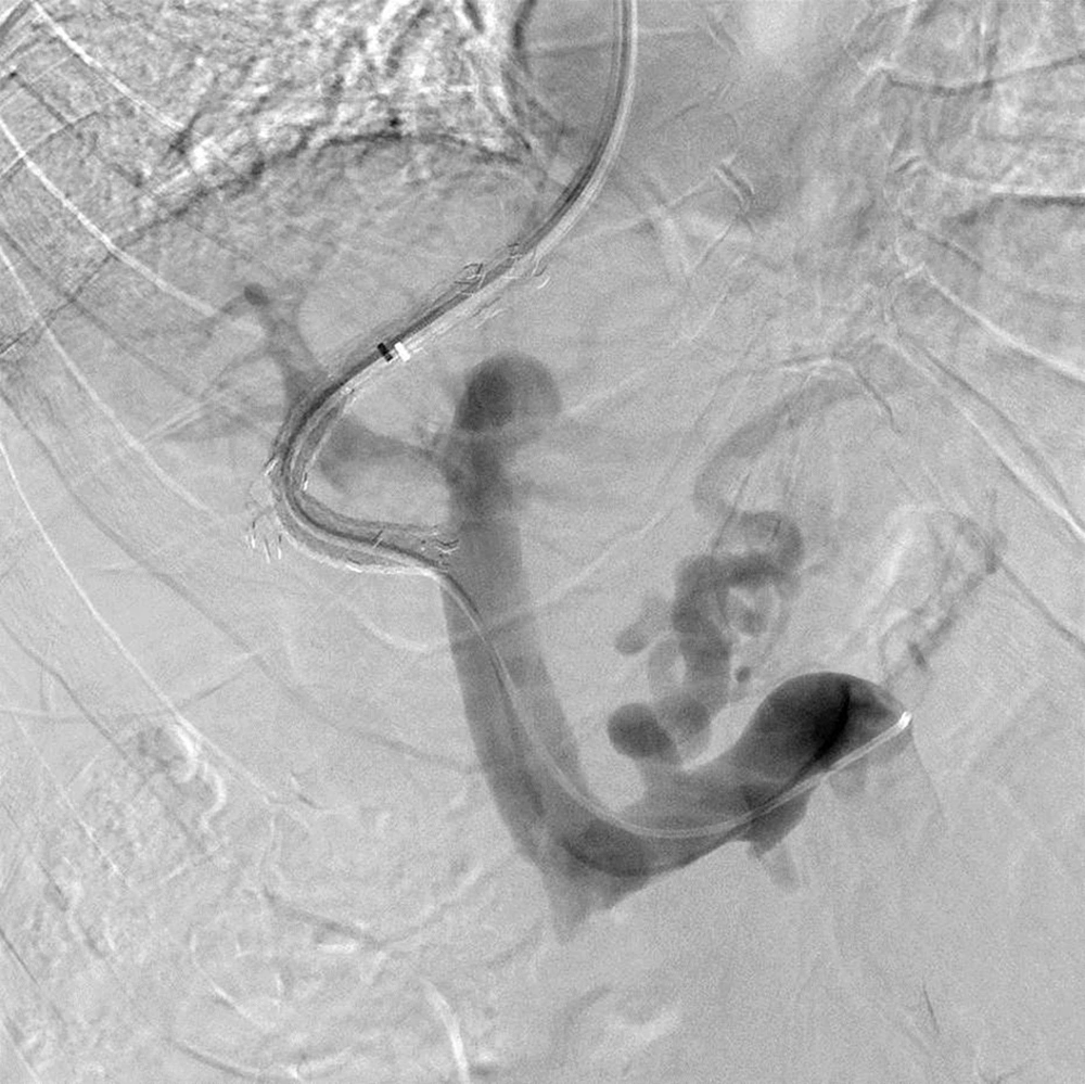
Venogram of the shunt revealed communication between the TIPS tract and the bile duct demonstrating a biliary-TIPS fistula combined with a focal stenosis of the stent at the hepatic venous segment of the TIPS tract (Figure 1). A very slow flow was detected within the TIPS tract and the right portal vein was not opacified. After negotiation of the occluded segment and the right portal vein with a 0.035 hydrophilic guide wire (Terumo, Japan) and 4F cobra catheter (Terumo, Japan), the catheter was placed in the main portal vein through the struts of the portal side bare segment. On the portal venogram, there was a partial filling defect, which extended from the proximal right portal vein to the posterior segmental portal vein that contained the stent graft. The increased portosystemic pressure gradient (27 mmHg) was measured. The occluded segment was dilated with an 8 mm balloon catheter, and an 8 mm × 10 cm PTFE-covered stent (S&G Biotech) was implanted in a stent-in-stent manner. The stent-graft was placed between the proximal right portal vein and the right hepatic vein near the opening of the inferior vena cava. Following the stent-graft placement, the venogram showed restoration of the blood flow in the TIPS tract, and the biliary fistula was no longer visualized. Slow flow in the peripheral portal veins and the coronary veins were observed without residual stenosis (Figure 2). Final portosystemic pressure gradient decreased to 15 mmHg. Moderate stenosis of the proximal right portal vein persisted as a filling defect on the completion portogram. Two days later, the patient was discharged from the hospital without any immediate complication. Doppler sonography six weeks after the TIPS revision showed patency of the TIPS tract. Flow velocity of the portal vein and the hepatic vein was measured to be 40 cm/second and 120 cm/second, respectively.
A 43-year-old cirrhotic man with TIPS occlusion complicated with biliary fistula. Venogram through the right internal jugular access. Total occlusion was observed at the parenchymal segment of the TIPS tract (open arrow). Transjugular Intrahepatic Portosystemic Shunt-biliary fistula with contrast leakage to the common bile duct was delineated (black arrow).
3. Discussion
TIPS is an effective treatment for reducing portal hypertension. It is applied for cases of recurrent variceal bleeding, refractory ascites, Budd-Chiari syndrome and clinically as a bridge to liver transplantation. Garicia-Pagan et al. reported that the use of early-TIPS for patients at high-risk of variceal bleeding resulted in a much lower incidence of bleeding control failure (3) and also reported that early TIPS was more effective than endoscopic treatment in preventing variceal re-bleeding and improving survival rate (3). However, long-term efficacy of TIPS is limited due to shunt stenosis or occlusion. LaBerge et al. reported that the rates of stenosis and occlusion within two years after TIPS were approximately 47% and 12%, respectively (4).
The TIPS procedure creates communication between the portal system and the systemic circulation via a percutaneous approach with penetration of the liver parenchyma. In the process, injury may develop in the structures adjacent to the targeted portal vein including the hepatic parenchyma or the portal triad, which consists of the portal vein, hepatic artery and bile duct. In cases of significant bile duct injury, TIPS-biliary fistula may develop resulting in bile leakage and ultimately TIPS occlusion (1).
The frequent locations of the TIPS stenosis are the hepatic venous end of the stent and the parenchymal portion (5). It has been reported that there is a histological difference between the two locations in case of stenosis due to different etiologies. Narrowings at the outflow tract of the hepatic vein were usually associated with intimal hyperplasia as a consequence of shear stress and turbulence from increased high-velocity blood flow (1, 6). In cases of mid-shunt stenosis, “pseudo-intimal hyperplasia” with the formation of granulation tissue and thickening of the neointima, composed of myofibroblasts and collagen, were usually observed. Szeet et al. reported that “pseudo-intimal hyperplasia” was closely related to bile duct injury (1). LaBerge et al. reported that biliary staining was associated with exuberant inflammation and granulation tissue replacement (6). Meanwhile, LaBerge et al. (6) suggested another theory on the association between bile leakage and the development of TIPS stenosis. Bile is considered thrombogenic because it is rich in bile acids, bile salts, cholesterol, and phospholipid and thus, by stimulating coagulation cascade, biliary fistula may lead to acute thrombosis. Accordingly, a case of acute TIPS occlusion nine hours after the TIPS procedure due to the identifiable biliary-to-TIPS fistula formation was reported (2).
In our case, stenosis developed at the hepatic venous segment and total occlusion with biliary fistula formation developed at the portal side of the stent-graft. The stenosis and the occlusion were each located at both ends of the covered segments of the stent. From this, it may be hypothesized that the insufficiently covered hepatic venous segment by the PTFE graft was related to stenosis and the exposure of the TIPS tract to bile acids due to the biliary fistula formation led to stent occlusion. Also, shortening of the stent-graft, which has a similar mesh structure with wallstents (Boston scientific), may have been related to the events; this explains the late development, eight months after the shunt creation, of the stenosis and the occlusion (7).
Shunt recanalization by angioplasty and re-stenting is regarded as a standard option in the revision of shunt occlusion. In case of biliary-to-TIPS fistula, there is no established recommendation regarding the type of stent yet generally covered stents are favored over bare stents. Tanaka et al. reported that with PTFE-covered stent grafts, the rate of in-stent stenosis or stent occlusion within the first year after TIPS dropped significantly compared to the bare metal stents (8). Jung et al. reported that TIPS created with expanded PTFE stent-grafts showed not only superior primary patency rates but also clinical outcomes defined as better control of bleeding and ascites compared with bare stents (9). These results may be related to the fact that covered stents can almost completely prevent bile from entering the stent lumen (10).
In summary, accumulating evidence supports the theory that biliary spillage is one of the major etiologies of the mid-shunt occlusion. In our case, stent occlusion complicated with clearly visualized biliary fistula was detected eight months after the placement with combined stenosis at the hepatic venous segment. The patient was successfully treated with angioplasty at the hepatic venous segment and implantation of a covered stent in a stent-in-stent manner. Revision using bare stents or angioplasty alone may be effective in some cases yet for the treatment of stent occlusion with proven biliary fistula, revision using covered stents seems to be advantageous in preventing permeation of the bile into the stent.

