
1. Background
Tongue trauma occurs in children by teeth due to falling on the ground, blowing, or an episode of convulsion. It is a rare event except in epileptic children. It may lead to panic attacks to the parents, an uncontrollable cry of the child, and the presence of blood or tissue debris in the mouth. The treatment of tongue trauma includes wound cleaning, foreign body removal, and suturing the wound under anesthesia (1).
There are various anesthesia techniques used for the surgical repair of tongue trauma. They may include monitored sedation, which imposes the risk of aspiration of blood and saliva (2) and more commonly, general anesthesia with an endotracheal tube. The latter, in spite of safety regarding aspiration risk, has many disadvantages in children including a cough, gagging, and reflex bronchospasm; it also takes a relatively long time for induction and recovery despite that it is a short surgical procedure (3, 4).
Laryngeal mask airway is widely used in anesthesia practice since its first appearance despite controversy to its use in certain types of surgeries (5). It has been used in a variety of pediatric surgeries and studied in some oral surgeries as tonsillectomy with success (6-8). The use of the endotracheal tube or laryngeal mask airway seems to be the same in ensuring adequate ventilation (9). However, laryngeal mask airway may have many advantages such as decreased stress response, decreased postoperative complications, and shortened recovery time (10, 11). On the other side, the use of laryngeal mask airway may have many disadvantages such as narrowing of the surgical field in head and neck surgeries and the possibility of intraoperative laryngospasm (12, 13).
2. Objectives
The use of flexible laryngeal mask airway instead of the traditionally used endotracheal tube for controlling the airway of children presented for elective surgical repair of tongue trauma may reduce the extubation time, decrease the hemodynamic response to the airway control, and decrease the probability of postoperative complications. This clinical study aimed to evaluate the extubation time (primary outcome), the hemodynamic response, and the safety (secondary outcomes) when flexible laryngeal mask airway was used instead of the endotracheal tube in tongue laceration surgical repair.
3. Methods
This clinical prospective randomized trial was approved by the Research Ethics Committee of the Faculty of Medicine, Tanta University (code 32321/05/2018). It was also registered before patient enrollment at the Pan African Clinical Trial Registry on 13 July 2018 (PACTR201807466395693). The study was conducted at the Department of Pediatric Surgery, Tanta University Hospitals from July 2018 to April 2019.
Children with tongue laceration admitted for elective repair under general anesthesia, aged 2 to 5 years, with American Society of Anesthesiologists (ASA) Class I or II, and in the fasting state were included in this study. The exclusion criteria consisted of patients with a full stomach (who underwent general anesthesia with endotracheal tube), active bleeding, tongue hematoma, predicted difficult airway, or the history of gastro-esophageal reflux.
In the preoperative period, the parents were asked about the cause of the tongue trauma and the medical history of their children, especially the history of epilepsy. Then, the general and local examination of the child was done with requesting laboratory investigations, especially coagulation studies. The purpose, advantage, procedure, and potential risks of this research work were adequately explained to the parents of children in detail with the reassurance that their children will receive the optimal and safe medical care. If they agreed to participate in the study, the guardian of each child would sign a written informed consent. The children were presented to surgery after fasting for 6 h from solid food and 2 h from clear fluids. The patients were randomly assigned into two groups according to the method used for securing airway through the aid of computer-generated software and closed sealed envelopes.
Endotracheal Tube (ETT) Group: In this group, the airway was secured by suitably sized cuffed right-angle endotracheal (RAE) tube according to the patient’s age and weight.
Flexible Laryngeal Mask Airway (LMA) Group: In this group, the airway of the patient was secured using flexible laryngeal mask airway of suitable size according to the weight of the patient.
The suitably sized laryngoscope and endotracheal tube and the suction device and catheter were prepared before the induction of anesthesia. A standardized anesthetic technique was used in all cases through the inhalational induction of anesthesia using sevoflurane 6% in 80% oxygen administrated through a facemask. An assistant helped in the establishment of intravascular access through the insertion of the 22-gauge peripheral venous cannula and the patient was attached to the basic 5 ASA monitoring (pulse oximeter, three-lead electrocardiogram, non-invasive blood pressure, end-tidal carbon dioxide, and temperature). All patients received 0.01 mg/kg of atropine intravenously. Fentanyl 1 μg/kg was then administrated intravenously.
The adequate depth of anesthesia was judged by the absence of increased heart rate or limb movement in response to jaw thrust. When it was achieved, the airway of the child was secured by the same expert anesthetist according to the group of the patient without the use of muscle relaxants. The presence of gagging or coughing during the trial to secure airway was managed by the restoration of face mask ventilation using sevoflurane inhalation until the achievement of the adequate depth of anesthesia. The airway was considered to be secure when there was bilateral chest elevation with positive capnograph wave during hand ventilation and movement of the bag during spontaneous ventilation. If patients in the laryngeal mask group had inadequate ventilation, the laryngeal mask was replaced by a suitably sized endotracheal tube and the patient was excluded from the study.
The anesthesia was maintained by inhalational anesthesia through sevoflurane 3.5% in oxygen to air of 1:1 and spontaneous ventilation. Any increase in the heart rate or mean arterial pressure during the surgery by more than 10% of the baseline values was managed by fentanyl 1 μg/kg intravenously. The surgical field was monitored adequately for the presence of excessive blood loss or tissue debris.
At the end of the surgery in the ETT group, sevoflurane was switched off with full awake extubation of the patient after careful suction of blood or secretions. In the LMA group, the mask was removed after careful suction while the patient was in deep anesthesia; then, the inhalational anesthetic was switched off. In both groups, face mask ventilation continued. When the modified Aldrete scale reached score 8 or more, the patient was transferred to the recovery room with the continuous monitoring of the patient for the modified Aldrete scale every 15 min. The patient was discharged from the recovery room when the modified Aldrete scale reached to score 10.
An assistant nurse, out of the research team, who was blinded to the study helped in the measurement of the following variables: intubation time that represented the time interval in seconds from the removal of the facemask until the insertion and securing of LMA or ETT (The assistant nurse who helped in measurement of the intubation time should be blind to the group of the patients and this could not be obtained as she can identify the method used for airway control in each patient (ETT or LMA) by naked eye. So, she was kept away from the patients and informed when the face mask was removed to start counting of time till she was informed that the airway is secured.); surgical time that was calculated as time in minutes from the start of the surgery until its end; total anesthesia time that represented the number of minutes elapsed from the start of anesthesia induction until the patient’s transfer to the recovery room; extubation time (primary outcome) that was the time in minutes elapsed from the end of the surgery to the transfer of the patient to the recovery room; and recovery time that represented the number of minutes from the arrival to the recovery room until discharge from it. Moreover, the hemodynamic data including heart rate and systolic arterial pressure were recorded before the induction of anesthesia, after the induction of anesthesia, after airway securing, at the beginning of the surgery, and at the end of the surgery.
At the end of the surgery, the surgeon was asked to evaluate the surgical exposure as 1 = extremely poor, 2 = poor, 3 = accepted, 4 = good, and 5 = optimal. Moreover, the incidence of perioperative adverse events as trauma to lip, gum, teeth, or larynx, gagging, coughing, laryngeal spasm or bronchospasm, stridor, and sore throat were recorded. Patients who developed laryngeal spasm were managed by increasing the inspired oxygen tension with the assistance of the ventilation while doing jaw thrust and the stimulation of the laryngeal notch. The patients who developed stridor were managed by oxygen supplementation with intravenous injection of dexamethasone 0.1 mg/kg and close observation. Additionally, inhaled bronchodilators and systemic corticosteroids were used in children who developed bronchospasm. All the patients that had developed adverse events were managed by conservative and medical treatment and none of them required re-intubation.
3.1. Statistical Analysis
A preliminary study was conducted on 10 patients (who were not included in the final study) presented for tongue trauma repair under general anesthesia with either the endotracheal tube (five patients) or the flexible laryngeal mask airway (five patients). The extubation time was significantly lower in the LMA group (7.34 ± 5.77 min) than in the ETT group (16.03 ± 5.54 min). As a result, at least 36 patients were required in each group to detect a significant difference by 5 min in the extubation time with the study power of 95% and the α value of 0.05. The dropout rate was assumed to be 20%; thus, 45 patients were required in each group. The SPSS computer program (SPSS Inc, Chicago, IL, USA) was used for the statistical analysis of the recorded data by either unpaired t-test for parametric data presented as means and standard deviations or Fisher’s exact test for numerical data presented as numbers and percentages. The Mann-Whitney U test was used to analyze the surgical exposure score expressed as medians with interquartile ranges. The values were considered statistically significant when the P values were less than 0.05.
4. Results
This clinical trial assessed the eligibility of 107 children presented for elective repair of tongue trauma under general anesthesia, from whom 17 patients were excluded due to either not meeting the inclusion criteria (8 patients) or unwillingness to participate (9 patients). The remaining 90 patients were randomly assigned to either the ETT group (45 patients) or the LMA group (45 patients). All the included patients received the allocated intervention and their data were successfully collected and analyzed (Figure 1). The differences in age, gender, body weight, and the ASA class of the studied patients were statistically insignificant between the two groups (P = 0.530, 0.671, 0.192, and 0.619, respectively) (Table 1).
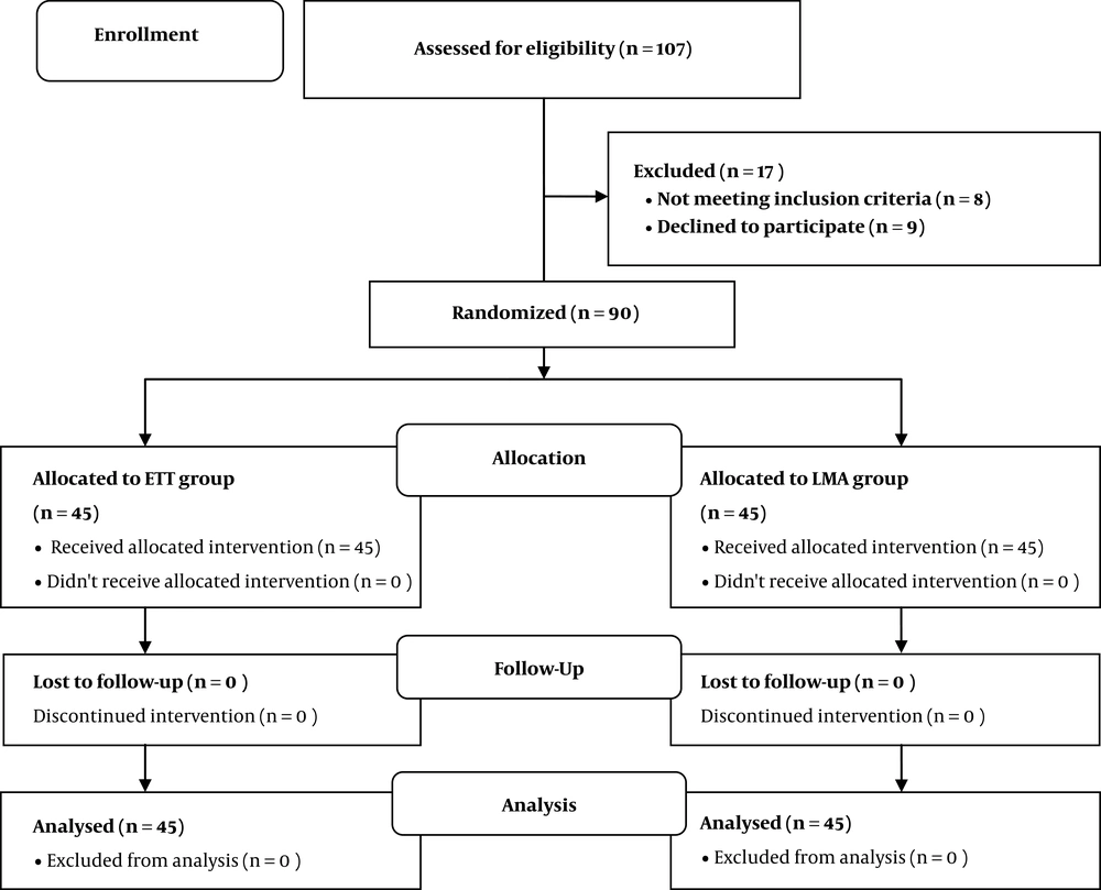
Figure 1.
CONSORT flow chart of the study
Table 1.
Demographic Data in the Studied Groupsa
| ETT Group | LMA Group | P Value | CI (95%) | |
|---|---|---|---|---|
| Age, y | 3.24 ± 0.857 | 3.13 ± 0.815 | 0.530 | -0.461 - 0.239 |
| Gender | ||||
| Male | 27 (60) | 24 (53.33) | 0.671 | 0.749 - 1.757 |
| Female | 18 (40) | 21 (46.67) | ||
| Body weight, kg | 15.33 ± 2.86 | 14.60 ± 2.42 | 0.192 | -1.842 - 0.376 |
| ASA class | ||||
| Class I | 36 (80) | 33 (73.33) | 0.619 | 0.707 - 2.096 |
| Class II | 9 (20) | 12 (26.67) | ||
| Perioperative complications | ||||
| Lip trauma | 1 (2.22) | 0 (0.00) | 1.00 | 1.639 - 2.496 |
| Gum trauma | 1 (2.22) | 0 (0.00) | 1.00 | 1.639; 2.496 |
| Teeth trauma | 0 (0.00) | 0 (0.00) | ||
| Larrynx trauma | 0 (0.00) | 0 (0.00) | ||
| Gagging | 10 (22.22) | 4 (8.89) | 0.144 | 1.028 - 2.340 |
| Cough | 11(24.44) | 3 96.67) | 0.039b | 1.212 - 2.544 |
| Laryngeospasm | 5 (11.11) | 1 (2.22) | 0.203 | 1.147 - 2.670 |
| Bronchospasm | 4 (8.89) | 1 (2.22) | 0.361 | 1.015 - 2.709 |
| Stridor | 8 (17.78) | 0 (0.00) | 0.006b | 1.746 - 2.814 |
| Sore throat | 10 (22.22) | 2 (4.44) | 0.027b | 1.305 - 2.643 |
Abbreviations: CI, confidence interval; ETT, Endotracheal Tube; LMA, laryngeal mask airway.
aData were presented as means ± standard deviation or as No. (%).
bSignificant difference between the two groups.
The time required for extubation was significantly shorter in the LMA group than in the ETT group (P < 0.0001). Moreover, the recovery time was significantly longer in the ETT group than in the LMA group (P = 0.001). However, the intubation time, surgical time, and total anesthesia time were comparable between the two studied groups (P = 0.874, 0.411, and 0.725, respectively) (Table 2).
Table 2.
Duration of Intubation, Extubation, and Recovery in the Studied Groups
| ETT Group | LMA Group | P | CI (95%) | |
|---|---|---|---|---|
| Intubation time, s | 46.98 ± 10.68 | 46.60 ± 11.73 | 0.874 | -5.078 - 4.323 |
| Surgical time, min | 37.89 ± 6.35 | 36.77 ± 6.41 | 0.411 | -3.784 - 1.562 |
| Total anesthesia time, min | 96.13 ± 11.79 | 95.20 ± 13.24 | 0.725 | -6.184 - 4.318 |
| Extubation time, min | 13.29 ± 2.263 | 7.47 ± 2.7 | < 0.0001b | 4.769 - 6.876 |
| Recovery time, min | 60.67 ± 11.16 | 52.67 ± 11.16 | 0.001b | 3.325 - 2.675 |
Abbreviations: CI, confidence interval; ETT, Endotracheal Tube; LMA, laryngeal mask airway.
aData were presented as means ± standard deviation or as No. (%).
bSignificant difference between the two groups.
The mean heart rate was significantly lower in the LMA group than in the ETT group both immediately after securing the airway and at the beginning of the surgery (P ≤ 0.0001 and 0.0017, respectively). However, the differences in the mean heart rate before anesthesia induction, before securing the airway, and at the end of the surgery were statistically insignificant between the two groups (P = 0.654, 0.718, and 0.4674, respectively) (Figure 2).
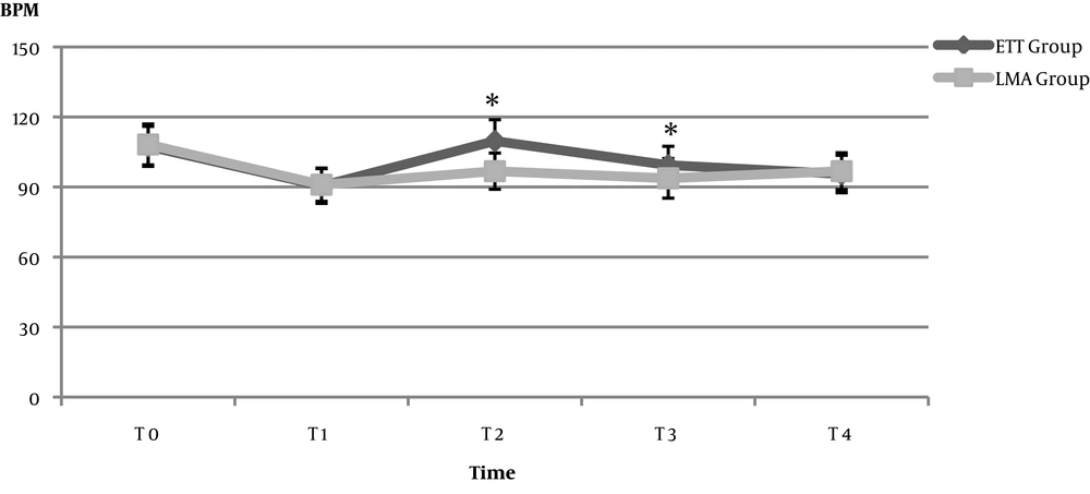
Figure 2.
Changes in the mean heart rate in the two groups. Data were presented as means ± standard deviation. *denotes a significant change. T0: before anesthesia induction, T1: after anesthesia induction and before airway securing, T2: after airway securing, T3: at the beginning of the surgery, T4: at the end of the surgery.
Moreover, systolic blood pressure was significantly higher in the ETT group than in the LMA group after airway securing and at the start of the surgery (P < 0.0001). However, systolic blood pressure was comparable between the two studied groups before anesthesia induction, before securing the airway, and at the end of the surgery (P = 0.119, 0.1503, and 0.2007, respectively) (Figure 3).
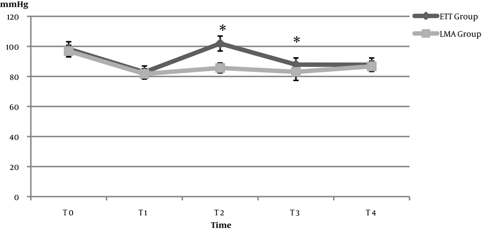
Figure 3.
Changes in the mean systolic blood pressure in the two groups. Data were presented as means ± standard deviation. *denotes a significant change. T0: before anesthesia induction, T1: after anesthesia induction and before airway securing, T2: after airway securing, T3: at the beginning of the surgery, T4: at the end of the surgery.
The incidence of coughing, stridor, and sore throat was significantly higher with the use of the endotracheal tube than with the use of laryngeal mask airway (P = 0.039, 0.006, and 0.027, respectively) while insignificant differences were observed between the two studied groups in the incidence of other perioperative adverse events including lip trauma, gum trauma, teeth trauma, laryngeal trauma, gagging, laryngospasm, and bronchospasm (P > 0.05) (Table 1).
The surgeons’ rating of the surgical exposure was statistically comparable between the two groups (P = 0.5005) as the scores in the two groups ranged from 1 to 4 with the interquartile range of 4 (95% CI = 0.0002 to 1.0000) (Figure 4).
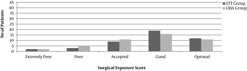
Figure 4.
Surgical exposure score in the two groups. Data were presented as the number of patients.
5. Discussion
This clinical trial showed that the use of flexible laryngeal mask airway instead of the endotracheal tube in the anesthetic management of children undergoing elective repair of tongue trauma significantly decreased the extubation time, the recovery time, the hemodynamic response after airway securing and at the beginning of the surgery, and the incidence of postoperative cough, stridor, and sore throat. However, it had no significant effect on the intubation time, surgical time, total anesthesia time, surgical exposure, and the incidence of other complications.
Since the introduction of laryngeal mask airway by Archie Brain and its approval by the Food and Drug Administration, it gained high popularity in anesthesia practice (14). The use of flexible laryngeal mask airway in the outpatient’s procedures has many advantages, especially in terms of the avoidance of the laryngoscope, decreased stress response, and decreased incidence of complications as coughing, sore throat, stridor, and laryngeal spasm (10, 15). Some authors suggested that it decreases the costs, as well (16). On the contrary, there are still disadvantages such as difficult visualization of the surgical field, leakage, and inability to control adequate ventilation that limit the use of flexible laryngeal mask airway (17).
A meta-analysis of 19 studies was conducted by Luce et al. to determine the incidence of perioperative respiratory complications with the use of laryngeal mask airway or endotracheal tube in the pediatric population. They concluded that the use of laryngeal mask airway in pediatric anesthesia practice decreased the incidence of perioperative desaturation, cough, laryngeal spasm, and breathe holding. The incidence of perioperative bronchospasm, sore throat, and aspiration was comparable between the groups (3). Moreover, a systematic review of 16 studies by Gómez et al. aimed to detect the safety and the effectiveness of laryngeal mask airway in adenotonsillectomy and found that flexible laryngeal mask airway is a safe and effective option for the anesthetic management of patients presented for adenotonsillectomy. In addition, the laryngeal mask had the advantage of rapid extubation and rapid recovery. They advised expert anesthesiologists to use laryngeal mask airway (8).
The use of flexible laryngeal mask airway or endotracheal tube during adenotonsillectomy was evaluated in the randomized clinical trials by Peng et al. (11) and Sierpina et al. (7) They found that the laryngeal mask airway was a safe and efficient alternative to the endotracheal tube in children undergoing adenotonsillectomy, as it significantly shortened the time for extubation. However, they noticed no significant difference in the incidence of laryngeal spasm and oxygen desaturation between the methods. In addition, the clinical trials by Fuentes-Garcia et al. (6) and Orfei et al. (18) concluded that the laryngeal mask airway was equally as safe and effective as the endotracheal tube in the airway management of children undergoing upper gastrointestinal endoscopy, in addition, its use was associated with a significant decrease in the extubation time and the recovery time. Moreover, Webster et al. noticed a lower hemodynamic response to the use of laryngeal mask airway than to the use of the endotracheal tube in adenotonsillectomy surgeries (19).
On the other hand, Ranieri et al. (20) and Hern et al. (17) demonstrated that the use of endotracheal tube in anesthetizing children for adenotonsillectomy surgery was safer than the use of the laryngeal mask, as the risk of oxygen desaturation, the need for intraoperative rescue endotracheal intubation, and the difficulties in the surgical access were higher with the use of laryngeal mask airway. In this study, the suitably sized endotracheal tube was prepared as a rescue in all patients of the LMA group. However, no rescue endotracheal intubation was used and the success rate of the use of the flexible LMA was 100%; thus, LMA represented as a good and safe alternative to ETT.
This clinical study was limited by the inability to perform all surgeries by the same surgeon. Additionally, the surgeon rating of surgical exposure was a subjective method. Moreover, the cuff pressure was not assessed. The depth of anesthesia and the need for ventilation assistance were not measured, which added to the study limitations.
We conclude that the flexible laryngeal mask airway is a good alternative to the endotracheal tube in children undergoing elective surgical repair of tongue trauma, as it significantly decreased the time of extubation and recovery, the incidence of postoperative cough, stridor, and sore throat, and the hemodynamic response to the intubation process and the start of the surgery.