
1. Background
Human papillomavirus (HPV) is a common sexually transmitted infection (STI) implicated in the pathogenesis of cervical cancer (1, 2). Cervical cancer is the fourth leading cause of cancer-induced mortality among women globally, with approximately 604 000 new cases diagnosed and 342 000 deaths recorded worldwide in 2020. High-risk HPV genotypes account for 2.5% of all cancers worldwide, 7.7% of all cancers in developing countries, and 2.2% of cancers in developed countries (3).
Recently, a study analyzing 5 176 cervical samples from 7 laboratories in Iran reported HPV positivity in 2 727 cases (53%), indicating an increasing prevalence of HPV infection in the Iranian population (4). According to GLOBOCAN 2012, mortality from cervical cancer in Iran is 1.2 per 100 000 individuals. A review reported a death rate of 42% from cervical cancer incidence (5). According to a recent study in Iran from 2008 to 2014, 5 304 women were diagnosed, 2 423 were followed, the mean age was 51.91 years, 65.91% were alive, and the 5- and 10-year survival rates were 58% and 50%, respectively. Older patients with squamous cell carcinoma (SCC) had better survival (6).
HPVs are small, nonenveloped viruses with double-stranded circular DNA genomes (7). More than 100 HPV types have been identified and are classified into low-risk and high-risk groups (8). The World Health Organization identifies 13 HPV types, including genotypes 16, 18, 31, 33, 35, 39, 45, 52, 56, 58, 59, 68, and 82, as high-risk variants. These types are characterized by oncogenic potential and a propensity for persistent infection, thereby contributing to cancer development (9). In contrast, other types, such as HPV types 6, 11, 40, 42, 43, 44, 54, 61, 70, 72, 81, and 89, are classified as low-risk and are associated with genital warts (10).
Cervical cancer screening is recommended for women aged 20 to 65 years and is typically performed every 3 years using the Pap smear test, which has been associated with a 41% to 92% reduction in cervical cancer mortality (11). Magnetic resonance imaging (MRI) is a highly accurate diagnostic modality for clinical staging of cervical cancer, demonstrating more than 94% accuracy in detecting tumor involvement across different anatomical regions of the cervix (12).
National guidelines in Iran, developed through a systematic review, recommend Pap smear screening every 5 years for women aged 30 to 69 years (13). For patients with atypical squamous cells of undetermined significance (ASCUS) lesions, medical protocols suggest immediate referral for colposcopy, repeat Pap smear testing after 1 year, and HPV genotype testing to determine the presence of high-risk HPV types (14).
In one study, approximately 10.9% of Pap smear specimens were identified as ASCUS. The distribution of high-risk HPV types varied among racial groups, ranging from 5.6% to 6.5%. Advances in molecular diagnostic tests have complemented traditional Pap smear procedures and improved detection capabilities (15).
Persistent infections with certain high-risk HPV types, particularly types 16 and 18, which express the E6 and E7 oncogenes, are known to lead to cervical cancer and cervical intraepithelial neoplasia (CIN) (16). Real-time PCR is a powerful tool that combines PCR amplification with computerized analysis to detect closely related sequences. It provides high sensitivity, and its ability to detect and genotype HPV DNA is particularly beneficial for inclusion in screening programs (17, 18).
Furthermore, SYBR Green-based PCR assays can detect and quantify specific nucleic acid sequences. This method is based on the fluorescence produced by the binding of SYBR Green dye to double-stranded DNA during PCR, with fluorescence intensity indicating the amount of target DNA present (19). The specificity of these fluorescence signals is assessed via melting curve analysis, which is directly related to the guanine-cytosine (GC) content and length of the PCR amplicons (20).
2. Objectives
This study aimed to molecularly profile HPV genotypes in Iranian women with ASCUS cytology using real-time PCR to improve the accuracy of viral detection and characterization. It also sought to determine the prevalence of high-risk HPV types, particularly those associated with cervical carcinogenesis, to inform risk stratification and clinical management. By highlighting the predominance of oncogenic genotypes, this research underscores the value of HPV genotyping as a crucial tool for the early diagnosis and prevention of cervical cancer in Iran.
3. Methods
3.1. Study Population
This cohort study enrolled 150 women with cytologically confirmed ASCUS at Shahid Beheshti Hospital, Tehran, between March and November 2023. Eligible participants were aged 25 to 65 years, had not undergone cervical treatment, such as loop electrosurgical excision procedure (LEEP) or cryotherapy, in the past year, were not pregnant, and were free of active genital infection at the time of sampling. All participants provided written informed consent, and the protocol was approved by the Ethics Committee of Islamic Azad University (IR.IAU.PS.REC.1402.142). Combined vaginal and exocervical specimens were collected using sterile flocked swabs, immediately placed in 1 mL of phosphate-buffered saline (PBS; pH 7.4), transported on wet ice within ≤ 2 hours, and processed within 4 hours. After vortexing and centrifugation (300 ×g, 5 min), the supernatants were aliquoted and stored at -20°C to preserve DNA integrity. No samples were excluded based on prespecified criteria, including insufficient volume, DNA < 10 ng/μL, or cytological ineligibility.
This study focused on the qualitative detection and genotyping of HPV DNA to assess genotype prevalence rather than quantitative viral load measurement.
3.2. DNA Extraction
The conventional phenol-chloroform-isoamyl alcohol technique was used to obtain HPV DNA from smear specimens. Briefly, cervical smear specimens were suspended in 500 μL of Tris-EDTA buffer (TE buffer; 10 mM Tris-HCl, 1 mM ethylenediaminetetraacetic acid [EDTA], pH 8.0) and homogenized by vigorous vortex mixing. An aliquot of the mixed specimen (100 μL) was then combined with proteinase K solution (10 μL; 20 mg/mL; Sigma-Aldrich, USA) and K buffer (250 μL) and incubated for 60 minutes at 45°C. DNA was extracted from the supernatant using 250 μL of alkaline phenol, followed by 250 μL of chloroform-isoamyl alcohol (24:1, v/v). The mixture was then precipitated with 500 μL of isopropanol. Next, 75% ethyl alcohol was used to wash the DNA at 10 000 g for 5 minutes at 4°C, followed by air-drying at 37°C and dissolution in 100 μL of distilled water.
3.3. Primer Design
Following genome analysis of the viruses and selection of conserved regions of the viral L1, E6, and E7 genes, primers were downloaded from the National Center for Biotechnology Information (NCBI) website to ensure specificity for the virus and humans. Primers were designed using NCBI (Table 1) and compared using Clustal Omega, GeneRunner, and the Basic Local Alignment Search Tool (BLAST). The primers were designed to achieve an optimal melting temperature, balanced GC content, and minimal self-complementarity and self-3' complementarity (21). The human β-globin gene was amplified as an internal control to confirm the presence of amplifiable cellular DNA and to exclude false-negative results due to sample degradation or PCR inhibition.
Table 1.
Primers
| Primers | Genotyping | Sequences | Amplicon Size | Target Gene |
|---|---|---|---|---|
| HPV 6/11 | Low-risk | F: 5'-gtatccaaagttgttgccacggat-3'; R: 5'-tgttaggatctggtaacacyacct-3' | 187 bp | L1 |
| HPV 16 | High-risk | F: 5'-cagatcatcaagaacacgtagagaa-3'; R: 5'-ccagctggaccatctatttcat-3' | 167 bp | E6-E7 |
| HPV 18 | High-risk | F: 5'-cagtgccattcgtgctgcaa-3'; R: 5'-ggaatttcattttgrggctctaaa-3' | 142 bp | E6-E7 |
| HPV 31 | High-risk | F: 5'-ttccacaacataggaggaaggtg-3'; R: 5'-ctccacgcatgtttacacttgggtt-3' | 90 bp | E6-E7 |
| HPV 33 | High-risk | F: 5'-tgcgtggaatgcaaaaamcctttgcaa-3'; R: 5'-acacagtttacatattccaaatgratt-3' | 111 bp | E6 |
| HPV 35 | High-risk | F: 5'-ttgtgtatactgcaaacaagaattaca-3'; R: 5'-catatggctggccttctctatat-3' | 95 bp | E6 |
| HPV 39 | High-risk | F: 5'-gcgaaggttgtcaatactgatga-3'; R: 5'-cctgcttgcgaccaccattc-3' | 127 bp | L1 |
| HPV 45 | High-risk | F: 5'-atttcacagcatagctggacagta-3'; R: 5'-ctatacttgtgtttccctacgtct-3' | 100 bp | E6 |
| HPV 52 | High-risk | F: 5'-gaagartcggtgcatgaaataag-3'; R: 5'-aggcacataatacacacgccatat-3' | 143 bp | E6 |
| HPV 56 | High-risk | F: 5'-gaatatgaattacaatttgtgtttcaacta-3'; R: 5'-aggttatagctgcacttttcacatat-3' | 178 bp | L1 |
| HPV 58 | High-risk | F: 5'-gaaaccacggacattgcatgatt-3'; R: 5'-atcgctgcaaagtctttttgcatt-3' | 98 bp | E6 |
| HPV 59 | High-risk | F: 5'-gtatggagaaacattagaggctgaa-3'; R: 5'-tgtcctgtatatattccagctatattat-3' | 156 bp | E6 |
| HPV 66 | Low-risk | F: 5'-ctgtgaacataaaagacgatttcattat-3'; R: 5'-ttggtactttaccatgcatggttata-3' | 127 bp | E6-E7 |
| HPV 68 | High-risk | F: 5'-ttaatcaccaccaacatgtactacta-3'; R: 5'-acgcttctactactagttcagt-3' | 111 bp | E7 |
| HPV 82 | High-risk | F: 5'-gttatacaaggtggggattacta-3'; R: 5'-gkgacactggtgcaggtggta-3' | 143/144 bp | L1 |
| Housekeeping gene | Housekeeping gene | F: 5'-GAAGAGCCAAGGACAGGTAC-3'; R: 5'-CAACTTCATCCACGTTCACC-3' | 268 bp | β-globin |
3.4. Real-time PCR
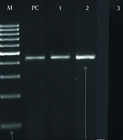
Real-time PCR was performed to detect 16 high-risk HPV genotypes in each clinical sample using genotype-specific primers listed in Table 1. The reaction mixture contained SYBR Green Master Mix (Applied Biosystems, USA), optimized concentrations of MgCl2 and primers (Table 2), and 2 μL of extracted DNA in a final volume of 20 μL. Thermal cycling was conducted on the ABI PRISM 7500 Sequence Detection System (Applied Biosystems, USA) under the standardized conditions shown in Table 3. Amplification was followed by HRM curve analysis, with fluorescence continuously monitored during a linear temperature ramp from 55°C to 95°C at 0.1°C/s. Although an approximate mean melting temperature (Tm) of 82 ± 1.5°C was initially observed across genotypes, genotype-specific Tm values were empirically determined during assay validation. To ensure genotyping accuracy, all SYBR Green-based calls were confirmed by Sanger sequencing of representative amplicons (n = 3 per genotype) and agarose gel electrophoresis (2%) to verify amplicon size and specificity.
Table 2.
Materials and Optimized Concentrations of Real-time PCR
| Material | Volume (μL) |
|---|---|
| SYBR Green Master Mix | 10 |
| DNA | 5 |
| Forward primer | 1 |
| Reverse primer | 1 |
| Distilled water | 3 |
| Total | 20 |
Table 3.
Real-Time PCR Program Used in a Thermal Cycler
| Stages and Steps | Temperature (°C) | Duration |
|---|---|---|
| Initial denaturation | 95 | 10 min |
| Amplification (45 cycles) | ||
| Denaturation | 95 | 20 s |
| Annealing | 60 | 25 s |
| Extension | 72 | 25 s |
| Final extension | 72 | 10 min |
| Melting curve analysis | 55 to 95 | 0.1°C/s (continuous fluorescence acquisition) |
A sample was considered HPV-positive if: 1) amplification occurred at a quantification cycle (Cq) ≤ 38 in at least 2 of 3 technical replicates; 2) the melt peak matched the expected Tm ± 0.5°C for the target genotype; and 3) no amplification was observed in negative controls. The Cq threshold of 38 was established based on the assay's limit of detection (LOD = 10 copies/reaction), determined using serial dilutions of quantified plasmid standards.
All reactions were run in technical triplicate (three PCR replicates per DNA extract); biological replicates were not applicable because each sample represented a unique patient. In cases of discordant replicate results (n = 4 samples), the run was repeated from the original DNA extract.
Each PCR plate included the following controls: 1) no-template controls (NTCs; nuclease-free water); 2) extraction blanks processed alongside clinical samples; and 3) positive controls consisting of plasmid DNA containing consensus sequences for HPV-16, -18, -31, and -45 (104 copies/reaction). All controls performed as expected across all runs.
Assay validation demonstrated 100% analytical specificity, with no cross-reactivity among 16 genotypes, an LOD of 10 copies/reaction, and intra- and interassay coefficients of variation < 3%.
3.5. Statistical Analysis
Because this study focused on the qualitative detection and genotyping of HPV DNA, based on presence/absence calls and Tm-based discrimination, rather than mRNA quantification, relative quantification methods such as the 2-ΔΔCt approach or Relative Expression Software Tool (REST), which are designed for reverse transcription quantitative PCR gene expression analysis, were neither appropriate nor applied, in accordance with the Minimum Information for Publication of Quantitative Real-time PCR Experiments (MIQE) guidelines.
HPV genotypes were identified by HRM-derived Tm profiles and confirmed by Sanger sequencing. A sample was considered positive for a given genotype if: 1) amplification occurred at a quantification cycle (Cq) ≤ 38 in ≥ 2 of 3 technical replicates; and 2) the melt peak fell within the genotype-specific Tm window (± 0.5°C).
Statistical analyses used frequencies (%) for qualitative variables, such as HPV prevalence and symptoms, and mean ± standard deviation (SD) for quantitative variables, such as age. Group comparisons employed Student t tests or analysis of variance (ANOVA) for continuous data and chi-square (χ2) or Fisher exact tests for categorical associations. All analyses were conducted in Stata software (version 14; StataCorp, USA), with P < 0.05 (two-sided) considered statistically significant.
4. Results
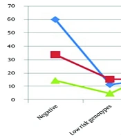
In this study of 150 women with ASCUS cytology (aged 21 - 63 years; mean, 35.65 ± 9.32 years), 46% reported vaginal discharge, 38.67% reported burning/itching, and 11.33% reported multiple sexual partners. Chi-square analysis revealed a significant association between age group and HPV risk type (χ2 = 18.42, P < 0.001), with high-risk genotypes predominating among those aged 25 to 50 years and low-risk types concentrated among those aged ≤ 25 years (P < 0.001 and P = 0.002, respectively). In addition, 38.67% had a history of genital warts. HPV DNA was detected in 100% of cases by PCR testing. The results showed a prevalence of 28.67% (43 cases) for low-risk HPV genotypes and 71.33% (107 cases) for high-risk HPV genotypes among the participants. According to PCR results, the highest prevalence of high-risk genotypes was observed in cases aged 25 to 50 years. In addition, 74.38% of low-risk genotypes were identified in individuals younger than 25 years (Figure 1).
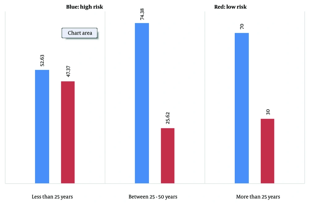
Figure 1.
HPV genotype prevalence was analyzed by age groups in the studied women based on PCR results.
Among women with high-risk genotypes, 39.25% experienced vaginal discharge, which was significantly higher than that among women with low-risk genotypes (P < 0.001). In addition, the prevalence of burning and itching among individuals with high-risk genotypes was significantly higher than that among women with low-risk genotypes (46.73% vs 18.60%; P = 0.001) (Table 4).
Table 4.
HPV Genotype Frequency Based on Real-Time PCR Across Variables in the Studied Women a
| Variables | High Risk (n = 107) | Low Risk (n = 43) | P Value |
|---|---|---|---|
| Genital discharge | |||
| Yes | 43 (25.39) | 4 (30.9) | < 0.001 |
| No | 65 (75.6) | 39 (70.9) | < 0.001 |
| Burning and itching | |||
| Yes | 51 (73.46) | 8 (60.18) | 0.001 |
| No | 57 (27.53) | 35 (40.8) | 0.001 |
| More than one sexual partner | |||
| Yes | 11 (35.9) | 7 (28.16) | 0.22 |
| No | 98 (65.9) | 36 (72.83) | 0.22 |
| History of HPV infection | |||
| Yes | 67 (68.61) | 26 (47.6) | 0.89 |
| No | 41 (32.38) | 17 (53.39) | 0.89 |
a Values are expressed as No. (%).
The prevalence of genotypes was as follows: HPV 16: 19 (12.66%), HPV 18: 14 (9.66%), HPV 31: 10 (6.66%), HPV 33: 8 (5.33%), HPV 35: 5 (33 3.3%), HPV 39: 6 (4%), HPV 45: 9 (6%), HPV 52: 6 (4%), HPV 56: 8 (5.33%), HPV 58: 4 (66/ 2%), HPV 59: 5 (3.33%), HPV 68: 6 (4%), and HPV 82: 7 (4.66%).
The highest frequencies among low-risk and high-risk genotypes were observed for HPV 11 (20%) and HPV 16 (12.67%), respectively.
5. Discussion
HPV, the primary cause of genital warts, is a major factor that increases the risk of cervical cancer, which is the fourth most common cancer and the fourth leading cause of cancer-related death among women worldwide (22). According to GLOBOCAN 2022 estimates, there were approximately 660 000 new cases and 350 000 deaths globally from cervical cancer in 2022 (24). In more developed regions, the incidence is notably lower, with approximately 15 600 new cases and 5 200 deaths reported annually (24). These figures underscore a major global health concern, particularly in low- and middle-income countries, where screening and vaccination programs are limited (23). More than 200 HPV genotypes have been identified and are categorized into low-risk groups, such as HPV 6 and 11, and high-risk groups, such as HPV 16, 18, and 31, based on their carcinogenic potential (25). Notably, HPV 16 and 18 are predominant oncogenic genotypes linked to most cervical cancers (26). Cytological examination of cervical smear samples is regarded as the most cost-effective method for diagnosing precursor lesions of cervical cancer. According to the Bethesda system for reporting cervical cytology, there are 2 types of squamous intraepithelial lesions: high-grade squamous intraepithelial lesions (HSIL) and low-grade squamous intraepithelial lesions (LSIL). Additionally, there are 2 subtypes of atypical squamous cells (ASC): ASCUS and atypical squamous cells that cannot rule out high-grade squamous intraepithelial lesions (27). ASCUS is the most frequently identified cytological abnormality, occurring in 1.6% to 9% of all smear test findings (28). HPV diagnosis relies on viral DNA detection using consensus PCR primer sets, such as GP5-GP6, SPF10, PGMY09-MY11, MY09-MY11, and LCR-E7, which offer broad genotype coverage (29-32). However, limitations of the commonly used MY09/11 system have been observed, including missed detection in 13.2% of patients infected with HPV and variable sensitivity across HPV types (17). Originally designed for types 11, 6, 16, 18, and 33, primer-template mismatches and a lengthy target sequence of approximately 450 bp increase the risk of false-negative results (33, 34). Our study addresses these issues by designing primers for products 90 to 187 bp in length (Table 1). Real-time PCR is essential for genotype-sensitive and rapid identification (35). HPV contributes to approximately 4.5% of head and neck cancers and nongenital cancers (36). In Iran, high-risk HPV types 18 and 16 predominate, constituting 77.5% and 32.4% of cervical and head and neck cancer cases (39). While low-risk HPV types are typically linked to benign anogenital warts (37), high-risk genotypes have also been identified in cases of genital warts (38). The incidence of HPV genotypes in Iran (77.5%) aligns with global patterns (39), and a comparable distribution of HPV 18, 16, and 11 is observed among Iranian women (37, 40).
In our study, PCR testing revealed a 28.67% incidence of low-risk HPV genotypes and a 71.33% incidence of high-risk HPV genotypes among participants. Notably, the highest frequencies were observed for low-risk genotype 11.6 (20%) and high-risk genotype 16 (12.67%). In a 2020 study on genital warts in 40 biopsy samples, HPV 6, HPV 16, and HPV 54 were prevalent at 77%, 15%, and 7.5%, respectively. No HPV 18 was detected, but coinfections of HPV 54 with HPV 6 and HPV 16 occurred (39). Ghobadi's 2023 study of 50 vaginal swab samples identified simultaneous high-risk HPV serotypes 16 and 18 in 10% of cases, along with various other genotypes (41). Manyere's 2020 study on genital warts, with participants' average age of 30.3 years, reported an overall HPV prevalence of 98%, with dominant low-risk genotypes (86%), including genotypes 11 (47%), 6 (42%), and 16 (14%) (42). Although only a limited number of studies have examined HPV genotype distribution among Iranian males, a recent investigation found an HPV prevalence of 55.7%, with HPV 6 identified as the most common genotype in Iran (40). In a 2019 study in Iran, females showed a prevalence of HPV 6 (43.3%) and HPV 11 (11.4%). High-risk HPV genotypes were observed more frequently, with HPV 16 at 16.6% and HPV 52 at 9.6% (43). HPV 16 (50%) and HPV 18 (12%) were prevalent in cervical carcinoma cases in women (44). In Yazd, Iran, HPV 16 (70%) and HPV 18 (16.7%) dominated cervical cancer genotypes (45). A Kerman study of 20 000 Pap smear samples identified HPV 16 and 18 as highly prevalent (46). Patients with cancer in Yazd and Mazandaran mostly had HPV 16 (45-47). In Tabriz, HPV 16 was the most common genotype. Guilan reported a frequency of 10.8% for genotype 16. Genotypes 16 and 18 were frequent in studies by Mobini Kesheh and Keyvani (44). These findings confirm those of our research.
Our study revealed a significant association between age group and HPV risk type (χ2 = 18.42, P < 0.001), with high-risk genotypes predominating in those aged 25 to 50 years and low-risk types concentrated in those aged ≤ 25 years (P < 0.001 and P = 0.002, respectively). In Alacam's (48) study, 36.3% of DNA samples were HPV-positive. The most common types of genital warts were HPV 16, 59, and 66. The highest HPV prevalence (44.1%) was in the age group of 17 to 34 years (48). A 2022 study reported 18.10% overall HPV prevalence, with rates of 22.94% in those aged ≤ 25 years and 21.25% in those aged 56 to 65 years (49).
Sexually transmitted infections pose a global health risk with high morbidity and mortality rates (50). More than half of infection-related malignancies stem from papillomavirus (51). This virus, linked to various cancers, manifests symptoms in women such as vaginal discharge, itching, and burning during urination (52, 53). In the present study, women with high-risk HPV genotypes had significantly higher rates of vaginal discharge (39.25%) and burning/itching (46.73%) than women with low-risk genotypes (P < 0.001 and P = 0.001, respectively). Lv's (54) study found a significant link between high-risk HPV positivity, cervical inflammation, and urinary incontinence. Zhang's (55) retrospective study of 1 330 women reported a 37.67% infection rate in vaginal secretions, with genotypes 16, 58, and 52 showing higher prevalence. Minhas (56) reported an overall HPV infection prevalence of 57%, with common genotypes correlating with women's general complaints and complications. Seyoum (57) emphasized the association between high-risk HPV genotypes and complications in women.
The type-specific primers generated comparable melting profiles, ensuring sensitive screening capability for various HPV genotypes.
5.1. Conclusions
In this study, HPV DNA was detected in 100% of Iranian women with ASCUS cytology, with high-risk genotypes accounting for 71.33% of infections, predominantly HPV 16 and 18. These oncogenic types were most prevalent among women aged 25 to 50 years. They were significantly associated with clinical symptoms such as vaginal discharge and genital burning or itching, suggesting a potential symptomatic burden beyond subclinical infection. To our knowledge, this is the first comprehensive molecular profiling of HPV genotypes in Iranian women presenting with ASCUS, revealing a striking predominance of high-risk HPV types, particularly HPV 16, which is strongly linked to cervical carcinogenesis. Although these findings cannot be generalized to all Iranian women, they provide critical insights into the virological landscape of a clinically relevant, at-risk subgroup referred for abnormal cytology. In settings with limited access to routine cervical screening, integrating molecular tools such as real-time PCR into the triage of ASCUS cases may improve risk stratification and guide timely intervention. These results support the development of genotype-informed public health strategies tailored to high-risk populations in Iran.
5.2. Limitations
This study focused on a well-defined cohort of 150 Iranian women with ASCUS cytology, all of whom were HPV-positive. Although this sample provides context-specific insights into the high prevalence of oncogenic HPV types, notably HPV 16 and 18, and their symptom associations in a setting where molecular data are scarce, it is not representative of the broader population because of its single-center design, modest size, and lack of random sampling. Consequently, broad public health recommendations should not be made from these findings alone; instead, validation through larger, multicenter, population-based studies is needed before HPV genotyping is integrated into routine clinical practice or policy in resource-limited settings.