
1. Background
Dyspepsia is defined as predominant epigastric pain lasting at least 1 month and is often accompanied by upper gastrointestinal symptoms such as early satiety, postprandial heartburn, nausea, and vomiting. It is highly prevalent and is among the most frequent reasons for referral to gastroenterology services (1). Dyspepsia affects 25%-50% of Western populations (2).
Dyspepsia is classified into 2 main subtypes. Organic dyspepsia is attributed to identifiable causes, such as peptic ulcer disease (PUD), gastroesophageal reflux disease (GERD), or malignancy, whereas functional dyspepsia lacks discernible structural or biochemical abnormalities (3). Functional dyspepsia accounts for approximately 75% of cases (3) and substantially affects quality of life, health care utilization, and productivity (4). Helicobacter pylori-associated gastritis is a major contributor to dyspepsia (1). A systematic review found that 30%-65% of patients with functional dyspepsia had H. pylori gastritis (5). In addition, patients with dyspepsia have a 2-fold higher risk of H. pylori infection than asymptomatic individuals (5). Although many individuals with H. pylori infection are asymptomatic, the infection is strongly associated with PUD and dyspepsia (5).
In Iran, meta-analyses have reported an H. pylori prevalence of 54%-63.8% in the general population (6), with rates ranging from 30%-90% among Iranian patients with dyspepsia (7-9). Studies from the Eastern Mediterranean Region have similarly documented variable but high infection rates of 30%-95% among patients with dyspepsia (10-13). Proposed pathophysiological mechanisms include gastric mucosal inflammation, altered acid secretion, and impaired motility (14). Moreover, eradication therapy may improve symptoms in some patients, supporting guidelines that recommend H. pylori testing and treatment for uninvestigated dyspepsia without alarm features (15). Recent therapeutic studies have also examined optimal eradication regimens in similar populations; however, this association remains complex (16).
Although H. pylori is a well-established risk factor for serious gastrointestinal diseases, its association with dyspeptic symptoms remains controversial. Furthermore, although prevalence data are available for the Iranian population, factors associated with H. pylori infection among Iranian patients with dyspepsia remain poorly characterized. These gaps may hinder targeted diagnostic and management strategies in clinical practice. The UBT is frequently used to diagnose H. pylori infection because of its high accuracy (sensitivity > 90% and specificity approximately 100%), noninvasiveness, and ability to detect active infection. Alternative diagnostic methods include serology, which detects antibodies but cannot distinguish active from previous infection; the stool antigen test, which is noninvasive and detects active infection; and endoscopic biopsy with rapid urease testing, histologic examination, or culture, which is invasive but permits antimicrobial susceptibility testing (17).
2. Objectives
This study aimed to determine the prevalence of H. pylori infection using the UBT and to identify associated factors among adults with dyspepsia referred to a clinic in Shiraz, southern Iran.
3. Methods
3.1. Study Design and Participants
This cross-sectional study included all adults aged ≥ 18 years with dyspepsia who underwent UBT at Motahari Clinic in Shiraz, southern Iran, during 2022 - 2023. The sample size was calculated using a type I error of 5% (α = 0.05) and a margin of error of 8%. Assuming an H. pylori prevalence of 67.1% among patients with dyspepsia based on previous studies (31) and using the following formula:
The initial sample size was 132. During the study period, 158 adults with dyspepsia were screened for eligibility. Of these, 13 were excluded: 8 did not follow the pretest drug-withdrawal protocol, 3 had a history of H. pylori eradication therapy, and 2 declined the telephone interview. The remaining 145 eligible patients were enrolled using a census method.
Patients were eligible if they had dyspepsia, defined as epigastric pain lasting ≥ 1 month with accompanying upper gastrointestinal symptoms such as early satiety, nausea, or heartburn (1); underwent UBT; discontinued proton pump inhibitors (PPIs) for ≥ 1 week; and discontinued antibiotics for ≥ 1 month before testing. The exclusion criteria were residence outside southern Iran, nonadherence to the pretest drug-withdrawal protocol, and previous eradication therapy.
The study protocol was approved by the Ethics Committee of Shiraz University of Medical Sciences (code: IR.SUMS.MED.REC.1403.484; October 15, 2024).
3.2. Data Collection
Data were extracted from medical records and supplemented by structured telephone interviews using a standardized data-collection form. The collected variables included demographic characteristics (age, sex, education level, and occupation); dyspeptic symptoms (epigastric pain, nausea, bloating, diarrhea, and weight loss); personal and family medical history (peptic ulcer, gastrointestinal bleeding, and gastric cancer); medication use (PPIs, nonsteroidal anti-inflammatory drugs [NSAIDs], and antibiotics); lifestyle factors (cigarette smoking and alcohol consumption); and UBT results.
The UBT was performed using the Helikit (Isodiagnostika, Canada) 13C-urea breath-test system. Breath samples were collected before and 30 minutes after the oral administration of 75 mg of 13C-urea. The test was considered positive when the delta-over-baseline value exceeded 5.0‰. No borderline or indeterminate results were observed. Previous studies have reported sensitivity > 90% and specificity of approximately 100% for this test (17).
3.3. Definition of Variables
All variables were obtained from medical records or structured telephone interviews. Occupation was categorized as housewife (unpaid domestic work), employed (regular salaried employment in the public or private sector), self-employed (eg, drivers, shopkeepers, or contract engineers), or other (retired, student, or unemployed). Nausea and diarrhea were recorded as present if reported during the previous 3 months. PPI and NSAID use referred to any use during the 4 weeks before UBT. Antibiotic use referred to any use during the 3 months before UBT. Cigarette smoking was defined as smoking at least 1 cigarette per day during the previous 3 months. Alcohol consumption was defined as any intake of alcoholic beverages during the previous 3 months, regardless of frequency or amount.
3.4. Statistical Analysis
Data were analyzed using SPSS version 26 (IBM Corp). Continuous variables were reported as mean ± SD when normally distributed, as verified using the Kolmogorov-Smirnov test, or as median and interquartile range (IQR) when nonnormally distributed. Categorical variables were reported as frequencies. Continuous variables were compared using independent t tests or Mann-Whitney U tests, and categorical variables were compared using chi-square or Fisher exact tests. Variables with P < 0.20 in the univariable analysis were included in the multivariable logistic regression model to identify independent predictors of H. pylori positivity. Statistical significance was defined as P < 0.05.
4. Results
4.1. Demographic Characteristics
Among the 145 patients with dyspepsia (110 women and 35 men; mean age, 43.38 ± 12.66 years), 69 (47.59%; 95% CI, 39%-55%) tested positive for H. pylori by UBT. Age was evaluated both as a continuous variable and categorically. UBT results were not significantly associated with age (P = 0.930), age group (P = 0.643), sex (P = 0.564), or education level (P = 0.961). However, occupation was significantly associated with UBT results (P = 0.041). Housewives accounted for 65.22% of UBT-positive cases and 53.95% of UBT-negative cases, whereas self-employed participants accounted for 4.35% and 15.79%, respectively (Table 1).
Table 1.
Association Between Urea Breath Test Results and Demographic Variables in Patients with Dyspepsia a
| Variables | Positive (n = 69) | Negative (n = 76) | P-Value b |
|---|---|---|---|
| Age, y c | 43.30 ± 11.18 | 43.48 ± 13.40 | 0.930 d |
| Age group, y | 0.643 | ||
| < 40 | 31 (44.93) | 32 (42.11) | |
| 40 - 60 | 33 (47.83) | 35 (46.05) | |
| > 60 | 5 (7.25) | 9 (11.84) | |
| Sex | 0.564 | ||
| Female | 54 (78.26) | 56 (73.68) | |
| Male | 15 (21.74) | 20 (26.32) | |
| Occupation | 0.041 | ||
| Housewife | 45 (65.22) | 41 (53.95) | |
| Employed | 6 (8.70) | 2 (2.63) | |
| Self-employed | 3 (4.35) | 12 (15.79) | |
| Other | 15 (21.74) | 21 (27.63) | |
| Education level | 0.961 | ||
| Illiterate | 3 (4.35) | 4 (5.26) | |
| Pre-university | 30 (43.48) | 32 (42.11) | |
| Diploma or higher | 36 (52.17) | 40 (52.63) |
a Values are expressed as mean ± SD or No. (%). Abbreviation: SD, standard deviation.
b Pearson chi-square test or Fisher exact test.
c Kolmogorov-Smirnov P > 0.05.
d independent t-test.
4.2. Clinical Symptoms
Nausea was significantly more frequent among H. pylori-positive patients than among H. pylori-negative patients (46.38% vs 26.32%; P = 0.015), whereas no other symptom was significantly associated with infection. Diarrhea was more frequent among UBT-positive patients, although the difference was not statistically significant (14.49% vs 5.26%; P = 0.090). The median duration of dyspepsia did not differ between the groups (12 months [IQR, 5.5 - 24] vs 12 months [IQR, 3 - 12]; P = 0.167) (Table 2).
Table 2.
Association Between Urea Breath Test Results and Signs and Symptoms in Patients with Dyspepsia a
| Variables | Positive (n = 69) | Negative (n = 76) | P-Value b |
|---|---|---|---|
| Epigastric pain | 0.691 | ||
| Yes | 55 (79.71) | 58 (76.32) | |
| No | 14 (20.29) | 18 (23.68) | |
| Fullness | > 0.99 | ||
| Yes | 41 (59.42) | 46 (60.53) | |
| No | 28 (40.58) | 30 (39.47) | |
| Halitosis | 0.221 | ||
| Yes | 27 (39.13) | 22 (28.95) | |
| No | 42 (60.87) | 54 (71.05) | |
| Nausea | 0.015 | ||
| Yes | 32 (46.38) | 20 (26.32) | |
| No | 37 (53.62) | 56 (73.68) | |
| Vomiting | 0.386 | ||
| Yes | 14 (20.29) | 11 (14.47) | |
| No | 55 (79.71) | 65 (85.53) | |
| Bloating | 0.246 | ||
| Yes | 38 (55.07) | 34 (44.74) | |
| No | 31 (44.93) | 42 (55.26) | |
| Reflux | 0.723 | ||
| Yes | 48 (69.57) | 50 (65.79) | |
| No | 21 (30.43) | 26 (34.21) | |
| Diarrhea | 0.090 | ||
| Yes | 10 (14.49) | 4 (5.26) | |
| No | 59 (85.51) | 72 (94.74) | |
| Constipation | 0.720 | ||
| Yes | 20 (28.99) | 25 (32.89) | |
| No | 49 (71.01) | 51 (67.11) | |
| Anorexia | 0.683 | ||
| Yes | 2 (2.90) | 4 (5.26) | |
| No | 67 (97.10) | 72 (94.74) | |
| Weight loss | 0.478 | ||
| Yes | 5 (7.25) | 3 (3.95) | |
| No | 64 (92.75) | 73 (96.05) | |
| Night sweats | 0.420 | ||
| Yes | 4 (5.80) | 2 (2.63) | |
| No | 65 (94.20) | 74 (97.37) | |
| Bloody diarrhea | 0.721 | ||
| Yes | 3 (4.35) | 5 (6.58) | |
| No | 66 (95.65) | 71 (93.42) | |
| Dyspepsia duration, mo c | 12 [5.5, 24] | 12 [3, 12] | 0.167 d |
a Values are expressed as No. (%) or median [IQR]. Abbreviations: IQR, interquartile range; SD, standard deviation
b Fisher exact test.
c Mann-Whitney U test.
d Kolmogorov-Smirnov P < 0.05.
4.3. Personal and Family Gastrointestinal History
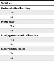
Personal and family gastrointestinal history did not differ significantly between the UBT-positive and UBT-negative groups. These factors included gastrointestinal bleeding (5.80% vs 11.84%; P = 0.252), peptic ulcer (13.04% vs 14.47%; P > 0.99), family history of gastrointestinal bleeding (7.25% vs 5.26%; P = 0.736), family history of gastric cancer (14.49% vs 11.84%; P = 0.806), and family history of peptic ulcer (17.39% vs 11.84%; P = 0.357) (Table 3).
Table 3.
Association Between Urea Breath Test Results and Personal and Family Gastrointestinal History in Patients with Dyspepsia a
| Variables | Positive (n = 69) | Negative (n = 76) | P-Value b |
|---|---|---|---|
| Gastrointestinal bleeding | 0.252 | ||
| Yes | 4 (5.80) | 9 (11.84) | |
| No | 65 (94.20) | 67 (88.16) | |
| Peptic ulcer | > 0.99 | ||
| Yes | 9 (13.04) | 11 (14.47) | |
| No | 60 (86.96) | 65 (85.53) | |
| Family gastrointestinal bleeding | 0.736 | ||
| Yes | 5 (7.25) | 4 (5.26) | |
| No | 64 (92.75) | 72 (94.74) | |
| Family gastric cancer | 0.806 | ||
| Yes | 10 (14.49) | 9 (11.84) | |
| No | 59 (85.51) | 67 (88.16) | |
| Family peptic ulcer | 0.357 | ||
| Yes | 12 (17.39) | 9 (11.84) | |
| No | 57 (82.61) | 67 (88.16) |
a Values are expressed as No. (%).
b Fisher exact test.
4.4. Lifestyle Factors and Medication Use
No significant associations were observed between H. pylori infection and lifestyle factors or medication use. Rates were similar for cigarette smoking (11.59% vs 11.84%; P > 0.99), alcohol consumption (10.14% vs 6.58%; P = 0.550), PPI use (27.54% vs 26.32%; P > 0.99), NSAID use (24.64% vs 19.74%; P = 0.549), and recent antibiotic use (2.90% vs 0%; P = 0.225) (Table 4).
Table 4.
Association Between Urea Breath Test Results and Lifestyle Factors and Medication Use in Patients with Dyspepsia a
| Variables | Positive (n = 69) | Negative (n = 76) | P-Value b |
|---|---|---|---|
| Cigarette smoking | > 0.99 | ||
| Yes | 8 (11.59) | 9 (11.84) | |
| No | 61 (88.41) | 67 (88.16) | |
| Alcohol consumption | 0.550 | ||
| Yes | 7 (10.14) | 5 (6.58) | |
| No | 62 (89.86) | 71 (93.42) | |
| PPI use | > 0.99 | ||
| Yes | 19 (27.54) | 20 (26.32) | |
| No | 50 (72.46) | 56 (73.68) | |
| NSAID use | 0.549 | ||
| Yes | 17 (24.64) | 15 (19.74) | |
| No | 52 (75.36) | 61 (80.26) | |
| Antibiotic use during the previous 3 mo | 0.225 | ||
| Yes | 2 (2.90) | 0 (0) | |
| No | 67 (97.10) | 76 (100) |
a Values are expressed as No. (%). Abbreviations: NSAID, nonsteroidal anti-inflammatory drug; PPI, proton pump inhibitor.
b Fisher exact test.
4.5. Multivariable Logistic Regression
In multivariable logistic regression adjusted for occupation, nausea, diarrhea, and dyspepsia duration, nausea was an independent predictor of H. pylori infection (aOR = 2.174; 95% CI, 1.050 - 4.500; P = 0.037). Employed participants had significantly higher odds of a positive UBT result than those in the reference occupational category (aOR = 5.979; 95% CI, 1.267 - 28.22; P = 0.024). Diarrhea showed a nonsignificant trend toward an association (aOR = 3.154; 95% CI, 0.902 - 11.028; P = 0.072), which should not be interpreted as clinically meaningful based on this statistical test alone. Dyspepsia duration was not predictive of infection (aOR = 1.002; 95% CI, 0.992 - 1.011; P = 0.732) (Table 5).
Table 5.
Multivariable Logistic Regression Predicting Helicobacter pylori Positivity by Urea Breath Test in Patients with Dyspepsia a
| Variables | aOR | SE | 95% CI | P-Value |
|---|---|---|---|---|
| Nausea | ||||
| Yes | 2.17 | 1.449 | 1.05 - 4.50 | 0.037 |
| No | Reference | - | - | |
| Diarrhea | ||||
| Yes | 3.154 | 1.894 | 0.90 - 11.02 | 0.072 |
| No | Reference | - | - | - |
| Dyspepsia duration | 1.002 | 1.002 | 0.99 - 1.01 | 0.732 |
| Occupation | ||||
| Housewife | 1.783 | 1.546 | 0.75 - 4.19 | 0.185 |
| Employed | 5.979 | 2.207 | 1.26 - 28.22 | 0.024 |
| Self-employed | 0.509 | 2.760 | 0.11 - 2.25 | 0.373 |
| Other | Reference | - | - | - |
a Abbreviations: aOR, adjusted odds ratio; CI, confidence interval; SE, standard error.
5. Discussion
This study evaluated the prevalence of H. pylori infection among patients with dyspepsia at a referral clinic in southern Iran during 2022 - 2023. The prevalence was 47.6% by UBT, and nausea and occupation were identified as independently associated factors. Diarrhea showed a nonsignificant trend toward association.
The prevalence observed in this study was similar to the 47.9% reported in a previous Shiraz study but was lower than the 66.6% - 86.8% reported in 3 studies from northern Iran (18-20) and the 60.9% - 69.1% reported in a recent Shiraz study by Moradi et al. (9). However, our prevalence was higher than the 31% reported by Niknam et al. (2). It was also lower than the 61.6% - 67.1% reported in 2 studies conducted in southern and southwestern Iran during 2006 - 2013 (21).
These differences may reflect (1) geographic and sociodemographic variation, (2) variation in diagnostic methods and their sensitivity, (3) differences in study setting, (4) temporal declines associated with improved sanitation, (5) heterogeneity in occupation, smoking, and alcohol consumption, and (6) differences in statistical power. The implementation of novel surveillance and telemedicine systems in the region, as described by Eilami et al. in the context of mpox preparedness, may also reflect an evolving health care infrastructure that could influence the epidemiology and detection of other infectious diseases, including H. pylori (22).
Occupational status was significantly associated with H. pylori infection. Employed participants had 5.979-fold higher odds of infection. This finding is consistent with a study from western Iran that reported higher infection rates in occupations involving poor hygiene practices, as well as a Ugandan study linking occupational stress to an increased risk of H. pylori colonization (23). The high infection rates reported among health care workers (24) further support the possibility of occupation-mediated exposure. However, the literature remains inconsistent. Aminde et al. (25) found a direct association with employment in Cameroon, whereas other studies reported no association (26, 27). H. pylori infection is generally associated with lower socioeconomic status. When an employment category does not reliably indicate higher socioeconomic status, a direct association between employment and H. pylori positivity may be observed; when employment improves living conditions, an inverse association may occur. Occupational risk assessment should therefore consider both hygiene-related exposure and socioeconomic context.
No significant association was observed between most dyspeptic symptoms and UBT results. This finding is consistent with the study by Yoshioka et al. (28), which included 3005 Japanese participants and found that H. pylori status did not affect upper gastrointestinal symptoms after patients with significant lesions were excluded. This may suggest that H. pylori infection does not directly drive dyspeptic symptoms in populations without serious underlying disease. Alternatively, the lack of symptom specificity may reflect the high background prevalence of H. pylori infection in the region (17), where long-standing host-pathogen coadaptation may attenuate symptom expression. This pattern partially resembles the African enigma, in which high infection prevalence coexists with low disease manifestation. Therefore, symptom-based screening for H. pylori in endemic areas may have limited predictive value, and epidemiologically informed test-and-treat strategies may be more appropriate than strategies based on symptoms alone.
Nausea was significantly associated with H. pylori infection, and patients with nausea had 2.46-fold higher odds of positivity. This finding was consistent with those of Correa Silva et al. (29) and Mwangi et al. (30). Potential mechanisms include impaired gastric motility caused by H. pylori-induced chronic gastritis (31), altered acid secretion due to corpus-predominant inflammation, and inflammation-mediated visceral hypersensitivity, whereby the bacterial immune response increases gastric mucosal sensitivity and manifests as nausea (32). Collectively, these mechanisms suggest that nausea among H. pylori-positive patients is a clinical marker of established infection-related mucosal changes rather than early colonization.
A borderline nonsignificant association between diarrhea and H. pylori infection was observed and should be interpreted cautiously. Wang et al. (33) reported an association between H. pylori infection and diarrhea-predominant irritable bowel syndrome. Although the mechanisms remain debated, potential explanations include H. pylori-induced changes in gut microbiota composition, increased bacterial adhesion to the intestinal mucosa, and immune-evasion strategies that may trigger postinfectious inflammatory responses in the lower gastrointestinal tract (34).
Neither age nor sex was significantly associated with H. pylori infection. These findings are consistent with those of Naja et al. (35). Although several studies have reported higher infection rates among older age groups (36), Wang et al. suggested that younger patients may have higher false-positive UBT rates because of lower baseline carbon dioxide production or altered urea hydrolysis rates (37). This age-related variation in testing may partly explain the lack of association in our cohort. Education level was also not significantly associated with H. pylori infection, in contrast to Iranian studies documenting an inverse association between education and prevalence (38). This discrepancy may reflect the relatively small sample size and limited power to detect modest associations. Larger studies are therefore warranted.
Neither a personal nor a family history of gastrointestinal disorders was significantly associated with H. pylori status, consistent with Shokrzadeh et al. (19). Although H. pylori is a major etiologic factor in peptic ulcer disease, a substantial proportion of cases, particularly in populations exposed to antibiotics, may have non-H. pylori causes (39). No significant associations were observed between H. pylori status and PPI use, NSAID use, smoking, or alcohol consumption. In contrast, PPIs may create a less favorable gastric environment for H. pylori (39, 40), and Chey et al. identified alcohol as an independent risk factor for H. pylori-associated intestinal metaplasia (41). The negative findings in the present study may reflect true population differences or limited power to detect modest effects.
5.1. Study Limitations
This study had several limitations. First, the relatively small sample size and cross-sectional design limited the generalizability of the findings. Second, the lack of comparable domestic studies using UBT among patients with dyspepsia limited direct comparisons. Third, the single-center design may have introduced selection bias. Future studies should use larger samples, longitudinal designs, and multicenter recruitment across diverse Iranian populations. The association with employment status was based on only 6 UBT-positive and 2 UBT-negative employed participants, resulting in a wide confidence interval (95% CI, 1.27 - 28.22). This unstable estimate warrants cautious interpretation.
5.2. Conclusions
The prevalence of H. pylori infection detected by UBT among patients with dyspepsia in southern Iran was 47.6%. Nausea and occupational status were independently associated with infection, whereas diarrhea showed a nonsignificant trend toward association. Clinicians should prioritize H. pylori testing among patients with dyspepsia who present with nausea, particularly in high-prevalence regions. Given the study limitations, these findings should be interpreted cautiously.