
1. Background
Coronaviruses (CoVs) are enveloped single-helix RNA viruses from the coronaviridae family (1). These viruses cause serious infections ranging from simple flu (influenza) to acute severe respiratory syndrome (SARS) (2). This virus can be symptom-free or cause diseases from simple flu-like symptoms (3) to severe respiratory, cardiac, gastrointestinal, and kidney disorders (4-7). Multisystemic inflammatory syndrome (MIS-C) with coronary artery aneurysm, fever, mucocutaneous symptoms, and hyperinflammatory symptoms that appear with findings similar to Kawasaki disease during the disease was defined in pediatric patients. These symptoms mostly develop because of immune system activation against viral infection (8-10). In addition, SARS-CoV-2 can cause serious complications, such as fulminant myocarditis and cardiogenic shock (11). Patients with congenital heart disease (CHD) are at high-risk for coronavirus disease 2019 (COVID-19) due to their limited cardiopulmonary reserves.
2. Objectives
This study aimed to evaluate the frequency, clinical characteristics, treatment, and follow-up of CHD in patients presented with COVID-19 to our hospital. Furthermore, we aimed to contribute to the literature by revealing the clinical course of COVID-19 patients with CHD.
3. Methods
This study retrospectively evaluated the patients referred to the Department of Pediatrics at the Inonu University Faculty of Medicine, Turkey, during March 2020-February 2021 due to COVID-19. The total number of patients admitted to our hospital and followed up was 232 due to COVID-19. Among these patients, 11 (4.7%) cases with CHD were identified. Only patients with underlying CHD were included in the study by total population sampling. Admission complaints, clinical findings, acute phase reactants, biochemical parameters, N-terminal prohormone of brain natriuretic peptide (NT-proBNP) levels, echocardiography and electrocardiography results, hospitalization times, intensive care requirements, treatments, and clinical follow-up findings were obtained from the patients' files.
The criteria for admission to the intensive care unit (ICU) were severe oxygen need (oxygen saturation below 90 despite receiving high-flow oxygen therapy), hypotensive course requiring inotropic support, circulatory disorders, the severity of organ involvement, and the clinical findings of the patients. This descriptive study presented categorical data as numbers and percentages, while numerical data were expressed as mean and range. The treatments of patients were planned according to the Ministry of Health guidelines and considering the individuals' clinical situation. The current research was conducted in accordance with the principles stated in the Declaration of Helsinki and following obtaining the consent of the patients’ parents. Ethics Committee of the Inonu University Scientific Research and Publication Ethics Board approved the study on 23.03.2021 with the code 2021/1771.
4. Results
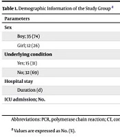
Eleven patients with CHD and COVID-19 referred during February 2020-March 2021 were included in the current study. We observed that 8 (72.7%) patients were male, and 3 (27.3%) were female. The mean age of patients was 5.3 years (range: 0.3 - 17). The most common reasons for admission to the hospital were fever and cough (63.6%), shortness of breath and fatigue (54.5%), loss of appetite, abdominal pain, vomiting, and diarrhea. It was found that fever generally started 2 - 6 days before admission to the hospital. The physical examination showed rales in the lungs in 63.6%, tenderness in the abdomen in 45.5%, and distension in 27.3% of the patients. Circulatory disorder due to dehydration was found in one person, and heart failure in three cases (Table 1).
Table 1.
Clinical Characteristics and Complaints of the Patients at Admission
| Symptoms and Signs of Patients | No. (%) (n=11) |
|---|---|
| Fever | 7 (63.6) |
| Cough | 7 (63.6) |
| Shortness of breath | 6 (54.5) |
| Weakness | 6 (54.5) |
| Loss of appetite | 5 (45.5) |
| Stomachache | 4 (36.4) |
| Vomiting | 3 (27.3) |
| Diarrhea | 2 (18.2) |
| Pneumonia | 7 (63.6) |
| Shock | 4 (36.4) |
| Heart failure | 4 (36.4) |
| Acute gastroenteritis | 2 (18.2) |
| Dehydration | 1 (9) |
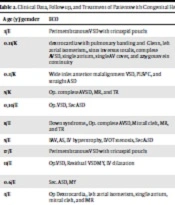
In terms of CHD, ventricular septal defect (VSD) was the most detected disorder, with 45.5% (n = 5) prevalence in participants. In addition, an operated complete atrioventricular septal defect (AVSD) was observed in two patients. Dextrocardia with pulmonary banding and Glenn shunt in one patient and left atrial isomerism, situs inversus totalis, complete AVSD, single atrium, single atrioventricular valve (AV), azygos vein continuity, operated dextrocardia were found in one patient. Left atrial isomerism, single atrium, mitral cleft, mitral regurgitation (MR) grade 1 - 2, aortic stenosis (AS), left ventricular outflow tract (LVOT) stenosis, left ventricular hypertrophy, and secundum atrial septal defect (ASD) were detected in one patient. Two patients who were followed up due to VSD were operated on and had residual pathologies. Two patients were followed up because of small VSD with a tricuspid pouch. One patient also had a large inlet anterior malalignment VSD and persistent left superior vena cava (PLSVC) combination. Both complete AVSD patients who were operated on had MR grade 1. One patient had simultaneous secundum ASD and MR grade 1-2. One of our patients had Down syndrome and had been operated on for complete AVSD, mitral cleft, MR grade 1 - 2, and TR grade 1. The cardiological diagnoses of patients are given in Table 2.
Table 2.
Clinical Data, Follow-up, and Treatment of Patients with Congenital Heart Disease
| Age (y) | Gender | ECO | Hospitalization (d) | IU (d) | Treatment/Duration | |
|---|---|---|---|---|---|---|
| 1 | 3 | M | Perimembranous VSD with tricuspid pouch | 10 | 0 | Steroid |
| 2 | 0.11 | F | dextrocardia with pulmonary banding and Glenn, left atrial isomerism, situs inversus totalis, complete AVSD, single atrium, single AV cover, and azygous vein continuity | 17 | 10 | Hydroxychloroquine, furosemide, aldactone, enalapril, dopamine, dobutamine, adrenaline, steroid |
| 3 | 0.3 | F | Wide inlet anterior malalignment VSD, PLSVC, and straight ASD | 0 | 2 | Intubation, captopril, furosemide, dopamine, dobutamine, steroid, IVIG |
| 4 | 5 | F | Op. complete AVSD, MR, and TR | 8 | 0 | Hydroxychloroquine and steroid |
| 5 | 0.10 | M | Op. VSD, Sec ASD | 19 | 5 | Intubation, IVIG, Azithromycin, Ceftriaxone, dopamine, dobutamine, and steroid |
| 6 | 8 | M | Down syndrome, Op. complete AVSD, Mitral cleft, MR, and TR | 32 | 18 | Intubation (MV), Favipiravir, hydroxychloroquine, and steroid |
| 7 | 5 | M | BAV, AS, LV hypertrophy, LVOT stenosis, Sec ASD | 9 | 6 | Intubation, Hydroxychloroquine and steroid |
| 8 | 17 | M | Perimembranous VSD with tricuspid pouch | 6 | 4 | Intubation, hydroxychloroquine, steroid |
| 9 | 11 | M | Op.VSD, Residual VSD MY, LV dilatation | 4 | 2 | Favipiravir, dopamine, dobutamine, steroid, high flow Oxygen, ACE inhibitor, and digoxin |
| 10 | 0.6 | M | Sec. ASD, MY | 6 | 0 | Steroid |
| 11 | 3 | M | Op Dextrocardia, left atrial isomerism, single atrium, mitral cleft, and MR | 0 | 0 | No treatment |
Abbreviations: F, female; M, male; VSD, ventricular septal defect; AVSD, atrioventricular septal defect; AV, atrioventricular ASD, atrial septal defect; PLSVC, persistent left superior vena cava; MR, mitral regurgitation; TR, tricuspid regurgitation; BAV, bicuspid aorta; AS, aortic stenosis; LV, left ventricle; LVOT, left ventricular outflow tract; IVIG, intravenous immunoglobulin
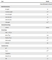
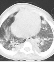
Laboratory findings of the patients obtained during hospital admission indicated white blood cell (WBC) elevation in three patients and low WBC count in three patients. C-reactive protein (CRP) above normal values was detected in all patients. Significant lymphopenia was detected in only two cases, and significant liver enzyme augmentation was detected in two patients. The COVID-19 PCR test results were positive in all patients except for one person. In the patient who was negative, a history of close contact and infiltrations compatible with COVID-19 were detected in the thorax computed tomography. Except for one patient, NT-proBNP values were above the normal reference range in all individuals (Table 3).
Table 3.
Laboratory Data of Patients
| 1 | 2 | 3 | 4 | 5 | 6 | 7 | 8 | 9 | 10 | 11 | |
|---|---|---|---|---|---|---|---|---|---|---|---|
| WBC (103/mm3) | 5.21 | 4.64 | 19.5 | 5.66 | 10.9 | 3.17 | 3.7 | 6 | 15.4 | 14.3 | 11.1 |
| HB (gr/dL) | 11.6 | 16.2 | 13.4 | 12.4 | 10.1 | 14 | 9.3 | 6.4 | 9.4 | 12.3 | 11.7 |
| HTC (%) | 35.0 | 55.3 | 41.4 | 37.8 | 30.2 | 41.1 | 26.1 | 23.1 | 27.2 | 33.2 | 31.4 |
| PLT (103/mm3) | 247 | 256 | 235 | 231 | 169 | 191 | 374 | 99 | 364 | 256 | 254 |
| Lymphocyte (10³/mm³) | 1.07 | 2.35 | 1.49 | 2.17 | 6.29 | 0.27 | 0.66 | 1.23 | 6.76 | 1.94 | 5.56 |
| CRP (mg/dL) | 7.89 | 1.1 | 3.5 | 4.41 | 1.34 | 8.65 | 17.5 | 6 | 3.8 | 1.41 | 1.3 |
| AST (u/L) | 19 | 29 | 26 | 177 | 81 | 125 | 21 | 34 | 87 | 20 | 30 |
| ALT (u/L) | 38 | 21 | 18 | 198 | 50 | 86 | 16 | 28 | 32 | 18 | 29 |
| PCR | + | + | + | + | + | - | + | + | + | + | + |
| NT-proBNP (pg/mL) | 370 | 190 | 30 | 110 | 35000 | 393 | 3310 | 598 | 25210 |
In the present study, 10 (91%) of 11 participants were hospitalized, and one patient was followed up as an outpatient. Moreover, 7 (73.6%) individuals were followed up and treated in the ICU. Five (45.5%) of the patients were intubated and mechanically ventilated. One patient was given high-flow oxygen therapy, and one was treated for heart failure and pneumonia. Out of the patients who were followed up in the service, the patient with perimembranous VSD and tricuspid pouch and the subject with secundum ASD and MR were given only steroid treatment. The patient with complete operated AVSD, MR, and TR who were followed up in the service received hydroxychloroquine and steroid treatment. No treatment was started for the patient with operated dextrocardia, left atrial isomerism, single atrium, mitral cleft, and MR due to good general condition. This patient was followed up as an outpatient.
Seven patients were followed up in the ICU, of whom the patient with Down syndrome and operated complete AVSD, mitral cleft, MR, and TR was initially followed up in the ward. Then high-current oxygen therapy was started in the ICU because of the increased oxygen demand. However, he was intubated due to low saturation and severe lung involvement. The patient was treated with favipiravir, hydroxychloroquine, and steroid and was discharged after 50 days, including 18 days in the ICU. Two months after discharge, the COVID-19 PCR result was positive again, and he was hospitalized for the second time due to pneumonia. Hydroxychloroquine treatment was started for the patient with tricuspid pouch VSD and pneumonia. The patient, who was intubated and mechanically ventilated, was extubated on the second day. Hydroxychloroquine treatment was started for the patient, followed up with bicuspid aortic valve (BAV), aortic stenosis, LVOT stenosis, LV hypertrophy, and secundum ASD. He was extubated on the fourth day.
The patient, who was three months old and had large inlet VSD, had severe dehydration and gastroenteritis. After hydration, intravenous immunoglobulin (IVIG) was given to the patient considering covid-related multisystemic inflammatory syndrome. Although IV inotrope treatment was started due to circulatory disorder and shock, he died on the second day of hospitalization. The IVIG treatment and steroid therapy were started because of severe lung involvement in another patient who was followed up with operated VSD, secundum ASD, pneumonia, and myocarditis. In addition, dopamine and dobutamine treatment was initiated for cardiac support. High-flow oxygen therapy was started for the patient with severe pneumonia and oxygen needs. However, the person whose general condition worsened in follow-up was intubated and subjected to a mechanical ventilator. The patient was lost on the 24th day of hospitalization. It is noteworthy that the NT-pro BNPs of both deceased cases were very high.
Hydroxychloroquine treatment was started in the patient who was followed up due to pulmonary banding and Glenn dextrocardia, left atrial isomerism, situs inversus totalis, complete AVSD, single atrium, single atrioventricular valve (AV), azygos vein continuity, and pneumonia, and had signs of heart failure. In addition, furosemide, aldactone, angiotensin-converting enzyme (ACE) inhibitor, dopamine, and dobutamine treatments were started as cardiac support treatment, followed by adrenaline infusion. The patient who was followed up due to operating VSD, residual VSD, MR, and pneumonia was given ACE inhibitor, digoxin, dopamine, dobutamine, and favipiravir, along with cardiac support treatment. Moreover, the patient in need of oxygen was given high-flow oxygen therapy.
For patients taking hydroxychloroquine and favipiravir, daily routine electrocardiograms were performed before and during treatment due to possible arrhythmia resulting from the side effects of the medications. In addition, the corrected QT interval (QTC) was calculated according to the Bazett formula. No arrhythmia or QTC prolongation due to hydroxychloroquine and favipiravir was detected in any patient.
5. Discussion
There are few studies on the relationship between COVID-19 coexistence, clinical findings, and disease severity in patients with CHD. Most reports are obtained from case studies or small case series. Therefore, scientific data about how COVID-19 affects CHD patients are limited. Past outbreaks caused by SARS and H1N1 influenza viral infections were more severe and had a higher mortality rate in patients with CHD than those without CHD (12, 13). Children who have CHD are considered high-risk because they have limited cardiopulmonary reserves. Complex cardiac pathologies, surgical repairs, arrhythmias, exercise capacities, significant residual shunts, valve stenosis-insufficiency, and hypoxemia conditions increase disease severity. In our opinion, complex CHDs are an important risk factor in the course of COVID-19. Patients with severe CHD have hypoxemia and refractory end-organ dysfunction, making them more vulnerable to the effects of COVID-19 (14).
Severe hypoxia from acute respiratory injury and myocardial involvement caused by COVID-19 may result in myocardial damage (15, 16). A study on adults reported that COVID-19 caused myocardial damage, and especially adult patients with underlying CHD were at higher risk for COVID-19 (17, 18). The most common symptoms at admission were fever and cough (63.6%), shortness of breath and fatigue (54.5%), loss of appetite, abdominal pain, vomiting, and diarrhea. In a study conducted in China, fever was reported in only 41.5% of patients (19). In another study in the U.S.A, fever was reported in 56%, cough in 54%, and shortness of breath in 13% of cases. The estimated rate of hospitalization in pediatric patients was reported to be 5.7 - 20%, and the admission rate to ICU was 0.58 - 2% (20). In a study carried out by Dong et al., 90% of pediatric patients had an asymptomatic, mild-, or moderate-intensity disease. Only 5% of these patients reported severe illness, and 0.6% had critically severe infection (3). In terms of comorbidities, 23% of hospitalized patients have underlying diseases. The research found that 40% of individuals had chronic lung diseases, 25% had cardiovascular diseases, and 10% had immunosuppressive disease (20). Similarly, some studies have reported that children with comorbidities have severe symptoms and require hospitalization in the ICU (21, 22). In our study, 10 (91%) of the 11 patients were hospitalized, and 7 (63.6%) of the hospitalized cases were followed up in the ICU. Our study showed that COVID-19 was more severe in children with underlying diseases, especially CHD. Dopamine and dobutamine treatments were started as inotropic support treatments for four patients who had developed severe hypotension and shock. Pneumonia was detected in most of our patients (63.6%), and four of these cases were intubated and followed up in the ICU. Non-invasive high-flow oxygen therapy was applied to one person.
Kawasaki-like disease may occur after a previous COVID-19 infection, documented by the presence of SARS-CoV-2 IgG antibodies or known contact with a confirmed COVID-19 case (23). The latter point indicates the presence of a post-infection immunity dysregulation in children who have previously been infected with COVID-19 or exposed to COVID-19 (16, 24). MIS-C developed in a three-month-old patient with large inlet VSD and severe dehydration due to gastroenteritis, and the patient died on the second day of hospitalization. Another patient who had been operated on because of VSD, secundum ASD, and pneumonia was followed up due to severe lung injury and myocarditis and was lost on the 24th day of hospitalization. Both cases were under the age of one year. The WBC, CRP, and biochemical parameters of our patients were like other COVID-19 patients. However, NT-pro BNP levels of both patients who died later were much higher than other patients. An investigation of adult patients reported that high NT-pro BNP was associated with mortality (25).
Our data on the clinical features and course of COVID-19 patients with underlying CHD are very valuable. The fact that 91% of the 11 patients we followed up were treated as inpatients, 63.6% were followed up in the ICU, and 45% were followed up on a mechanical ventilator suggests that care should be taken in CHD patients infected with this disease. Although the patient population was small, the death of two cases indicates that they are at high risk for mortality.
5.1. Conclusions
According to the findings of the current research, COVID-19 is more clinically severe in patients with underlying CHD despite being milder in children. Therefore, we suggest that more care should be taken in the follow-up and treatment of patients with underlying CHD, and proper guidelines should be prepared for these people. COVID-19 in patients with CHD requires more serious and careful follow-up. These patients should be protected by an appropriate vaccination program in the following processes.
In this retrospective study, conducted in our hospital, the small number of patients, the absence of some data, or the lack of records in the patients' files were the limitations. Furthermore, patients with more severe conditions were admitted because our hospital is a third-level hospital. This limits our study as patients with milder CHD, and COVID-19 comorbidity may not have been included.