1. Background
Neonatal respiratory distress syndrome (NRDS), also known as neonatal lung hyaline membrane disease, is mainly observed in premature infants. Pulmonary surfactant (PS) synthesis and secretion normally begin during the last trimester of fetal lung development. However, in premature infants, lung immaturity results in insufficient PS production, leading to increased alveolar surface tension, alveolar collapse, and impaired gas exchange (1, 2). With the continuous advancement of perinatal medical technologies, the survival rate of neonates has progressively increased. Nevertheless, the incidence of NRDS has not shown a corresponding decline and remains one of the leading causes of neonatal mortality and long-term complications (3). In the treatment of NRDS, PS replacement therapy and mechanical ventilation constitute the cornerstones of treatment. Pulmonary surfactant, secreted by type II alveolar epithelial cells, is primarily composed of phospholipids and proteins, among which dipalmitoyl phosphatidylcholine plays a critical role. This component forms a monomolecular layer at the alveolar air-liquid interface, effectively reducing surface tension (4, 5). Although conventional mechanical ventilation can improve overall oxygenation to a certain extent, it is also associated with ventilator-induced lung injuries, such as barotrauma and volutrauma, which may adversely affect long-term pulmonary development in neonates. High-frequency oscillatory ventilation (HFOV), characterized by low tidal volume, low airway pressure, and high frequency, has been proposed as an alternative ventilation strategy with the potential to minimize lung injury caused by mechanical ventilation, thereby offering new therapeutic perspectives for the treatment of NRDS (6).
In recent years, the combined application of HFOV and PS therapy has gradually emerged as a research focus in the treatment of NRDS, driven by ongoing advances in neonatal intensive care. Existing studies suggest that this combined approach may provide significant benefits in improving oxygenation, shortening the duration of mechanical ventilation, and reducing complication rates (7). However, the optimal timing, dosage, and overall clinical efficacy of HFOV-assisted PS therapy remain controversial, and uniform clinical standards or consensus guidelines have not yet been established.
2. Objectives
The present study was designed to evaluate the effect of HFOV-assisted PS therapy in neonates with NRDS.
3. Methods
3.1. Subjects
This study was designed as a retrospective analysis of real-world clinical data because prospective randomization of ventilation strategies was not feasible in the emergency management of NRDS. Therefore, the sample size was determined by the number of eligible patients meeting the inclusion criteria during the study period. The final cohort of 100 neonates was comparable in size to those reported in previous single-center studies evaluating HFOV- or PS-based interventions in NRDS and was considered appropriate for exploratory comparative analyses.
A total of 100 neonates diagnosed with NRDS and admitted to our hospital between March 2022 and February 2024 were retrospectively enrolled in this study. Patients were assigned to the control or observation group according to the ventilation strategy actually applied during routine clinical management rather than by randomization. Specifically, neonates who received PS therapy alone were included in the control group (n = 50), whereas those who received HFOV-assisted PS therapy were included in the observation group (n = 50). In the control group, there were 31 boys and 19 girls, with a postnatal age of 6 - 9 hours (7.21 ± 1.20 hours) and a birth weight of 1 - 3 kg (1.95 ± 0.11 kg). According to chest X-ray grading, 27 cases were classified as grade I NRDS and 23 cases as grade II NRDS. The observation group included 30 boys and 20 girls, with a postnatal age of 6 - 9 hours (7.19 ± 1.19 hours) and a birth weight of 1 - 3 kg (1.93 ± 0.13 kg). Chest X-ray grading showed 26 cases of grade I NRDS and 24 cases of grade II NRDS. Baseline characteristics were comparable between the 2 groups, and no statistically significant differences were observed (P > 0.05), indicating suitability for comparative analysis. Although retrospective group assignment may introduce selection bias, this risk was partly mitigated by the application of uniform inclusion and exclusion criteria and the comparability of baseline characteristics between groups; nevertheless, residual confounding cannot be completely excluded.
This study was approved by the Ethics Committee of the hospital, and informed consent was obtained retrospectively from the parents or legal guardians of the neonates during hospitalization or follow-up in accordance with institutional ethical requirements. This study was conducted and reported in accordance with the Strengthening the Reporting of Observational Studies in Epidemiology (STROBE) guidelines for observational studies.
The inclusion criteria were as follows: (1) Diagnosis of NRDS confirmed by chest computed tomography (CT) and/or other imaging examinations; (2) birth weight ≥ 1600 g; (3) postnatal age < 48 hours; (4) grade I - II NRDS based on chest X-ray findings; and (5) written informed consent obtained from parents or legal guardians.
The exclusion criteria were as follows: (1) Severe hepatic or renal diseases, including renal failure; or (2) leukemia or other severe systemic immune diseases. Retrospective assignment to groups may introduce selection bias, which was mitigated by matched baseline characteristics.
3.2. Pulmonary Surfactant Intervention Method for the Control Group
Pulmonary surfactant therapy was administered as follows. Neonates were placed in a neonatal intensive care unit (NICU) incubator and continuously monitored using an electrocardiogram (ECG) monitor for heart rate, respiratory rate, and oxygen saturation. The type and dosage of PS were calculated according to body weight. Calf Pulmonary Surfactant for Injection (CR Double-Crane Pharmaceuticals Co., Ltd.; approval no. NMPN H20052128; specification, 70 mg) was used. Before administration, airway secretions were aspirated, and a drug delivery catheter, sterile syringe, and appropriate endotracheal intubation equipment were prepared. The reconstituted PS solution was drawn into a sterile syringe at an initial dose of 100 - 200 mg/kg, and the dosage was double-checked to ensure accuracy. The syringe was then connected to the endotracheal tube, and PS was slowly and evenly administered by bolus injection, with the entire dose delivered within 10 - 15 seconds. Afterwards, endotracheal intubation was performed under aseptic conditions using a tube appropriate for the infant’s weight and age. Pulmonary surfactant was administered into the trachea in 2 - 4 aliquots, with the infant’s head slightly elevated and alternately tilted. Manual positive-pressure ventilation using a resuscitation bag was immediately applied after each aliquot to facilitate uniform pulmonary distribution of the surfactant. Ventilation parameters were adjusted based on blood gas results, targeting PaO₂ of 60 - 80 mmHg and PCO₂ of 35 - 45 mmHg.
3.3. High-Frequency Oscillatory Ventilation-Assisted Pulmonary Surfactant Intervention Method for the Observation Group
In the observation group, HFOV-assisted PS therapy was applied. After vital sign monitoring, endotracheal intubation, and PS administration identical to those used in the control group, the endotracheal tube was promptly connected to a high-frequency oscillatory ventilator. Initial ventilator parameters were set according to body weight and clinical condition as follows: Oscillation frequency, 10 - 15 Hz; amplitude, 20 - 40 cmH₂O, adjusted to achieve visible chest vibration; mean airway pressure (MAP), initially 2 - 5 cmH₂O above conventional levels and generally maintained at 10 - 20 cmH₂O; inspiratory time, 0.3 - 0.5 seconds, representing 30% - 50% of the oscillatory cycle; and fraction of inspired oxygen (FiO₂), 0.6 - 1.0, adjusted based on oxygen saturation and blood gas results. Oxygen saturation and ventilator parameters were continuously monitored, and oscillation frequency and amplitude were adjusted as needed. Ventilator settings, including MAP, oscillation frequency, amplitude, and FiO₂, were recorded every 15 - 30 minutes, and dynamic parameter charts were generated. Arterial blood gas analysis was reassessed every 4 - 6 hours or more frequently if clinically indicated, and ventilator parameters were individually optimized according to the results. High-frequency oscillatory ventilation parameter adjustments were guided by predefined clinical criteria, primarily based on arterial blood gas targets and oxygenation status: MAP and FiO₂ were titrated to maintain PaO₂ within 60 - 80 mmHg and adequate oxygen saturation, whereas oscillation amplitude and frequency were adjusted to maintain PCO₂ within 35 - 45 mmHg and ensure effective chest vibration. Detailed parameter ranges and optimization principles are provided in the Supplementary File.
3.4. Observation Indicators and Assessment Criteria
Clinical treatment indicators, including duration of mechanical ventilation and total length of stay, were recorded and compared between groups.
Blood gas parameters were measured before and after treatment using a blood gas analyzer and included PaO₂ (normal range, 55 - 80 mmHg), PCO₂ (normal range, 35 - 45 mmHg), and SaO₂ (normal range, 95% - 98%). Complications, including barotrauma, pulmonary air leak, and infection, were recorded. Outcome assessments were conducted by attending physicians using predefined clinical criteria. Owing to the retrospective design and the nature of the ventilation strategies, outcome assessors were not blinded to treatment allocation. The complication rate was calculated as follows: (Number of cases with barotrauma + number of cases with pulmonary air leak + number of cases with infection) / total number of cases × 100%.
3.5. Statistical Analysis
Data were analyzed using Statistical Package for the Social Sciences (SPSS) version 26.0 software. Count data, including complication rates, were expressed as n (%) and analyzed using the χ² test. For continuous variables, normality was assessed using the Shapiro-Wilk test, and homogeneity of variance was evaluated using Levene’s test. Continuous data that satisfied these assumptions were expressed as mean ± standard deviation (x̄ ± SD). Independent-sample t-tests were used for intergroup comparisons, and paired t-tests were applied for intragroup comparisons before and after treatment. In addition to P-values, effect sizes were calculated using Cohen’s d to quantify the magnitude of differences, and corresponding 95% confidence intervals (CIs) were reported. P < 0.05 indicated statistical significance.
4. Results
4.1. Clinical Treatment Indicators
In the between-group comparison after treatment, the observation group exhibited a significantly shorter duration of mechanical ventilation than the control group (mean difference, -1.30 days; 95% CI, -2.03 to -0.57; Cohen’s d = 0.70; P = 0.001). Similarly, the total length of hospital stay was significantly reduced in the observation group compared with the control group (mean difference, -2.17 days; 95% CI, -3.38 to -0.96; Cohen’s d = 0.70; P = 0.001) (Table 1).
Table 1.
Clinical Treatment Indicators (x̄ ± SD, d) a
| Groups | Duration of Mechanical Ventilation | Length of Stay |
|---|---|---|
| Observation (n = 50) | 12.45 ± 1.92 | 29.44 ± 2.94 |
| Control (n = 50) | 13.75 ± 1.78 | 31.61 ± 3.22 |
| Mean difference b | -1.30 (-2.03 to -0.57) | -2.17 (-3.38 to -0.96) |
| Effect size c | 0.70 | 0.70 |
| t | 3.511 | 3.519 |
| P-value d | 0.001 | 0.001 |
a Values are presented as mean ± standard deviation.
b Mean differences were calculated as the observation group minus the control group, with corresponding 95% CIs.
c Effect sizes are expressed as Cohen’s d.
d P-values were derived from independent-sample t-tests.
4.2. Levels of Blood Gas Indicators
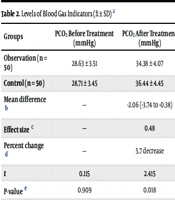
No statistically significant differences were observed in blood gas parameters between the 2 groups before treatment (P > 0.05). In the between-group comparison after treatment, the observation group showed lower PCO₂ (mean difference, -2.06 mmHg; 95% CI, -3.74 to -0.38; Cohen’s d = 0.48) and lower SaO₂ (mean difference, -3.29%; 95% CI, -5.12 to -1.46; Cohen’s d = 0.71), whereas PaO₂ (mean difference, 3.22 mmHg; 95% CI, 1.33 to 5.11; Cohen’s d = 0.67) was increased compared with the control group (all P < 0.05) (Table 2).
Table 2.
Levels of Blood Gas Indicators (x̄ ± SD) a
| Groups | PCO₂ Before Treatment (mmHg) | PCO₂ After Treatment (mmHg) | SaO₂ Before Treatment (%) | SaO₂ After Treatment (%) | PaO₂ Before Treatment (mmHg) | PaO₂ After Treatment (mmHg) |
|---|---|---|---|---|---|---|
| Observation (n = 50) | 28.63 ± 3.51 | 34.38 ± 4.07 | 73.11 ± 3.37 | 92.93 ± 4.91 | 49.21 ± 5.29 | 62.78 ± 4.89 |
| Control (n = 50) | 28.71 ± 3.45 | 36.44 ± 4.45 | 73.21 ± 3.41 | 96.22 ± 4.42 | 49.75 ± 5.32 | 59.56 ± 4.67 |
| Mean difference b | — | -2.06 (-3.74 to -0.38) | — | -3.29 (-5.12 to -1.46) | — | 3.22 (1.33 to 5.11) |
| Effect size c | — | 0.48 | — | 0.71 | — | 0.67 |
| Percent change d | — | 5.7 decrease | — | 3.4 decrease | — | 5.4 increase |
| t | 0.115 | 2.415 | 0.147 | 3.521 | 0.509 | 3.367 |
| P-value e | 0.909 | 0.018 | 0.883 | 0.001 | 0.612 | 0.001 |
Abbreviations: PCO₂, partial pressure of carbon dioxide; SaO₂, arterial oxygen saturation; PaO₂, arterial partial pressure of oxygen.
a Values are presented as mean ± standard deviation.
b Mean differences were calculated based on posttreatment values as the observation group minus the control group, with corresponding 95% CIs.
c Effect sizes are expressed as Cohen’s d.
d Percent change represents the relative difference in posttreatment values between groups, calculated as (control - observation)/control × 100%.
e P-values were derived from independent-sample t-tests.
4.3. Complication Rate
In the between-group comparison after treatment, the overall complication rate was significantly lower in the observation group than in the control group (2.0% vs. 14.0%; relative risk, 0.14; 95% CI, 0.02 - 1.08; P = 0.027) (Table 3).
| Groups | Barotrauma (N) | Lung Air Leak (N) | Infection (N) | Complication Rate (%) |
|---|---|---|---|---|
| Observation (n = 50) | 1 | 0 | 0 | 2.00 |
| Control (n = 50) | 2 | 3 | 2 | 14.00 |
| χ² | — | — | — | 4.891 |
| P-value | — | — | — | 0.027 |
| Relative risk c | — | — | — | 0.14 (0.02 - 1.08) |
a Any complication refers to the occurrence of at least 1 complication event.
b Group comparisons for overall complication rates were performed using the χ² test.
c Relative risk and 95% CI were calculated based on overall complication rates in the observation and control groups.
5. Discussion
Neonatal respiratory distress syndrome is a severe respiratory disorder and remains a major focus in neonatal medicine and clinical practice. Epidemiological studies have demonstrated a global increase in the proportion of premature births, and NRDS continues to be one of the leading causes of morbidity, mortality, and long-term complications in premature infants, severely affecting survival quality and prognosis (8, 9). The pathological basis of NRDS lies primarily in pulmonary immaturity, resulting in insufficient synthesis and secretion of PS. Severe PS deficiency may lead to hypoxemia and respiratory failure. Exogenous PS replacement therapy is therefore a cornerstone of NRDS management and has been shown to significantly improve respiratory function and reduce mortality. However, mechanical ventilation is still required in critically ill neonates (10).
Mechanical ventilation plays a central role in NRDS treatment, and HFOV represents a novel ventilation strategy. By maintaining airway pressure through high-frequency oscillation with extremely low tidal volumes, HFOV can reduce ventilator-induced lung injury, improve gas exchange, facilitate uniform PS distribution, lower alveolar surface tension, enhance alveolar compliance and stability, and ultimately improve therapeutic efficacy (11). Recent systematic reviews and meta-analyses have evaluated the role of HFOV in neonatal respiratory failure, with mixed results. A 2022 Cochrane meta-analysis comparing HFOV with conventional ventilation in preterm infants with respiratory distress syndrome (RDS) reported no consistent reduction in mortality but suggested a potential benefit in reducing air leak syndromes, particularly when lung-protective strategies were applied (12). Similarly, a 2023 meta-analysis focusing on early HFOV initiation indicated improved oxygenation indices without a clear survival advantage (13). These findings suggest that the benefits of HFOV are likely context-dependent rather than universally generalizable.
In the present study, 100 neonates with NRDS were retrospectively analyzed to evaluate the efficacy of HFOV-assisted PS therapy. The results demonstrated that SaO₂ and PCO₂ were significantly decreased, whereas PaO₂ was increased in the observation group compared with the control group (P < 0.05). These findings are consistent with recent randomized controlled trials reporting improved gas exchange parameters when HFOV is combined with early or repeated PS administration compared with PS therapy alone (14, 15). Mechanistically, exogenous PS rapidly reduces alveolar surface tension, stabilizes alveolar structure, prevents end-expiratory alveolar collapse, and increases functional residual capacity. High-frequency oscillatory ventilation, in turn, maintains alveolar recruitment under relatively low tidal volumes and stable MAP, creating optimal conditions for sustained alveolar patency and homogeneous PS distribution. However, these mechanistic advantages should be interpreted within the context of the specific patient population and treatment timing examined in this study.
Additionally, an appropriately elevated effective MAP increases the proportion of alveoli recruited for gas exchange, thereby expanding the effective oxygen diffusion surface and contributing to higher PaO₂ levels. Through these complementary mechanisms, HFOV-assisted PS therapy facilitates correction of hypoxemia and supports the maintenance of SaO₂ within a physiological range (16, 17). Compared with conventional ventilation, HFOV enables efficient gas exchange at substantially lower tidal volumes, reduces dead-space ventilation, and enhances carbon dioxide elimination, thereby maintaining PaCO₂ within normal limits. Pulmonary surfactant supplementation further optimizes alveolar mechanics and gas exchange efficiency, which may synergistically promote CO₂ excretion (18). Although these mechanisms provide a plausible explanation for the observed physiological improvements, they do not necessarily imply equivalent clinical benefits across all NRDS populations or care settings.
Consistent with these mechanisms, the present study demonstrated significantly shorter durations of mechanical ventilation and hospital stay in the observation group. However, not all studies have reported unequivocal benefits of HFOV combined with PS. Several studies have shown neutral effects on major clinical outcomes, such as mortality or bronchopulmonary dysplasia, particularly when HFOV was applied late or in less severe cases of NRDS (19, 20). These discrepancies highlight the importance of patient selection, early intervention, and ventilation parameter optimization and caution against extrapolating our findings beyond similar clinical contexts.
Importantly, the complication rate was significantly lower in the observation group (P < 0.05). This observation aligns with recent evidence indicating that lung-protective ventilation strategies, including HFOV, may reduce the incidence of barotrauma and air leak syndromes compared with conventional ventilation (21, 22). High-frequency oscillatory ventilation minimizes alveolar overdistension and pressure fluctuations, while PS supplementation enhances alveolar stability, together reducing susceptibility to air leak and pressure-related lung injury (23-25). Emerging studies also suggest that PS participates in pulmonary immune defense, regulating immune cell activity, enhancing phagocytosis, and inhibiting pathogen adhesion and invasion, whereas HFOV ensures a stable and unobstructed airway, promotes secretion clearance, and reduces infection risk associated with secretion retention (26, 27).
This study also has several novel and clinically relevant aspects. Although the combination of HFOV and PS therapy is well established, our work extends existing evidence by focusing on a well-defined subgroup of neonates with early-stage NRDS, defined as radiographic grade I - II disease, treated within the first hours after birth in a real-world NICU setting. Unlike prior trials and meta-analyses that enrolled heterogeneous populations or initiated HFOV at later disease stages, we evaluated early HFOV-assisted surfactant therapy using standardized ventilatory parameters. Notably, improvements in gas exchange were accompanied by clinically meaningful short-term benefits, including shorter durations of mechanical ventilation and hospitalization, as well as lower complication rates, underscoring the practical relevance of this strategy in routine NICU care.
Beyond its novelty, the potential implications of these findings for clinical practice and resource allocation merit consideration. Our results suggest that early HFOV-assisted surfactant therapy may support a more stratified ventilation approach for neonates with early-stage NRDS, complementing existing lung-protective ventilation protocols. Because HFOV was implemented using standard settings within a routine NICU environment, its scalability appears feasible in centers with established HFOV capability, and the observed reductions in ventilation duration, length of stay, and complications may partially offset associated resource demands.
However, several limitations of this study should be acknowledged. First, the retrospective and nonrandomized design is inherently subject to selection bias and residual confounding. In particular, the lack of multivariable adjustment precluded full control of potential confounders, such as gestational age and Apgar scores, thereby limiting causal inference. Second, the absence of blinding of outcome assessors, due to the retrospective nature of the study and the distinct ventilation modalities applied, may have introduced observer bias, particularly in the assessment of subjective clinical outcomes. Third, this study was conducted at a single center over a limited 2-year period, and the relatively small sample size may restrict the generalizability of the findings to other clinical settings or populations. Finally, although key short-term clinical outcomes were evaluated, long-term respiratory and neurodevelopmental outcomes, such as postdischarge or 6-month respiratory status, were not assessed. Future multicenter prospective studies with larger sample sizes, appropriate multivariable adjustment, and extended follow-up are warranted to further validate these findings.
5.1. Conclusions
High-frequency oscillatory ventilation-assisted PS therapy significantly improves blood gas parameters, shortens the duration of mechanical ventilation and hospitalization, and reduces complication rates in neonates with NRDS, demonstrating considerable clinical value.