To the best of our knowledge, this is the first study that investigated the possible mechanisms of MDMA hepatotoxicity and protective effects of MDMA+ghrelin administration on the rat liver tissues. In our study, MDMA could transiently increase liver enzymes, an indicative of MDMA-induced hepatotoxicity. MDMA could also remarkably reduce hepatic TNF-α level with less influence on programed cell death and apoptosis-related gene expression despite the focal hepatic necrosis. Ghrelin administration post MDMA challenge neither suppress MDMA-induced hepatic necrosis and liver enzyme rise nor alleviate MDMA immunosuppressive effects, in spite of significant DNA fragmentation and unchanged apoptosis- related genes expressions.
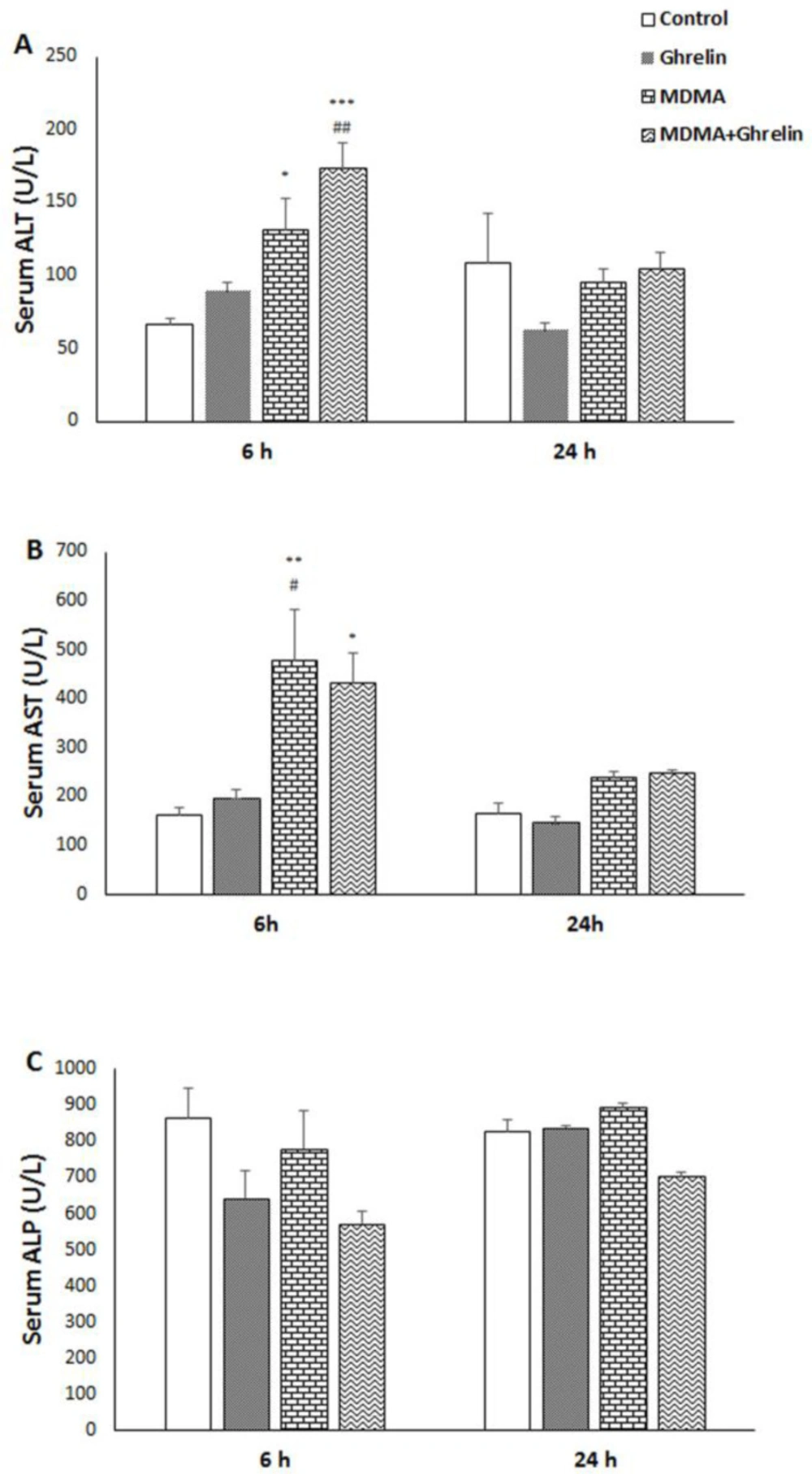
Significant transient increases in serum ALT and AST levels in MDMA and MDMA in combination with the ghrelin groups as compared to the control group were observed. Additionally, ghrelin showed more elevation of serum ALT and less elevation of serum AST versus those elevations induced by MDMA. Increased AST and ALT activities as indices of MDMA-induced cytotoxicity in rat hepatocytes have been previously demonstrated (
37). It has also been reported that MDMA-induced hepatotoxicity is associated with hepatic transaminases elevation in humans and laboratory animals (
3,
6 and
38). Transaminases rises are indicative of necrosis, since ALT and AST are two of the most reliable indicators of hepatic injury and necrosis (
39). Our results showed that co-administration of MDMA and ghrelin increases ALT level, as a specific marker for hepatic injury, and decreased AST levels as compared to the control. In fact, ghrelin could not prevent MDMA-induced hepatic injury. Since AST is also present in other tissues (
39), ghrelin-induced reduction in AST may be indicative of the beneficial effect of ghrelin on the other tissues. It should be noted that when baseline values were compared between treated and control groups, ghrelin was not able to completely normalize MDMA-induced AST elevation.
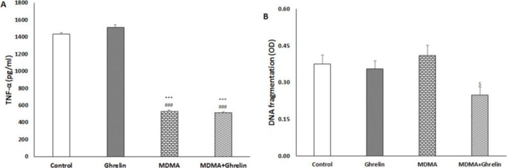
In the present study, MDMA could reduce liver TNF-α level when compared to the control group. Ghrelin did not alter MDMA-induced TNF-α reduction. MDMA inhibitory effect on TNF-α level has been reported in the presence of pre and post immune challenge, but surprisingly, in this study for the first time we saw that single dose MDMA could suppress TNF-α production in the absence of any immune challenge. Interestingly, several contradictory studies are found in the literature. For instance, Cerretani
et al. reported a strong positive expression for TNF-α production post MDMA use (
31). However, Connor et al observed no change in TNF-α levels following MDMA administration within 2 h to 24 h (
16-
19,
40). One possible mechanism refers to vagus nerve and peripheral nicotinic acetylcholine receptors that mediate MDMA- induced TNF-α inhibition (
17). Reportedly, ghrelin has a wide array of anti-inflammatory activities in endotoxin shock and sepsis models (
41,
42). In our study, MDMA seems to have strong inhibitory effects on TNF-α production that cannot be further potentiated by ghrelin administration (
Figure 2A).
The pro-inflammatory cytokine TNF-α is regarded as an important signaling molecule in initiating and coordinating a range of immune-related responses against pathogenic agents (
18). Therefore, MDMA-induced TNF-α reduction may affect host resistance to infection (
43-
47). Additionally, it has been suggested that immunosuppressive property of MDMA is at least partly responsible for MDMA-induced hepatotoxicity (
4). Since, liver is an important organ involved in detoxification and likely to be injured by ingested toxins or drugs, it is highly capable of regeneration for its survival (
48). Several studies have depicted TNF-α role in tissue regeneration (
49,
50). Thus, in our study reduced TNF-α level may indicate MDMA-induced regeneration defect following toxic injury. Anti-TNF-α agents have shown to be toxic for liver tissues with unknown underlying mechanisms (
51).
Despite
in-vitro MDMA pro-apoptotic effects on rat hepatocytes, in our
in-vivo study, we could not show MDMA pro-apoptotic activities. This finding may be due to different MDMA dose, way of administration, duration of exposure and
in-vivovs.in-vitro disparities on apoptosis induction (
1,
19 and
31). Apoptotic effect of MDMA may occur in
in-vivo setting if MDMA is used for repeated doses or for chronic purposes. In support of this claim, Warren
et al. has shown that apoptosis of the liver occurred just 72 h after MDMA administration (
52).
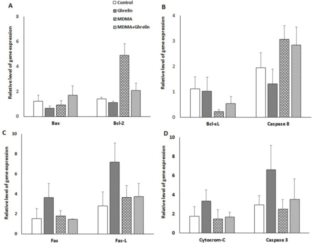
In the present study, real- time RT-PCR approach was used to assess molecular mechanisms that may be involved in the apoptotic process. Gene expressions varied between the groups; however, it failed to reach a significant level. It has been reported that an up-regulation of bcl-2 gene can prevent methamphetamine-induced apoptosis of immortalized neuron cells (
53). MDMA-treated animals demonstrated a 3.5-fold increase of Bcl-2 and a 0.8-fold decrease of Bax expression, probably indicating a defensive mechanism of the liver cells to resists MDMA-induced programed cell death. As far as we know, no study has examined the effects of MDMA on Fas/Fas-L expression in liver. The Fas ⁄Fas-L system is an important pathway for programed cell death in the hepatocytes (
54). Another interesting finding of this study was that the gene expression of Fas/Fas-L in the liver remained unchanged following MDMA expousure. Despite no significant results for MDMA-induced apoptosis, we observed a significant reduction of apoptosis in MDMA+Ghrelin as compared to MDMA group. These effects could be related in part to the fact that expression of anti-apoptotic cell regulator Bcl-xl decreased (non-significant) by 20% in the MDMA and restored (non-significant) to 50% of the control with MDMA+Ghrelin treatment. It has been demonstrated that ghrelin treatment following irradiation increase the Bcl-xl protein expression levels which was followed by apoptosis reduction (
30). Ghrelin has shown to prevent apoptosis of cardiomyocytes and endothelial cells through stimulating anti-apoptotic intracellular signalling pathways, such as activation of extracellular-signal-regulated kinase-1 and -2, protein kinase B (Akt), and tyrosine phosphorylation of intracellular proteins (
29). This may indicate that ghrelin via post-translational modifications exerts anti-apoptotic effect in liver. Previously, it has been hypothesized that apoptosis plays a major role in MDMA-induced hepatotoxicity; however, our study shows that ghrelin, being an anti-apoptotic agent fails to diminish MDMA-induced hepatic injury.
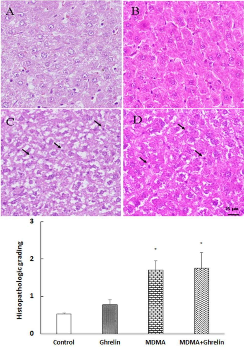
Tweny four hours follwing MDMA administration, histological analysis showed a significant increase in necrosis of hepatocytes and ghrelin administration did not improve the tissue histology (
Figure 4). Several clinical studies have suggected a more prominent role of necrosis than apoptosis in causing MDMA-induced hepatotoxity (
55). Additionaly, previous
in-vitro studies have demonstreted that MDMA triggers necrosis rather than apoptosis by increasing the temperature of the medium (
55,
56). The rise in temperature has been shown to induce oxidative stress and deplete cellular ATP which in turn may lead to hepatic necrosis (
55). Since, necrosis and apoptosis are key features of liver injury (
57), it is assumed that MDMA-induced necrosis may be attributed to transient increases in liver enzymes. However, further studies are warranted to clarify the role of necrosis in MDMA-induced liver injury. In the present study ghrelin did not show any hepatoprotective effect in MDMA-induced hepatotoxicity since, ghrelin administration did not decrease nighter liver enzymes and necrosis elevation nor TNF-α reduction.
Serum (A) ALT, (B) AST and (C) ALP levels in control animals, ghrelin (10 nmol/kg, i.p. with 3 h apart) injected animals, MDMA (20 mg/kg, i.p.) injected animals and MDMA in combination with ghrelin. Levels of enzyme were measured 6 h and 24 h after intervention. Statistical comparison between groups was performed using analysis of variance followed by the Tukey test. *p < 0.05, **p < 0.01, ***p < 0.001 as compared to control group; #p < 0.05, ##p < 0.01 as compared to ghrelin group
(A) TNF-α and (B) DNA fragmentation in control animals, ghrelin (10 nmol/kg, i.p. with 3 h apart) injected animals, MDMA (20 mg/kg, i.p.) injected animals and MDMA in combination with ghrelin at the same dosage exposure animals. Level of TNF-α was measured in liver tissue 6 h after intervention. DNA fragmentation was measured in liver tissue 24 h after intervention. Statistical comparison between groups was performed using analysis of variance followed by the Tukey test. ***p < 0.001 as compared to control group, ###p < 0.001 as compared to ghrelin group, $p < 0.05 as compared to MDMA group
The relative gene expression level of (A) Bax and Bcl-2, (B) Bcl-xL and caspase 8, (C) Fas and Fas-L, (D) cytochrome c and caspase 3 in control animals, ghrelin (10 nmol/kg, i.p. with 3 h apart) exposure animals, MDMA (20 mg/kg, i.p.) exposure animals and MDMA in combination with ghrelin. The relative expression level of each gene was determined by real-time polymerase chain reaction. Statistical comparison between groups was performed using analysis of variance followed by the Tukey test. There were no statistical differences between groups
Histological sections of the rat liver from (A) control, (B) ghrelin (10 nmol/kg, i.p. with 3 h apart) injected animals, (C) MDMA (20 mg/kg, i.p.) injected animals and (D) MDMA in combination with ghrelin at the same dosage exposure animals were stained with Hematoxylin and Eosin stain. Magnification 400x. The histological changes were scored according to the following criteria: 0, absent; 1, mild; 2, moderate; and 3, severe hepatocellular necrosis. Statistical comparison between groups was performed using analysis of variance followed by the Tukey test. *p < 0.05 as compared to control group
| mRNA | Forward (5’-3’) | Reverse (5’-3’) |
|---|
| Bax | CTCAAGGCCCTGTGCACTAAA | GGGGGTCCCGAAGTAGGAA |
| Bcl-2 | CATCGCTCTGTGGATGACTGA | CTGGGGCCATATAGTTCCACAA |
| Bcl-xl | GCAGTCAGCCAGAACCCTATC | GGGCTCAACCAGTCCATTGT |
| Cytocrom c | CTTGGGCTAGAGAGCGGGA | GTGGCACTGGGCACACTTTT |
| Caspase 3 | GAGCTTGGAACGCGAAGAAA | GAGTCCATCGACTTGCTTCCA |
| Caspase 8 | AAAGCCCAGGTTTCTGCCTA | ATCAAGCAGGCTCGAGTTGTC |
| Fas | AGGGCATGGTTTAGAAGTGG | GTGCAAGGCTCAAGGATGTC |
| Fas-L | ACTCCGTGAGTTCACCAACC | TAAGTGGGCCACACTCCTTG |
| GAPDH | TCTCTGCTCCTCCCTGTTCTA | GGTAACCAGGCGTCCGATAC |