
Introduction
Experimental
| Group | Treatment |
|---|---|
| N-C | Normal control |
| S-C | Cigarette smoking control + vehicle |
| FVT-1 | Smoke-exposed + plant extract (FVBM) (50 mg/rat/day) |
| FVT-2 | Smoke-exposed + plant extract (FVBM) (100 mg/rat/day) |
| CT | Smoke-exposed + bioactive compound (F18) (1 mg/rat/day) |
Values are mean (grams) SD from 5 rats in each group.
Significantly different from N-C at p < 0.001.
Significantly different from S-C at p < 0.001.
Significantly different from S-C at p < 0.05.
| Group* | Haemoglobin | Carbon monoxide | Nicotine |
|---|---|---|---|
| N-C | 14.86 0.764 | 3.26 0.089 | 0.998 0.012 |
| S-C | 11.60 0.515 | 5.78 0.095 | 2.16 0.033 |
| FVT-1 | 12.35 0.617 | 5.16 0.080 | 2.11 0.024 |
| FVT-2 | 14.62 0.721 | 4.07 0.091 | 1.48 0.042 |
| C-T | 14.74 0.732 | 4.76 0.078 | 1.59 0.035 |
| A-T | 13.99 0.677 | 3.97 0.043 | 1.98 0.022 |
Values are mean SD from blood of 5 rats in each group.
Significantly different from N-C at p < 0.01.
Significantly different from S-C at p < 0.001
Significantly different from S-C at p < 0.01.
Significantly different from S-C at p < 0.01.
Non-significantly different from S-C at p > 0.05.
| Group*/Ratio† | HDL-C/TC | HDL-C/LDL-C | TC/HDL-C | LDL-C/ HDL-C |
| N-C | 0.35 ± .015 | 0.70 ± 0.031 | 2.85 ± 0.105 | 1.43 ± 0.057 |
| S-C | 0.08 ± 0.003 | 0.12 ± 0.006 | 12.51 ± 0.46 | 8.38 ± 0.35 |
| FVT-1 | 0.18 ± 0.008 | 0.30 ± 0.014 | 5.55 ± 0.24 | 3.33 ± 0.186 |
| FVT-2 | 0.30 ± 0.013 (+3.8 f) a | 0.58 ± 0.026 | 3.33 ± 0.135 | 1.73 ± 0.079 |
| C-T | 0.33 ± 0.016 | 0.65 ± 0.029 | 3.04 ± 0.091 | 1.53 ± 0.061 |
| A-T | 0.33 ± 0.015 | 0.64 ± 0.028 | 3.03 ± 0.129 | 1.56 ± 0.064 |
For the calculation of ratios, data is taken from Figure 1.
Values are mean (ratio) SD from plasma of 5 rats in each group.
Significantly different from N-C at p < 0.001.
Significantly different from S-C at p < 0.001.
| Group* | CD | LOOH | MDA | Total antioxidants |
|---|---|---|---|---|
| N-C | 2.86 ± 0.141 | 1.02 ± 0.022 | 1.24 ± 0.015 | 85.47 ± 3.46 |
| S-C | 7.52 ± 0.325 | 2.88 ± 0.104 | 2.68 ± 0.125 | 44.21 ± 2.04 |
| FVT-1 | 5.28 ± 0.216 | 1.96 ± 0.083 | 1.95 ± 0.098 | 56.42 ± 2.79 |
| FVT-2 | 3.21 ± 0.110 | 1.29 ± 0.034 | 1.46 ± 0.062 | 81.53 ± 3.16 |
| C-T | 2.91 ± 0.108 | 1.08 ± 0.028 | 1.28 ± 0.042 | 79.18 ± 3.73 |
| A-T | 5.74 ± 0.209 | 2.28 ± 0.026 | 2.14 ± 0.044 | 51.79 ± 2.92 |
Values are mean (µmole/dL) ± SD from plasma of 5 rats in each group.
Significantly different from N-C at p < 0.001.
Significantly different from S-C at p < 0.001.
Significantly different from S-C at p < 0.01.
Significantly different from S-C at p < 0.05.
| Group* | MDA | MDA release(percent) |
|---|---|---|
| N-C | 5.67 ± 0.24 | 22.45 ± 0.92 |
| S-C | 12.74 ± 0.64 | 46.6 ± 2.13 |
| FVT-1 | 9.78 ± 0.46 | 34.26 ± 1.92 |
| FVT-2 | 6.82 ± 0.31 | 29.87 ± 1.12 |
| C-T | 5.82 ± 0.23 | 23.68 ± 1.02 |
| A-T | 9.54 ± 0.32 | 36.81 ± 1.24 |
Values are mean (µmole/dL) ± SD from packed erythrocytes of 5 rats in each group.
Significantly different from N-C at p < 0.001.
Significantly different from S-C at p < 0.001.
Significantly different from S-C at p < 0.01.
Significantly different from S-C at p < 0.05.
| Group | LDL oxidation+ | ||||
|---|---|---|---|---|---|
| Conjugated diene formation* | MDA content# | ||||
| Basal | Maximal | Lag Phase** | Basal | Maximal¥ | |
| N-C | 178.49 | 945.86 | 88 | 5.43±0.212 | 15.26 |
| S-C | 264.36 | 1348.56 | 68 | 8.64±0.364a | 23.65 |
| FVT-1 | 234.72 | 1264.35 | 75 | 7.43±0.202 b | 20.26 |
| FVT-2 | 192.41 | 1048.95 | 85 | 6.24±0.244 a | 18.45 |
| C-T | 187.32 | 989.35 | 87 | 5.87±0.231 a | 16.6 |
| A-T | 244.54 | 1294.28 | 73 | 7.56±0.292 c | 20.43 |
The CD values are expressed as nmole MDA equivalents/mg protein. Basal conjugated diene represent the in-vivo status of oxidized LDL.
The lag phase is defined as the interval between the intercept of the tangent of the slope of the curve with the time expressed in minutes.
The maximum in-vitro oxidation of LDL was achieved after 12 h of incubation with CuSo4 in each group,
Percent increase with respect to basal value in N-C,
Percent decrease with respect to basal value in S-C,
Percent decrease with respect to lag phase value in N-C,
Percent increase with respect to lag phase value in S-C,
Percent increase with respect to maximal value in N-C,
Percent decrease with respect to maximal value in S-C, Significantly different from N-C at
P < 0.001.
Values are obtained from LDL subpopulation, isolated from plasma of 5 rats in each group.
p < 0.01, significantly different from S-C at
p < 0.05.
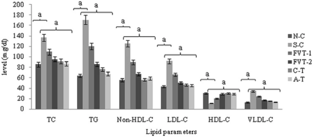
Effect of FVBM extract, F18 bioactive compound and atorvastatin on plasma triglycerides, total cholesterol, non-HDL-cholesterol, LDL-C, HDL-C and VLDL-C in cigarette smoke-exposed rats after 4 weeks of treatment.
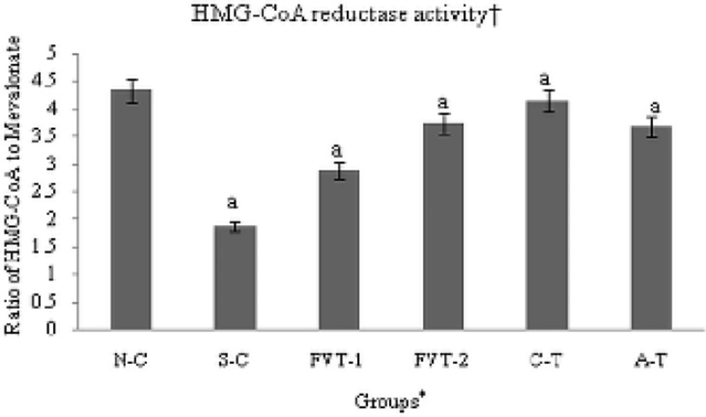
In-vivo regulation of hepatic HMG-CoA reductase activity in cigarette smoke-exposed rats treated with FVBM extract, F18 bioactive compound and atorvastatin for 4 weeks of treatment.
