Chemicals and Animals
Crocin (Cat NO-17304) was purchased from Sigma (USA). Male Wistar rats (body weight 130-160g) were purchased from the animal house of Ahvaz Jundishapur University of Medical Sciences. The animals were fed on conventional diets and had free access to tap water. They were maintained under standard conditions of humidity, temperature (22 ± 2 ºC) and light/dark cycle (12 h:12 h). The animals were deprived of food but not water 16 h before the experiment. All experiments were carried out in accordance with ethics committee of Ahvaz Jundishapur University of Medical Sciences (PRC144).
Animal grouping and surgical procedures
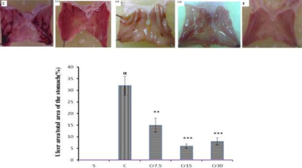
Forty male Wistar rats were randomly assigned to one of 5 groups (n=8): Sham, control (gastric ischemia-reperfusion; I/R injury) and 3 crocin-pretreated groups. Gastric I/R injury was induced according to the method of Wada (
13). Briefly, under a sodium pentobarbital anesthesia (50 mg/kg, i.p.), the rats underwent a midline laparatomy and the celiac artery was carefully isolated from its adjacent tissues. The celiac artery was then clamped by a ligature for 30 min to induce ischemia and the ligature was removed to allow reperfusion for 3 h. Sham-operated rats underwent laparatomy without inducing I/R injury. To investigate the gastroprotective effect of crocin against mucosal damage induced by I/R injury, 3 groups of animals received crocin (i.p.) at doses of 7.5, 15 or 30 mg/kg 30 min prior to I/R injury. At the end of experiment, animals were killed by cardiac exsanguination. In order to calculate the gastric mucosal lesions, the stomachs of animals were removed, opened along the greater curvature, rinsed with physiological saline and pinned out in ice-cold saline. To calculate the degree of gastric lesions, the total area of mucosal lesions were measured by Image J software. The lesion area is expressed as a percentage of the total area of the glandular stomach except for the fundus using following formula : UI(%)=[Ulcerated area/total stomach area expect fundus]×100 (
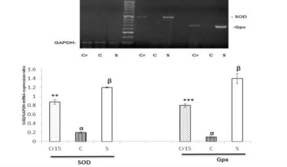
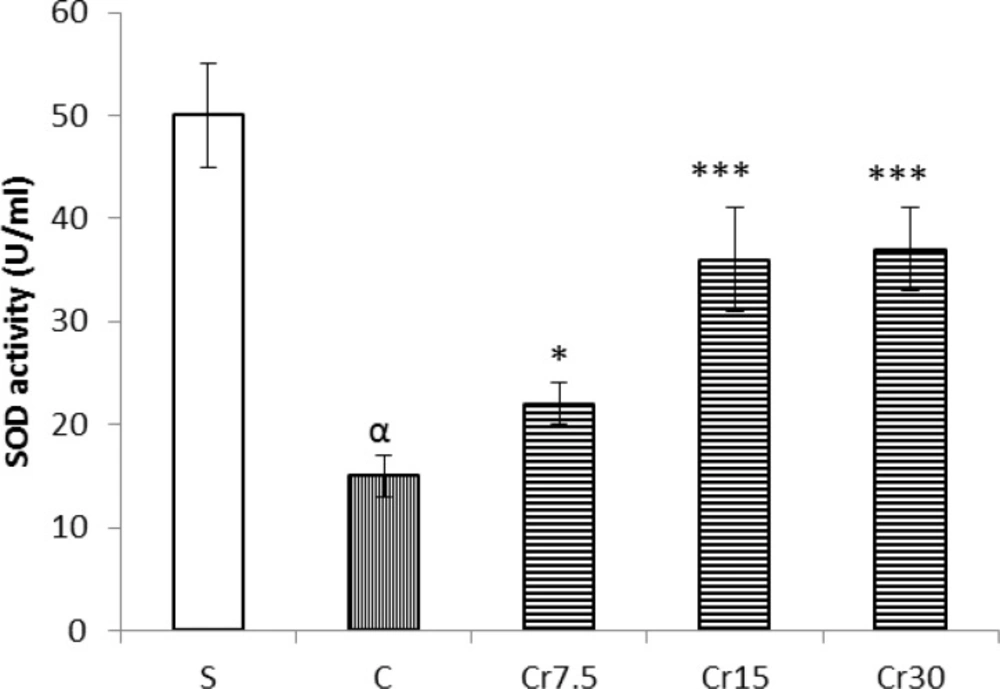
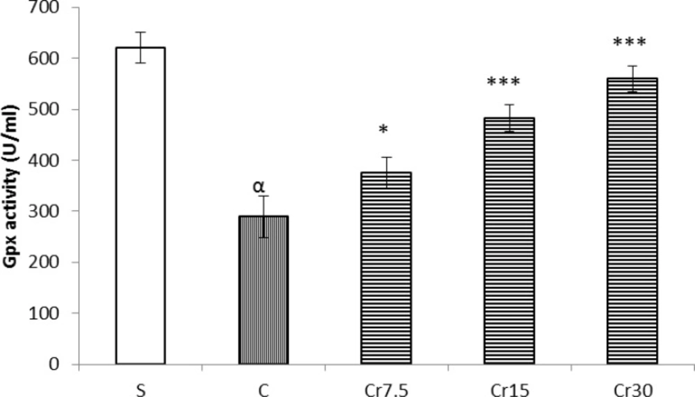
14). Immediately after taking photo of the stomachs for measurement of the surface area of gastric lesions, two samples of gastric mucosal tissue (50 mg in each) including the lesions area and the surrounding ulcer margin were quickly excised, snap-frozen and stored in liquid nitrogen for molecular analysis, determination of enzyme activity and lipid peroxidation. The macroscopic evaluation of gastric mucosal lesions showed that the optimal protective dose of crocin against I/R injury was 15 mg/kg. Therefore, the molecular analysis was carried out in animals that received the optimal dose.
RNA extraction and cDNA synthesis
The total RNA was extracted from the frozen tissue samples using RNeasy mini plus kit (Qiagen, USA). The concentration and purity of the total RNA was determined spectrophotometerically at 260 and 280 nm wavelength (Eppendorf, BioPhotometer Plus, Germany). The cDNA was synthesized from one microgram of the total RNA by using Quantitect Reverse Transcription kit (Qiagen, USA) according to the manufacturer´s instruction.
Reverse transcriptase PCR
All PCR amplifications were performed in final volume of 25 µL containing 1µg cDNA, 50 nm of specific primers, 2.5 µL of 10X PCR buffer, 1 uDNA Taq polymerase and 50 nm of dNTP. The mRNA levels of SOD, Gpx and the housekeeping gene glyceraldehyde-3-phosphate dehydrogenase (GAPDH) were measured by RT-PCR using
Master Cycle Personnel (Eppendorf AG, Hamburg, Germany). The specific primers (Bioneer, Daejoun, South Korea) used in this study are listed in
Table1. The thermal cycling conditions for the amplification of GAPDH, SOD and Gpx genes were as follows: initial denaturation at 94 °C for 5 min followed by 40 cycles of 1 min at 94 °C; annealing time 60 s and the temperature was at 53 °C for GAPDH, 54 °C for SOD and 55 °C for Gpx and the elongation time was 1 min at 72 °C. A. final elongation cycle at 72 °C for 5 min was also performed. The PCR products were analyzed on a 2% agarose gel and the density of each band was measured with Image J software. The levels of the target studied genes; SOD and Gpx; were determined by calculating the density ratio of each studied mRNA/GAPDH mRNA.
| Primer sequence |
|---|
| SOD |
| F: 5´-GCAGAAGGCAAGCGGTGAAC-3´ |
| R: 5´-TAGCAGGACAGCAGATGAGT-3´ |
| Gpx |
| F: 5´-CTCTCCGCGGTGGCACAGT-3´ |
| R: 5´-CCACCACCGGGTCGGACATAC-3´ |
| GAPDH |
| F: 5´-TGC-TGG-TGC-TGA-GTA-TGT-CGT-G-3´ |
| R: 5´-CGG-AGA-TGA-TGA-CCC-TTT-TGG-3´ |
The activity of superoxide dismutase (SOD) and glutathione peroxidase (GPx) in the homogenates of gastric mucosal tissue were measured using a commercial kit (Biocore Diagnostik Ulm GmbH, Veltlinerweg 29, Deutschland) according to the manufacturer’s instructions.
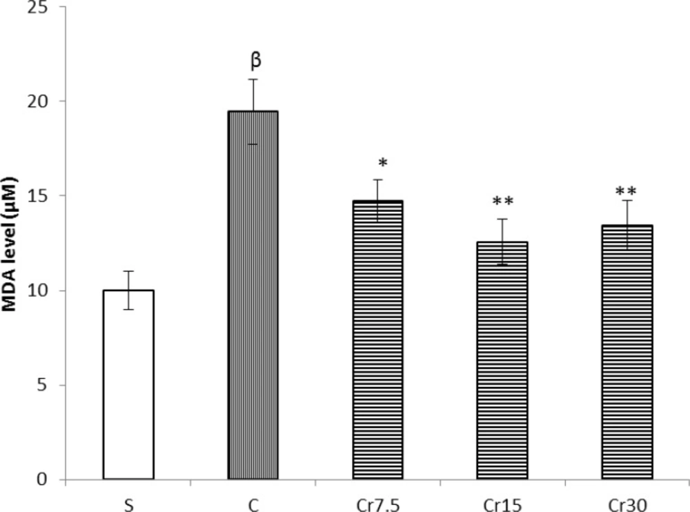
Determination of lipid peroxidation
MDA levels were measured to show the level of lipid peroxidation. Briefly, in this method MDA reacts with thiobarbituric acid (TBA) as a thiobarbituric acid reactive substance (TBARS) to generate a red-colored complex that has peak absorbance at 532 nm. Three mL phosphoric acid (1 %) and 1 mL TBA (0.6 %) was added to 0.5 mL of homogenate and the mixture was heated for 60 min in a boiling water bath. Then, twenty-five µL HCl was added to the ice-cooled mixture and vortexed. At the end, 3.5 mL of n-butanol was added the mixture and incubated for 5 min and centrifuged at 15000 rpm for 10 min to separate n-butanol phase. The supernatant was transferred to a new tube and its absorbance was measured at 532 nm. The standard curve of MDA was constructed over the concentration range of 0-40 μM (
9).
Statistical analysis
Data are shown as mean ± S.E.M. Statistical analysis was performed by one-way ANOVA and followed by post hoc Tukey’s test. Significance was set at a P<0.05 level.