Mitochondria are important organelles with major role in cellular function, ATP production, regulating energy expenditure, apoptosis signaling, and production of reactive oxygen species (
20). Recently, analysis of mitochondrial function is used in diagnosis of many diseases including the cancer, diabetes, cardiovascular disease, oxidative stress, and the age-related neurodegenerative diseases (
20). Therefore, it decided to evaluated toxicity mechanism of cigarette smoke in fetus in pregnant mothers. Epidemiological reports suggested that cigarette smoke contains many known potent carcinogens, that increases the oxidative burden of the cell, which when persisted may lead to many pathological conditions (
22,
23).
It has been reported that exposure to environmental tobacco smoke for pregnant women also results in the reduction of the fetal bi parietal diameter during weeks 20–24 of gestation , low birth weight and an increased risk of spontaneous abortion (
22,
23). Our previous studies in cigarette smoke suggested that mitochondria play a key role in the apoptosis of cells by CSE via decline in the antioxidant activity and increasing of ROS formation accompanied with the loss of mitochondrial membrane potential and decreasing of ATP level (
7,
11). These results are warranting data for public health which smoking cessation or avoidance of exposure with cigarette smoke in crowded places. Our key finding in previous study suggested internal tissue (brain and heart) have high sensitivity compared with external tissue (skin) (
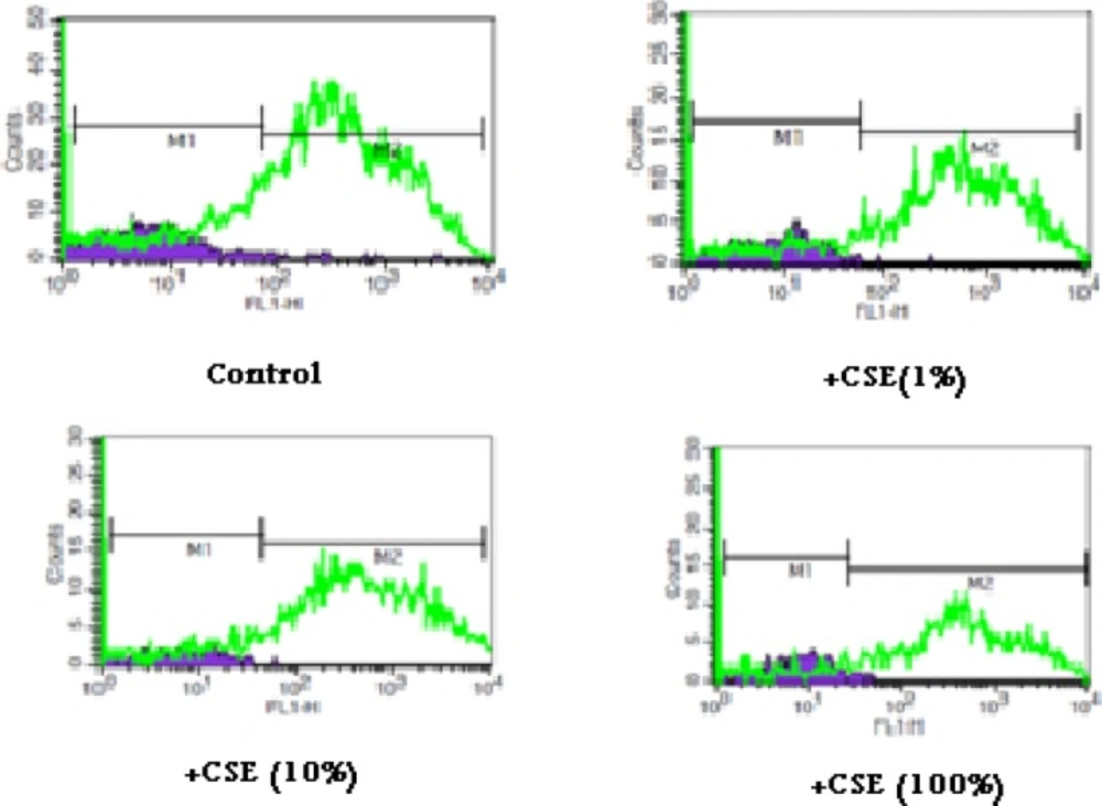
6). In present study, rapid increase in ROS formation following addition of CSE on isolated fetus mitochondria confirmed the probable involvement of mitochondrial ROS in CSE induced toxicity mechanisms (
Figure 1 and
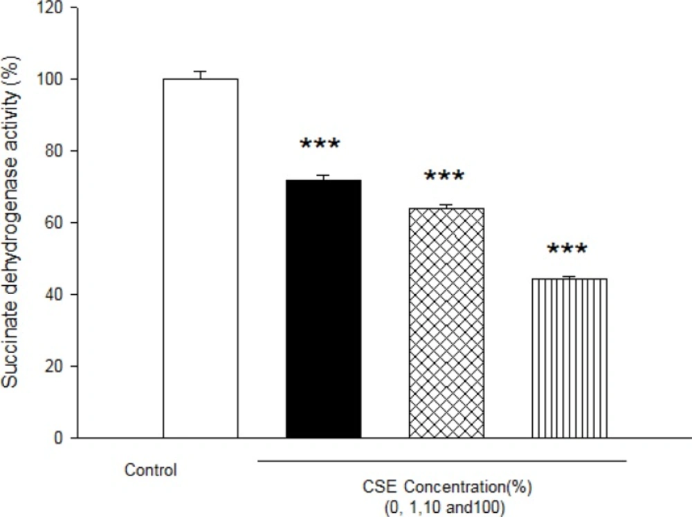
Table 2). Furthermore, significant reduction in complex II activity as indicator of decreased mitochondrial oxidative capacity (
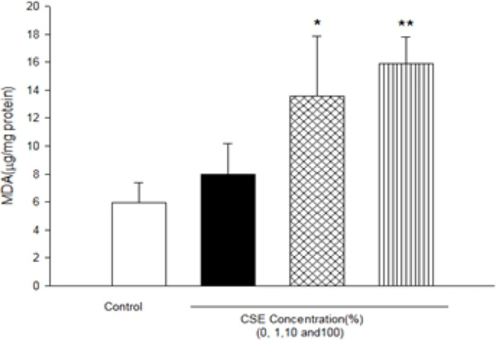
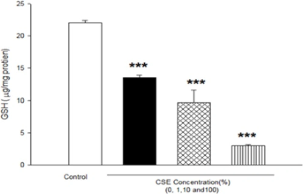
24), increasing of LPO and glutathione reduction in fetus mitochondria represented oxidative stress as one of the pathophysiological mechanisms of cigarette smoke toxicity. Our data in fetus mitochondria also corroborated with the previous findings, which showed that free radicals and ROS generated by CSE may play a key role in the instigation of membrane LPO (
6,
7). It suggested that oxidation of mitochondrial lipid membranes could result in disruption of mitochondrial electron transfer chain and consequently collapse of mitochondrial membrane potential and mitochondrial swelling which both events lead to increased ROS formation and disturbance in mitochondrial electron transfer. Besides, GSH (reduced glutathione) oxidation in MPT pores could damage to mitochondrial membrane integrity and opening of MPT pores, leading to cell death signaling (
15,
19). Therefore, decreasing of GSH level in present study caused impairing in protection of cellular antioxidant system and direct action on mitochondrial ATPase activity and cytochrome C oxidase rather than through oxygen free radical injury (
12,
15,
24 and
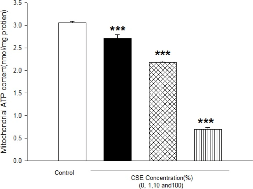
25). Finally, addition of CSE to isolated fetus mitochondria will inevitably decrease the proton motive force in mitochondria and then inhibition of ATP synthesis and inhibition of cytochrome oxidase (
25,
26). Our data also showed similar results with previous studies which related in impairment of ETC in ability of ATP synthesis. In addition, depletion in ATP levels promotes a switch from apoptotic to necrotic cell death, however, ATP depletion more than 80% leads to necrosis due to down regulation of an ATP synthase subunit (
24,
25, and
26). Mitochondrial depolarization or MPT (mitochondrial permeability transition) pores opening would be expected to cause ATP depletion due to conversion of mitochondrial ATP syntase to ATPase, followed by substantial mitochondrial swelling.
The previous studies about precise mechanism of cigarette extract toxicity suggested that oxidative stress plays a key role in vital tissues. Present investigation also confirmed impairment in electron transfer chain leading to increased ROS production, failure of oxidative phosphorylation, rapid increase in mitochondrial membrane lipid peroxidation, mitochondrial swelling and finally decline of cellular ATP which can then promote cell death signaling. Besides, it seems this is unique study to address CSE -induced oxidative stress and its consequences on isolated fetus mitochondria which can help to better understanding of CSE toxicity mechanism.